

doi: 10.1371/journal.ppat.1012203.
eCollection 2024 May.
Protecting the endothelial glycocalyx in COVID-19
Affiliations
- PMID: 38753622
- PMCID: PMC11098429
- DOI: 10.1371/journal.ppat.1012203
Item in Clipboard
Protecting the endothelial glycocalyx in COVID-19
PLoS Pathog.
.
No abstract available
Conflict of interest statement
The authors have declared that no competing interests exist.
Figures
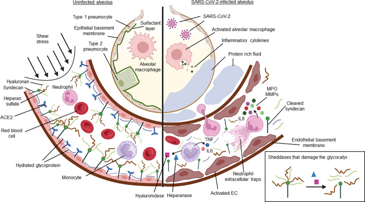
Uninfected condition (left panel), in an uninfected condition, the EG regulates the mechanotransduction of flow-induced shear stress in the vascular lumen, promoting a homeostatic environment. SARS-CoV-2 infection (right panel), SARS-CoV-2 infection activates the immune defence to eliminate the pathogen. In the alveolus, activated alveolar macrophages release inflammatory cytokines, driving a local inflammatory response. If unresolved, this inflammation may damage the alveolus. Damaged to the (EG) lining promotes neutrophil migration to the site of infection. Activated neutrophils release antimicrobials such as myeloperoxidase (MPO) and neutrophil extracellular traps, which can damage the glycocalyx. Other sheddases including matrix metalloproteinase (MMP), heparanase, and hyaluronidase, are also released and may further disrupt the glycocalyx integrity. These events damage the pulmonary vasculature, which can lead to fluid buildup in the lungs (pulmonary edema) and respiratory failure. Importantly, a damaged EG also affects mechanotransduction in the circulatory system, potentially altering endothelial cell functions in various organs. This may give rise to multiorgan failure, which is fatal in severe COVID-19 cases. Created with Biorender.
Similar articles
-
Vascular endothelial injury exacerbates coronavirus disease 2019: The role of endothelial glycocalyx protection.Microcirculation. 2021 Apr;28(3):e12654. doi: 10.1111/micc.12654. Epub 2020 Aug 30. Microcirculation. 2021. PMID: 32791568 Free PMC article. Review.
-
Pathogenesis of COVID-19 described through the lens of an undersulfated and degraded epithelial and endothelial glycocalyx.FASEB J. 2022 Jan;36(1):e22052. doi: 10.1096/fj.202101100RR. FASEB J. 2022. PMID: 34862979 Review.
-
Glycocalyx as Possible Limiting Factor in COVID-19.Front Immunol. 2021 Feb 22;12:607306. doi: 10.3389/fimmu.2021.607306. eCollection 2021. Front Immunol. 2021. PMID: 33692785 Free PMC article. Review. No abstract available.
-
Heparin prevents in vitro glycocalyx shedding induced by plasma from COVID-19 patients.Life Sci. 2021 Jul 1;276:119376. doi: 10.1016/j.lfs.2021.119376. Epub 2021 Mar 27. Life Sci. 2021. PMID: 33781826 Free PMC article.
-
Is It All About Endothelial Dysfunction and Thrombosis Formation? The Secret of COVID-19.Clin Appl Thromb Hemost. 2021 Jan-Dec;27:10760296211042940. doi: 10.1177/10760296211042940. Clin Appl Thromb Hemost. 2021. PMID: 34693754 Free PMC article. Review.
Cited by
-
Mesenchymal stromal cell secretome reduces lung injury and thrombo-inflammation induced by SARS-CoV-2 spike protein.Stem Cell Res Ther. 2025 Jul 1;16(1):324. doi: 10.1186/s13287-025-04472-6. Stem Cell Res Ther. 2025. PMID: 40597299 Free PMC article.
-
The Crucial Triad: Endothelial Glycocalyx, Oxidative Stress, and Inflammation in Cardiac Surgery-Exploring the Molecular Connections.Int J Mol Sci. 2024 Oct 10;25(20):10891. doi: 10.3390/ijms252010891. Int J Mol Sci. 2024. PMID: 39456673 Free PMC article. Review.
-
Dysregulated autoantibodies targeting AGTR1 are associated with the accumulation of COVID-19 symptoms.NPJ Syst Biol Appl. 2025 Jan 13;11(1):7. doi: 10.1038/s41540-025-00488-z. NPJ Syst Biol Appl. 2025. PMID: 39805853 Free PMC article.
References
MeSH terms
LinkOut - more resources
Full Text Sources
Medical
