. 2024 May 16;19(5):e0303296.
doi: 10.1371/journal.pone.0303296. eCollection 2024.

Sphingosine-1-phosphate promotes liver fibrosis in metabolic dysfunction-associated steatohepatitis

Affiliations

Sphingosine-1-phosphate promotes liver fibrosis in metabolic dysfunction-associated steatohepatitis

Yosuke Osawa et al. PLoS One. .

Abstract

Aim: Metabolic dysfunction-associated steatohepatitis (MASH) is one of the most prevalent liver diseases and is characterized by steatosis and the accumulation of bioactive lipids. This study aims to understand the specific lipid species responsible for the progression of liver fibrosis in MASH.

Methods: Changes in bioactive lipid levels were examined in the livers of MASH mice fed a choline-deficient diet (CDD). Additionally, sphingosine kinase (SphK)1 mRNA, which generates sphingosine 1 phosphate (S1P), was examined in the livers of patients with MASH.

Results: CDD induced MASH and liver fibrosis were accompanied by elevated levels of S1P and increased expression of SphK1 in capillarized liver sinusoidal endothelial cells (LSECs) in mice. SphK1 mRNA also increased in the livers of patients with MASH. Treatment of primary cultured mouse hepatic stellate cells (HSCs) with S1P stimulated their activation, which was mitigated by the S1P receptor (S1PR)2 inhibitor, JTE013. The inhibition of S1PR2 or its knockout in mice suppressed liver fibrosis without reducing steatosis or hepatocellular damage.

Conclusion: S1P level is increased in MASH livers and contributes to liver fibrosis via S1PR2.


Conflict of interest statement

Tatsuya Kanto has personal financial interests from Gilead Sciences for Lecture fees. Hideo Shindou was collaborated with ONO Pharmaceutical company, but the collaboration is not related to this manuscript. This does not alter our adherence to PLOS ONE policies on sharing data and materials.

Figures

Fig 1
Fig 1. Changes in bioactive lipids in MASH liver.
C57BL/6J male mice were fed a normal diet or CDD for 8 weeks and were euthanized. (A) S1P levels in the plasma or liver were measured by ELISA or LC/MS/MS. (B) mRNA expression of SphK1 and SphK2 in the livers was measured using quantitative real-time RT-PCR. (C) Expression of SphK1 was examined by immunohistochemistry with FITC-conjugated anti-SphK1 antibody (original magnification; ×100). The levels of ceramides and sphingosine (D), eicosanoids and PAF (E) in the livers were determined by LC/MS/MS. (F) The levels of cholesterol ester and free cholesterol in the liver were measured. (G) Plasma FFA levels were determined. Results are representative of at least 3 independent experiments. Results are shown as the means ± SD. *P < 0.05 based on a 2-tailed Student’s t-test. ND; normal diet. CDD; choline-deficient diet.
Fig 2
Fig 2. SphK1 mRNA expression in the liver of MASLD patients.
(A) The levels of mRNA expression of SphK1 in the livers from MASLD patients were measured using quantitative real-time RT-PCR and compared to those from control livers. Correlations of the SphK1 expression with the indicated values and indexes are shown. (B) Expression of SphK1 in the cirrhotic liver from MASH patients was examined by immunohistochemistry with FITC-conjugated anti-SphK1 antibody (original magnification; ×100). Results are shown as the means ± SD. *P < 0.05 based on a 2-tailed Student’s t-test. The values for Spearman’s correlation coefficient are indicated.
Fig 3
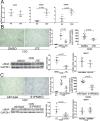
Fig 3. The effects of inhibition or knockout of S1PR2 on MASH.
Wild-type or S1PR2KO mice were fed a normal diet or CDD for 8 weeks with or without JTE013 (JTE) treatment and were euthanized. (A) mRNA expression of S1PR1, S1PR2, and S1PR3 in the livers of CDD-fed mice was measured using quantitative real-time RT-PCR and compared to ND-fed mice. (B, C) The effects of S1PR2 inhibition (B) or S1PR2KO (C) on liver fibrosis were determined. Collagen deposition was assessed by Sirius Red staining (upper left panels, original magnification: 40×; graph, upper middle panel) and hydroxyproline content (upper right panels). Liver protein extracts were separated on SDS-PAGE gels, and then immunoblotting was performed with antibodies against αSMA and GAPDH (lower left panels; graph, lower middle panel). mRNA expression of SphK1 in the livers of CDD-fed mice was measured using quantitative real-time RT-PCR and compared to control mice (lower right panel). Results are representative of at least 5 independent experiments. Results are shown as the means ± SD of data collected from at least 5 independent experiments. *P < 0.05 based on a 2-tailed Student’s t-test. ND; normal diet. CDD; choline-deficient diet.

References

    1. Tokushige K, Ikejima K, Ono M, Eguchi Y, Kamada Y, Itoh Y, et al.. Evidence-based clinical practice guidelines for nonalcoholic fatty liver disease/nonalcoholic steatohepatitis 2020. Hepatol Res. 2021; 51(10): 1013–1025. doi: 10.1111/hepr.13688 . - DOI - PubMed
    1. Bellentani S. The epidemiology of non-alcoholic fatty liver disease. Liver International. 2017; 37(S1): 81–84. doi: 10.1111/liv.13299 - DOI - PubMed
    1. Bedossa P. Pathology of non-alcoholic fatty liver disease. Liver International. 2017; 37(S1): 85–89. 10.1111/liv.13301. - DOI - PubMed
    1. Angulo P, Kleiner DE, Dam-Larsen S, Adams LA, Bjornsson ES, Charatcharoenwitthaya P, et al.. Liver Fibrosis, but No Other Histologic Features, Is Associated With Long-term Outcomes of Patients With Nonalcoholic Fatty Liver Disease. Gastroenterology. 2015; 149(2): 389–397.e310. doi: 10.1053/j.gastro.2015.04.043 . - DOI - PMC - PubMed
    1. Musso G, Cassader M, Paschetta E, Gambino R. Bioactive Lipid Species and Metabolic Pathways in Progression and Resolution of Nonalcoholic Steatohepatitis. Gastroenterology. 2018; 155(2): 282–302.e288. doi: 10.1053/j.gastro.2018.06.031 . - DOI - PubMed

MeSH terms