. 2024 Jun 21;384(6702):eadn6354.
doi: 10.1126/science.adn6354. Epub 2024 Jun 21.

AlphaFold2 structures guide prospective ligand discovery

Affiliations

AlphaFold2 structures guide prospective ligand discovery

Jiankun Lyu et al. Science. .

Abstract

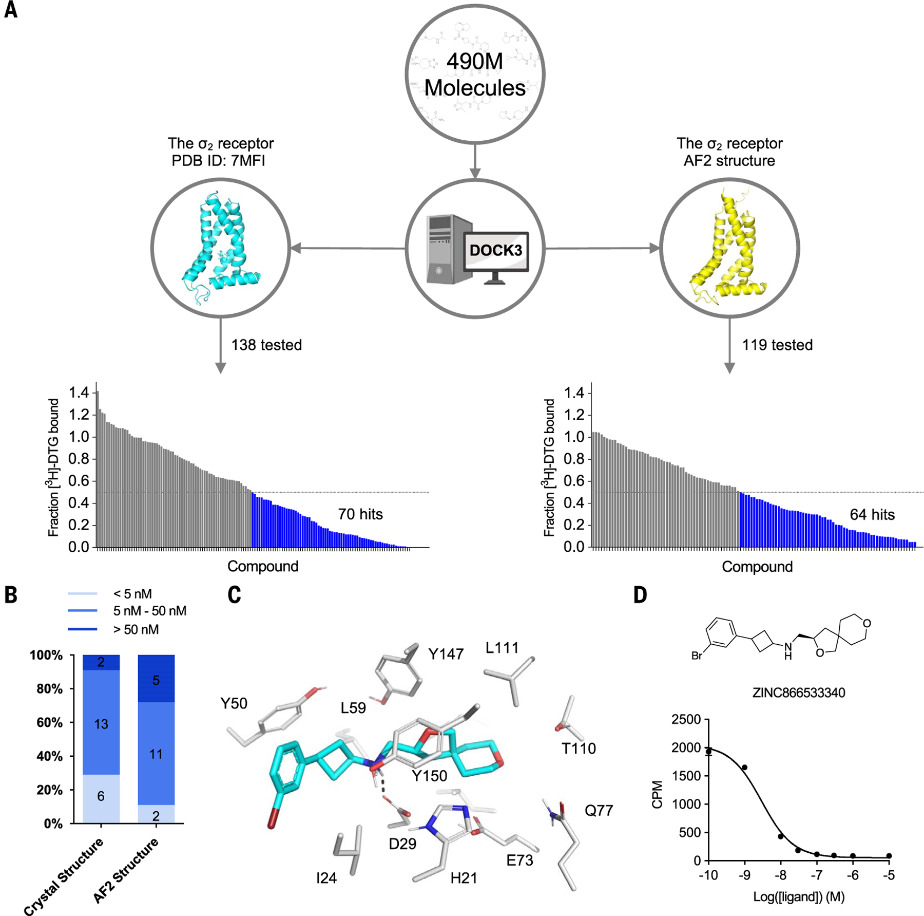
AlphaFold2 (AF2) models have had wide impact but mixed success in retrospective ligand recognition. We prospectively docked large libraries against unrefined AF2 models of the σ2 and serotonin 2A (5-HT2A) receptors, testing hundreds of new molecules and comparing results with those obtained from docking against the experimental structures. Hit rates were high and similar for the experimental and AF2 structures, as were affinities. Success in docking against the AF2 models was achieved despite differences between orthosteric residue conformations in the AF2 models and the experimental structures. Determination of the cryo-electron microscopy structure for one of the more potent 5-HT2A ligands from the AF2 docking revealed residue accommodations that resembled the AF2 prediction. AF2 models may sample conformations that differ from experimental structures but remain low energy and relevant for ligand discovery, extending the domain of structure-based drug design.


Conflict of interest statement

Competing interests: B.K.S. is co-founder of BlueDolphin LLC, Epiodyne, and Deep Apple Therapeutics, Inc.; serves on the scientific review boards of Genentech, Schrodinger LLC, and Vilya Therapeutics; and consults for Levator Therapeutics, Hyku Therapeutics, and Great Point Ventures. J.J.I. co-founded Deep Apple Therapeutics, Inc., and BlueDolphin LLC. B.L.R is a co-founder of Epiodyne and Onsero and is on the scientific advisory boards of Onsero, Epiodyne, Levator, Escient, and Septerna. A.C.K. is a co-founder and consultant for biotechnology companies Tectonic Therapeutic and Seismic Therapeutic and for the Institute for Protein Innovation, a nonprofit research institute; A.C.K. is also a member of the board of directors of Tectonic Therapeutic. X.B.-Á. is now a senior scientist at Tectonic Therapeutics.

Figures

Fig. 1.
Fig. 1.. Structural comparisons of the AF2 predicted structure and experimental structure for the σ2 and 5-HT2A receptors.
The σ2 receptor is shown in the left column and the 5-HT2A receptor in the right column. (A and B) The experimental structure (7) (cyan) is overlaid with the AF2 predicted structure (yellow). The RMSD values were calculated based on backbone atoms. The ligand binding site residues were selected within a 5-Å distance from the ligand. In (B), two residues with large conformational differences between the AF2 and experimental structures used in docking, L229 and F234, are highlighted in orange for the 5-HT2A receptor. (C) The full-atom RMSD values of the binding site residues between the AF2 and the experimental structures. Single-letter abbreviations used in the figures for the amino acid residues are as follows: C, Cys; D, Asp; E, Glu; F, Phe; G, Gly; H, His; I, Ile; L, Leu; M, Met; N, Asn; Q, Gln; S, Ser; T, Thr; V, Val; W, Trp; and Y, Tyr.
Fig. 2.
Fig. 2.. Comparison of prospective screens against the crystal and AF2 structures of the σ2 receptor.
(A) The same 490 million molecules from ZINC20 were screened against both the crystal and AF2 structures of the σ2 receptor. From these, 138 molecules from the crystal docking campaign and 119 from the AF2 docking campaign were synthesized and tested in a radioligand displacement assay. The campaign involving the crystal structure has already been published. The data shown on the left were replotted based on the previously published dataset. Displacement of the radioligand [3H]-DTG by each tested molecule occurs at 1 μM (mean ± SEM of three technical replicates). The horizontal dashed lines indicate 50% radioligand displacement. Bars below the dashed lines represent confirmed binders, which are colored in blue. (B) The distribution of binding affinity levels among the hits from both the AF2 and crystal structure screens. We measured competition binding curves for the top 21 docking hits from the crystal structure screen and the top 18 hits from the AF2 structure screen. These hits are categorized into three affinity ranges: <5 nM, 5 to 50 nM, and >50 nM. (C) The docked poses of the best binder from the screen against the AF2 model. (D) The competition binding curve of the best binder from (C) against the σ2 receptor. The data are represented as mean ± SEM from three technical replicates. Mean values and standard error are available in data S1.
Fig. 3.
Fig. 3.. Comparison of prospective screens against the cryo-EM and AF2 structures of the 5-HT2A receptor.
(A) The same set of 1.6 billion molecules from ZINC22 were docked against the cryo-EM and AF2 structures of the 5-HT2A receptor. Of these, 223 molecules were prioritized from the cryo-EM docking campaign (left) and 161 from the AF2 docking campaign (right). (B) Displacement of the radioligand [3H]-LSD by each molecule at 10 μM (mean ± SEM of three independent replicates). The horizontal dashed lines indicate 50 and 90% radioligand displacement, respectively. (C) The Ca2+ mobilization functional assay in agonist mode. Each compound was tested at a concentration of 3 μM. the horizontal dashed lines indicate agonism equivalent to 10% 5-HT activity. Data are presented as mean ± SEM from three biological replicates. (D) The Ca2+ mobilization functional assay in antagonist mode. Each compound was tested at a concentration of 3 μM. The dashed lines indicate antagonism equivalent to 20% clozapine activity. Data are presented as mean ± SEM from three biological replicates. Mean values and standard error are available in data S2.
Fig. 4.
Fig. 4.. Dose-response curves of the top agonists against the 5-HT2A, 5-HT2B, and 5-HT2C receptors.
Top agonists from both docking campaigns were tested at the 5-HT2A (blue), 5-HT2B (red), and 5-HT2C (green) receptors. (A) Data from functional assays measuring calcium mobilization (top) and Gαq protein dissociation or β-arrestin2 recruitment (bottom) for the top four agonists from the cryo-EM docking campaign. Fit parameters for each compound can be found in table S5. (B) Data from functional assays measuring calcium mobilization (top) and Gαq protein dissociation or β-arrestin2 recruitment (bottom) for the top five agonists from the AF2 docking campaign. For (A) and (B), the chemical structure of each compound is displayed below its respective dose-response curve. Data are presented as mean ± SEM from three biological replicates.
Fig. 5.
Fig. 5.. Structural characterization of the 5-HT2A receptor in complex with lisuride and Z7757.
(A and B) Maps of lisuride (A) and Z7757 (B) active-state heterotrimers. Models of the compounds built into the electron density are shown. (C) Overlay of the lisuride and Z7757 structures, which superpose to 0.78-Å Cα-RMSD. (D and E) Interactions between lisuride and the receptor (D) and between Z7757 and the receptor (E). (F) Overlay of Z7757 and lisuride in the orthosteric pocket. (G) Comparison of the experimental Z7757 structure and the predicted structure from the AF2 docking screen. (H) Predicted interactions from the docked pose of Z7757 in the AF2 structure. (I) Aligned lisuride cryo-EM structure, AF2-model, Z7757 cryo-EM structure, and the lisuride crystal structure (PDB ID 7WC7) highlighting residues that showed the biggest difference between the AF2 model and lisuride cryo-EM structures and that were used in the docking studies. In (D), (E), and (H), dashed lines indicate potential modeled interactions between the ligand and the receptor.

Update of

Comment in

  • Assessing accuracy of AlphaFold2.
    Crunkhorn S. Crunkhorn S. Nat Rev Drug Discov. 2024 Jul;23(7):499. doi: 10.1038/d41573-024-00090-8. Nat Rev Drug Discov. 2024. PMID: 38822115 No abstract available.

References

    1. Sadybekov AV, Katritch V, Computational approaches streamlining drug discovery. Nature 616, 673–685 (2023). doi: 10.1038/s41586-023-05905-z; - DOI - PubMed
    1. Wang Y et al. , Structures of the entire human opioid receptor family. Cell 186, 413–427.e17 (2023). doi: 10.1016/j.cell.2022.12.026; - DOI - PubMed
    1. Xu P et al. , Structural insights into the lipid and ligand regulation of serotonin receptors. Nature 592, 469–473 (2021). doi: 10.1038/s41586-021-03376-8; - DOI - PubMed
    1. Lyu J et al. , Ultra-large library docking for discovering new chemotypes. Nature 566, 224–229 (2019). doi: 10.1038/s41586-019-0917-9; - DOI - PMC - PubMed
    1. Stein RM et al. , Virtual discovery of melatonin receptor ligands to modulate circadian rhythms. Nature 579, 609–614 (2020). doi: 10.1038/s41586-020-2027-0; - DOI - PMC - PubMed

Publication types

MeSH terms

LinkOut - more resources