. 2024 May 16;19(5):e0292152.
doi: 10.1371/journal.pone.0292152. eCollection 2024.

Leishmania Ribosomal Protein (RP) paralogous genes compensate each other's expression maintaining protein native levels

Affiliations

Leishmania Ribosomal Protein (RP) paralogous genes compensate each other's expression maintaining protein native levels

Francisca S Borges et al. PLoS One. .

Abstract

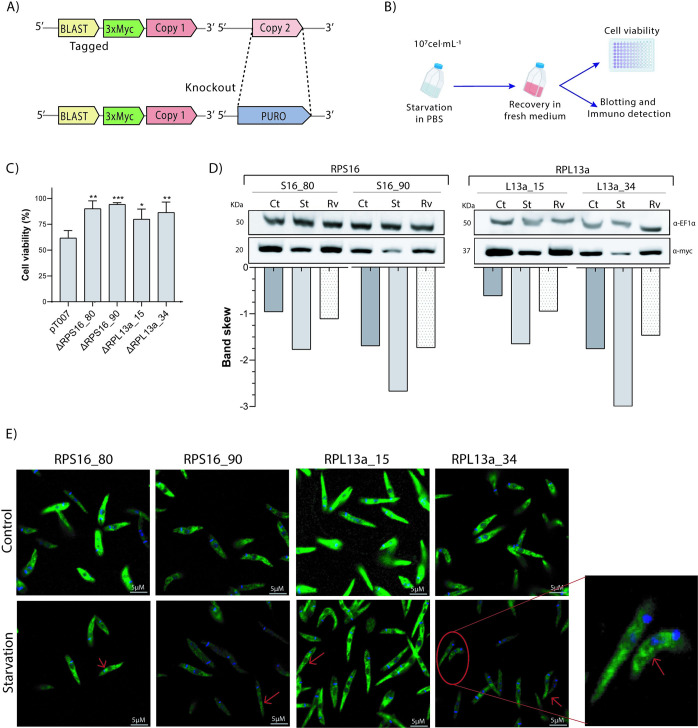
In the protozoan parasite Leishmania, most genes encoding for ribosomal proteins (RPs) are present as two or more copies in the genome. However, their untranslated regions (UTRs) are predominantly divergent and might be associated with a distinct regulation of the expression of paralogous genes. Herein, we investigated the expression profiles of two RPs (S16 and L13a) encoded by duplicated genes in Leishmania major. The genes encoding for the S16 protein possess identical coding sequences (CDSs) and divergent UTRs, whereas the CDSs of L13a diverge by two amino acids and by their UTRs. Using CRISPR/Cas9 genome editing, we generated knockout (Δ) and endogenously tagged transfectants for each paralog of L13a and S16 genes. Combining tagged and Δ cell lines we found evidence of differential expression of both RPS16 and RPL13a isoforms throughout parasite development, with one isoform consistently more abundant than its respective copy. In addition, compensatory expression was observed for each paralog upon deletion of the corresponding isoform, suggesting functional conservation between these proteins. This differential expression pattern relates to post-translational processes, given compensation occurs at the level of the protein, with no alterations detected at transcript level. Ribosomal profiles for RPL13a indicate a standard behavior for these paralogues suggestive of interaction with heavy RNA-protein complexes, as already reported for other RPs in trypanosomatids. We identified paralog-specific bound to their 3'UTRs which may be influential in regulating paralog expression. In support, we identified conserved cis-elements within the 3'UTRs of RPS16 and RPL13a; cis-elements exclusive to the UTR of the more abundant paralog or to the less abundant ones were identified.


Conflict of interest statement

The authors have declared that no competing interests exist.

Figures

Fig 1
Fig 1. Paralog genes and proteins alignments.
(A) 40S ribosomal protein S16 genes are found in tandem on the minus strand of chromosome 26 and (B) 60S ribosomal protein L13a genes are found on distinct chromosomes (15 and 34) and in opposite directions. (C) RPS16 and (D) RPL13a gene alignments, highlight the divergences in the UTR sequences. (E) RPL13a amino acid sequence alignment showing the two amino acid substitutions (*).
Fig 2
Fig 2. Protein levels and subcellular localization of duplicated S16 and L13a ribosomal proteins.
(A) Strategy for 3xmyc tag insertion in only one of the copies of the RP genes using CRISPR/Cas9. (B) Procyclic promastigotes were cultivated until the log phase from which metacyclic forms were purified by Ficoll gradient (see methods). Their morphological differences were confirmed by scanning electron microscopy. (C) Western blots probed with α-myc show that, for both RPs, the levels of protein for one paralog are consistently reduced in comparison to the other, and moreover, abundance is further reduced in metacyclic compared to procyclic promastigotes. (D) Immunofluorescence analysis confirms the cytoplasmic localization for all RP (green) proteins in procyclic promastigotes (nucleus [N] and kinetoplast [K] are indicated and stained in blue using Hoechst).
Fig 3
Fig 3. Identification of proteins interacting with the 3’UTRs of the RPS16 and RPL13a paralogs mRNA.
(A) For the pulldown assays, fragments from the 3’UTRs of each gene were amplified and cloned into PUC-54-4xS1m; the position of the primers and lengths of the fragments are indicated. (B) Gene ontology analysis of proteins identified in vitro interacting with individual 3’UTR of each RPS16 and RPL13a paralog genes (shades of blue), and proteins interacting with both paralogs (red). (C) Specific and shared proteins for each duplicated gene were identified, with total of 24 and 18 for RPS16_80 and 90, respectively. For RPL13a paralogues, similar numbers of specific proteins were obtained: 30 and 31 proteins for RPL13a_15 and 34, respectively. (D) All 3’UTRs of ribosomal protein duplicated genes bound to proteins involved in ribosome pathways, when considered the five most relevant p-values (D). All the analyses were based on the results from three independent assays.
Fig 4
Fig 4. Nutritional stress response and expression levels of RPs under starvation.
(A) After tagging one of the paralogues, the other one was deleted using CRISPR/Cas9. (B) Procyclic promastigotes in exponential growth were exposed to total starvation in PBS for 4h at 27°C, followed by viability assays, blotting and immunofluorescence analysis. (C) Cell viability was quantified by MTT assay at 24h post-starvation, to examine the recovery capacity of the mutant cells compared to parental line (pT007). (D) Expression levels of RPs were lower under starvation (St), particularly for RPS16_90 and RPL13a_34, with general recovery of parental levels after 24h of recovery in fresh medium (Rv) in comparison to the non-starved cells (control–Ct)–detection by western blotting with α-myc. (E) Immunofluorescence revealed no significant difference between the RPs distribution (green) under stress, but a subtle accumulation can be observed by increased signal in some regions of the cytoplasm (arrows) after 4h of total starvation (D).
Fig 5
Fig 5. Knockout of RPL13a paralogs decreases 60S, 80S and polysomes.
(A) RPL13a Δ mutants and the parental cell lines were subjected to sucrose gradient fractionation and polysomal profile determination, where FP: free polysomes fraction, PP: pre-polysomes, LP: light polysomes and HP: heavy polysomes. 254nm absorbance values were set arbitrarily and the relative intensity of the 80S and 40S peaks are shown in the dashed lines. Accumulation of both RPL13a isoforms in light polysomes fraction was determined by western blotting after cycloheximide treatment. (B) Additionally, both RPL13a isoforms were found in light polysome fraction (LP), even after ribosome dissociation by puromycin treatment. Experiments were performed in biological duplicate and the polysome profiles correspond to the average of each sample.
Fig 6
Fig 6. Compensatory expression of RP paralogs in knockout cells.
(A) Expression levels in procyclic and metacyclic promastigotes were analyzed by western blotting using α-myc. The presence and absence (Δ) of the correspond RPS16 and RPL13a paralogues are indicated by + and -, respectively. Band intensity was quantified in ImageJ software and the results were plotted in the corresponding graph with the increment of band color indicated. (B) RT-qPCR was used to quantify the relative expression of RPL13a transcripts in the parental (WT) and Δ promastigotes. Expression of the gene of interest was normalized to the G6PDH housekeeping gene.

Similar articles

Cited by

References

    1. Peña C, Hurt E, Panse VG. Eukaryotic ribosome assembly, transport and quality control. Nat Struct Mol Biol. 2017;24(9):689–99. doi: 10.1038/nsmb.3454 - DOI - PubMed
    1. Komili S, Farny NG, Roth FP, Silver PA. Functional Specificity among Ribosomal Proteins Regulates Gene Expression. Cell. 2007;131(3):557–71. doi: 10.1016/j.cell.2007.08.037 - DOI - PMC - PubMed
    1. Soto M, Requena JM, Garcia M, Gomez LC, Navarrete I, Alonso C. Genomic organization and expression of two independent gene arrays coding for two antigenic acidic ribosomal proteins of Leishmania. J Biol Chem. 1993;268(29):21835–43. - PubMed
    1. Dean S, Sunter J, Wheeler RJ, Hodkinson I, Gluenz E, Gull K. A toolkit enabling efficient, scalable and reproducible gene tagging in trypanosomatids. Open Biol. 2015;5(1). doi: 10.1098/rsob.140197 - DOI - PMC - PubMed
    1. Sim EU-H, Er C-M. Extra-Ribosomal Functions of the Ribosomal Protein, RPS3 as Predicted by In Silico Analysis. Borneo J Resour Sci Technol. 1970;4(2):62–9.

MeSH terms