

Updated understanding of the protein-DNA recognition code used by C2H2 zinc finger proteins
- PMID: 38754172
- PMCID: PMC12264921
- DOI: 10.1016/j.sbi.2024.102836
Updated understanding of the protein-DNA recognition code used by C2H2 zinc finger proteins
Abstract
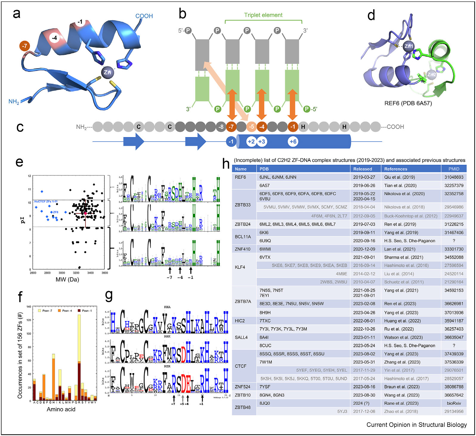
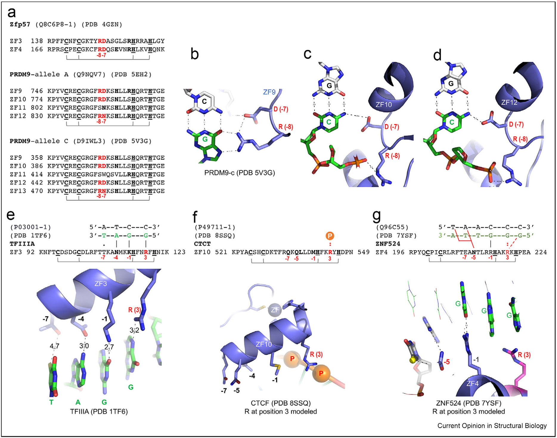
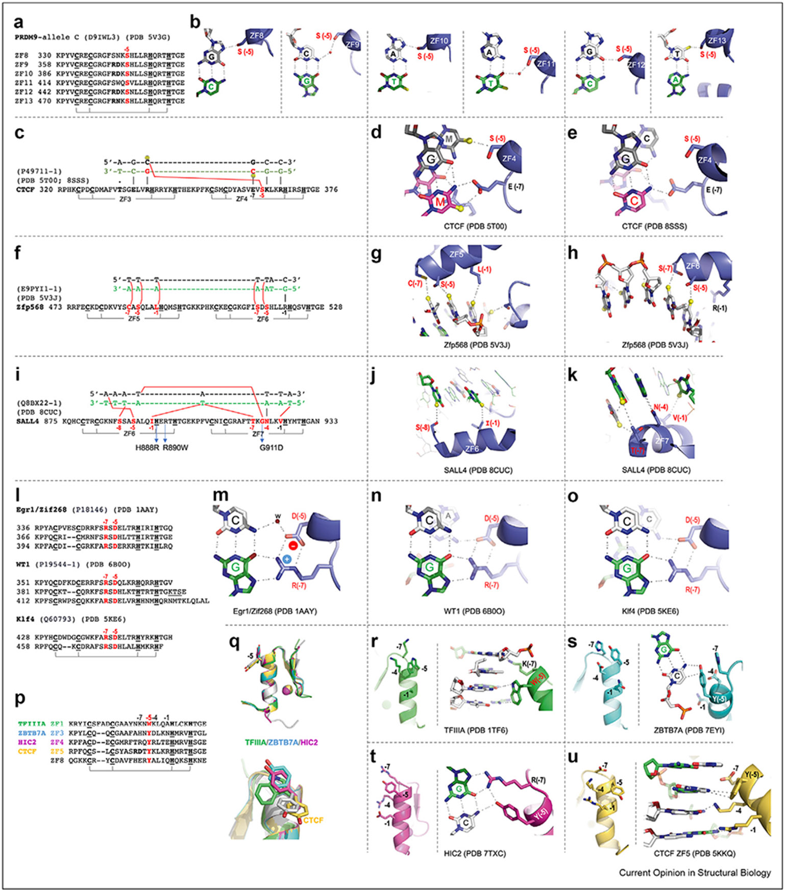
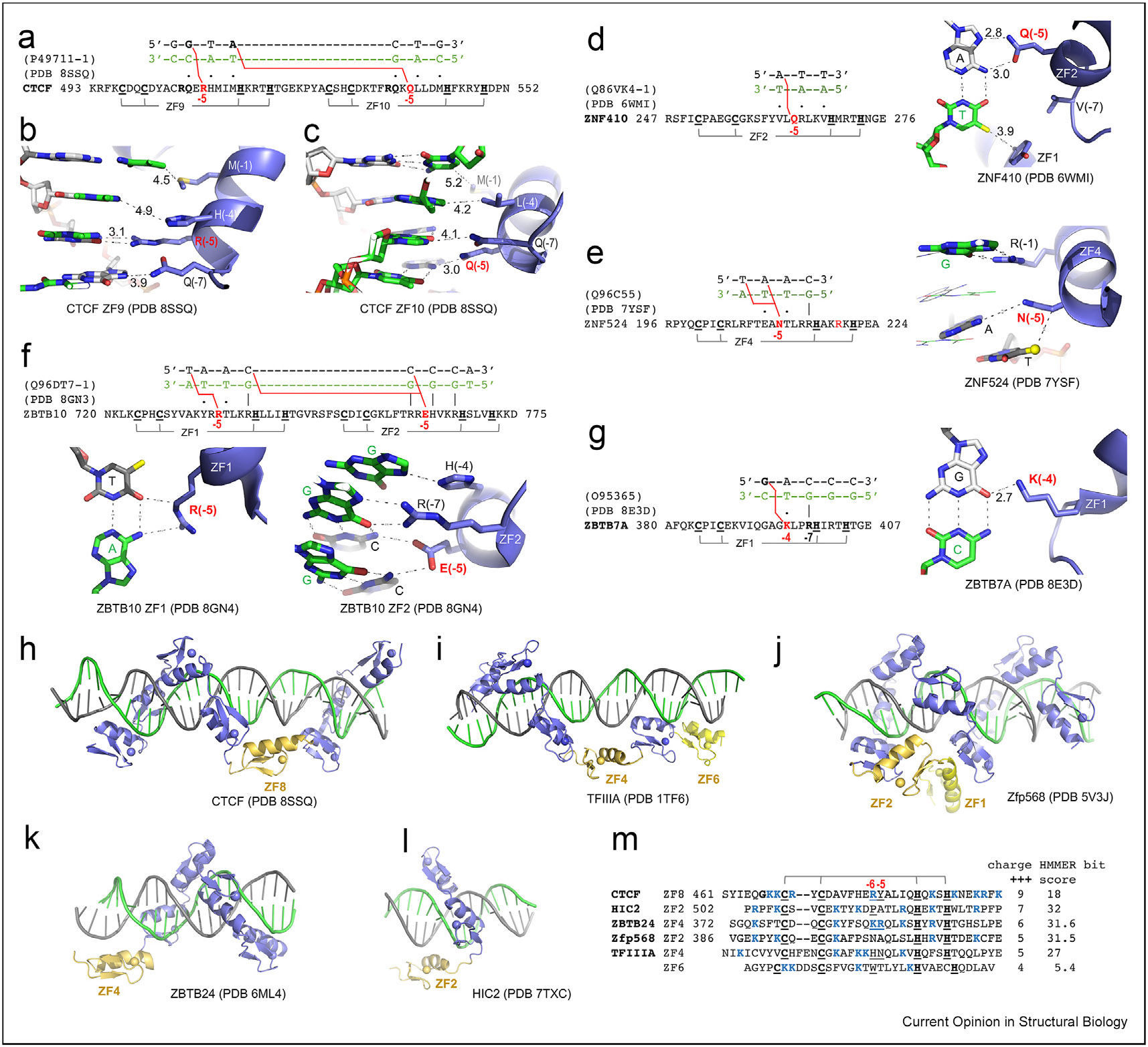
C2H2 zinc-finger (ZF) proteins form the largest family of DNA-binding transcription factors coded by mammalian genomes. In a typical DNA-binding ZF module, there are twelve residues (numbered from -1 to -12) between the last zinc-coordinating cysteine and the first zinc-coordinating histidine. The established C2H2-ZF "recognition code" suggests that residues at positions -1, -4, and -7 recognize the 5', central, and 3' bases of a DNA base-pair triplet, respectively. Structural studies have highlighted that additional residues at positions -5 and -8 also play roles in specific DNA recognition. The presence of bulky and either charged or polar residues at these five positions determines specificity for given DNA bases: guanine is recognized by arginine, lysine, or histidine; adenine by asparagine or glutamine; thymine or 5-methylcytosine by glutamate; and unmodified cytosine by aspartate. This review discusses recent structural characterizations of C2H2-ZFs that add to our understanding of the principles underlying the C2H2-ZF recognition code.
Keywords: C2H2 zinc fingers; DNA sequence-specific recognition; protein-DNA interactions; transcription factors.
Copyright © 2024 The Author(s). Published by Elsevier Ltd.. All rights reserved.
Conflict of interest statement
Declaration of competing interest The authors declare that they have no known competing financial interests or personal relationships that could have appeared to influence the work reported in this paper.
Figures
Similar articles
-
DNA-binding proteins from MBD through ZF to BEN: recognition of cytosine methylation status by one arginine with two conformations.Nucleic Acids Res. 2024 Oct 28;52(19):11442-11454. doi: 10.1093/nar/gkae832. Nucleic Acids Res. 2024. PMID: 39329271 Free PMC article.
-
Solution structure of the Z0 domain from transcription repressor BCL11A sheds light on the sequence properties of protein-binding zinc fingers.Protein Sci. 2025 Apr;34(4):e70097. doi: 10.1002/pro.70097. Protein Sci. 2025. PMID: 40099876
-
DNA-binding affinity and specificity determine the phenotypic diversity in BCL11B-related disorders.Am J Hum Genet. 2025 Feb 6;112(2):394-413. doi: 10.1016/j.ajhg.2024.12.012. Epub 2025 Jan 10. Am J Hum Genet. 2025. PMID: 39798569 Free PMC article.
-
Systemic pharmacological treatments for chronic plaque psoriasis: a network meta-analysis.Cochrane Database Syst Rev. 2021 Apr 19;4(4):CD011535. doi: 10.1002/14651858.CD011535.pub4. Cochrane Database Syst Rev. 2021. Update in: Cochrane Database Syst Rev. 2022 May 23;5:CD011535. doi: 10.1002/14651858.CD011535.pub5. PMID: 33871055 Free PMC article. Updated.
-
The effect of sample site and collection procedure on identification of SARS-CoV-2 infection.Cochrane Database Syst Rev. 2024 Dec 16;12(12):CD014780. doi: 10.1002/14651858.CD014780. Cochrane Database Syst Rev. 2024. PMID: 39679851 Free PMC article.
Cited by
-
Conformational dynamics in specialized C2H2 zinc finger domains enable zinc-responsive gene repression in S. pombe.Protein Sci. 2025 Feb;34(2):e70044. doi: 10.1002/pro.70044. Protein Sci. 2025. PMID: 39865413 Free PMC article.
-
C(P)XCG Proteins of Haloferax volcanii with Predicted Zinc Finger Domains: The Majority Bind Zinc, but Several Do Not.Int J Mol Sci. 2024 Jun 28;25(13):7166. doi: 10.3390/ijms25137166. Int J Mol Sci. 2024. PMID: 39000272 Free PMC article.
-
Structural insights into a highly flexible zinc finger module unravel INSM1 function in transcription regulation.Nat Commun. 2025 Mar 4;16(1):2162. doi: 10.1038/s41467-025-57478-2. Nat Commun. 2025. PMID: 40038295 Free PMC article.
-
Novel Compound Heterozygous Variants in ZNF526 Causing Dentici-Novelli Neurodevelopmental Syndrome: A Case Report and Literature Review.Mol Genet Genomic Med. 2025 Apr;13(4):e70089. doi: 10.1002/mgg3.70089. Mol Genet Genomic Med. 2025. PMID: 40197775 Free PMC article. Review.
-
"Disguise strategy" to bacteria: A multifunctional hydrogel with bacteria-targeting and photothermal conversion properties for the repair of infectious bone defects.Bioact Mater. 2025 Feb 12;47:343-360. doi: 10.1016/j.bioactmat.2025.02.002. eCollection 2025 May. Bioact Mater. 2025. PMID: 40026823 Free PMC article.
References
-
- Boumpas P, Merabet S, Carnesecchi J: Integrating transcription and splicing into cell fate: transcription factors on the block. Wiley Interdiscip Rev RNA 2023, 14, e1752. - PubMed
-
- Hecker M, Wagner AH: Transcription factor decoy technology: a therapeutic update. Biochem Pharmacol 2017, 144:29–34. - PubMed
-
- Radaeva M, Ton AT, Hsing M, Ban F, Cherkasov A: Drugging the ‘undruggable’. Therapeutic targeting of protein-DNA interactions with the use of computer-aided drug discovery methods. Drug Discov Today 2021, 26:2660–2679. - PubMed
Publication types
MeSH terms
Substances
Grants and funding
LinkOut - more resources
Full Text Sources
