Review
. 2024 May 15;112(10):1595-1610.
doi: 10.1016/j.neuron.2024.04.027.

Toward an interventional science of recovery after coma

Affiliations
Review

Toward an interventional science of recovery after coma

Nicholas D Schiff. Neuron. .

Abstract

Recovery of consciousness after coma remains one of the most challenging areas for accurate diagnosis and effective therapeutic engagement in the clinical neurosciences. Recovery depends on preservation of neuronal integrity and evolving changes in network function that re-establish environmental responsiveness. It typically occurs in defined steps: it begins with eye opening and unresponsiveness in a vegetative state, then limited recovery of responsiveness characterizes the minimally conscious state, and this is followed by recovery of reliable communication. This review considers several points for novel interventions, for example, in persons with cognitive motor dissociation in whom a hidden cognitive reserve is revealed. Circuit mechanisms underlying restoration of behavioral responsiveness and communication are discussed. An emerging theme is the possibility to rescue latent capacities in partially damaged human networks across time. These opportunities should be exploited for therapeutic engagement to achieve individualized solutions for restoration of communication and environmental interaction across varying levels of recovery.


Conflict of interest statement

Declaration of interests N.D.S. is a listed inventor on a patent application WO2023/043786 describing detailed methods of integrating magnetic resonance imaging, biophysical modeling, and electrophysiological methods for localization and placement of DBS electrodes in the CL/DTTm of the human thalamus as described in the paper cited in the review, Schiff et al.(21) N.D.S. is also a listed inventor on a related patent application WO2021/195062 and an issued US Patent 9,9592,383. All patents have been assigned by N.D.S. to Cornell University.

Figures

Figure 1:
Figure 1:. Toward an interventional science of coma recovery
Possible therapeutic entry points at varying stages of recovery of consciousness following coma induced severe brain injuries are represented on a two-dimensional axis. The horizontal axis plots the level of recovery of cognitive function (organized conceptually, but corresponding to changes in objective measurement scales, see); the vertical axis plots recovery of measured motor function. The functional equivalence of absence of cognition in both coma and vegetative state (VS) is reflected in their placement to the left of the vertical interrupted blue line. No behavioral evidence of consciousness is present in either coma or vegetative state; VS differs from coma by presence of intermittent eyes open periods that typically do not reflect electrographic sleep cycles. Guidelines for nomenclature differ and VS is substituted by term “unresponsive wakeful state, UWS” or “VS/UWS” as will be used here (see,). A dark grey oval between coma-VS/UWS and minimally conscious state (MCS) indicates a transition zone where fragments of behavior untied to sensory stimuli may be observed prior to the unequivocal but potentially intermittent behavioral evidence of consciousness demonstrated by MCS patients. MCS spans a wide range of expressed behaviors from non-reflexive movements such as visual tracking to inconsistent gestural communications; recovery of consistent accurate communication of situationally contingent information or goal-directed behaviors marks the emergence from MCS above the interrupted green line into the range of the confusional state (CS) in which patients are disoriented and exhibit a limited range of cognitive function]. CS patients cannot be formally tested using standard neuropsychometric measures. To the extreme right of the figure, Locked-in state (LIS) designates a condition that is typically associated with brainstem strokes in which normal conscious awareness and cognitive function are present but severe motor impairment is limits communication channels typically to vertical eye movements. The green bracket with question marks spanning the region corresponding to the subset of MCS patients who show evidence of language comprehension, also known as MCS+, see, and fully intact cognition indicates the range of possible cognitive function in the condition cognitive motor dissociation (CMD). CMD patients show behavioral examinations consistent with coma, VS, or limited non-reflexive behaviors seen in MCS- patients, but nonetheless demonstrate command following as assessed using neuroimaging or electrophysiological techniques. As marked by the bracket, the underlying cognitive capacity present in such patients remains uncertain without a behavioral response to allow for direct assessments. CMD patients may retain a level of cognitive function across a range consistent with that observed in MCS+ to the complete locked-in state (CLIS) in which no communication is evident. Determination of level of cognitive capacity in a CMD patient requires methods allowing the patient to both initiate and respond to communication. The four potential entry points for novel therapeutics considered in this review include: 1) restoration of communication in persons with cognitive motor dissociation (CMD); 2) restoration of communication in inconsistently responsive patients in the minimally conscious state; 3) late restoration of cognition in outcomes of moderate to severely disabilty following coma; and 4) future development of possible controlled ‘protective down-regulated states in humans [9] that might allow rescuing of good outcomes in some patients with prolonged coma before withdrawal of life sustaining therapies (WLST).
Figure 2:
Figure 2:. Brain computer interfaces for potential ‘clearinghouse’ matching of patterns of injury in cognitive motor dissociation.
The complexity of structural brain injuries in CMD will likely require individualized and highly customized approaches to support chronic BCI based communication. A “clearinghouse” of CMD patients with known injury patterns and demonstration of reliable command following with proxy fMRI or EEG methods is envisioned to match with usable interfaces. Such a clearinghouse registry might be created in the future to allow the best mix and match of approaches and to ensure fail-safe back up strategies if one device were to be effective but then fail. Existing methods include: A. high performance BCI implantations in motor, speech and parietal cortex.., image from with permission. B. novel ultra-fine and large array microelectrode systems (Figure elements from,(bottom)). C. Electrode systems cannulated within the central venous system.
Figure 3:
Figure 3:. Example of BCI “readiness” in a CMD subject
A. Longitudinal structural remodeling identified by DTI in a CMD patient over 3.5 years at two points (left early, right late) of operation of a ‘one-way’ communication system (cf.) using a single downward eye-movement [48]. Diffusion tensor imaging shows increased fractional anisotropy arising within the pars opercularis and pars triangularis (Broca’s area). Fluorodeoxyglucose imaging measured across the same two time points revealed increased left frontal lobe metabolism. B. Functional magnetic resonance imaging of the same subject in response to “keep opening and closing your right hand” showing activation of the left hand motor region (unpublished data, Nicholas Schiff and Henning Voss). C. Corresponding location of placement of the BRAINGATE system in left motor hand region.
Figure 4:
Figure 4:. Mesocircuit hypothesis
A. Mesocircuit theory for recovery of anterior forebrain function with CL/DTTm DBS in msTBI (adapted from). Schematic diagram illustrating mesocircuit model for alteration of function following coma and moderate to severe brain injury and restoration of function with CL/DTTm. Left figure element: Healthy normative function of corticothalamic system. Projections of central lateral thalamic neurons to anterior forebrain mesocircuit and posterior medial complex. CL co-activates frontal-parietal corticocortical connections and modulates their feed-forward and feedback connectivity via layer-specific effects within cortical columns. CL specifically targets supragranular and infragranular cortical layers avoiding projections into the input layers; these anatomical specializations support a proposed selective role in modulation of long-range corticocortical functional connectivity. CL projections to the striatum strongly activate this structure via projections to medium spiny neurons, MSNs and act via AMPA receptors, whereas Cm-Pf afferents act via NMDA receptors, see. Middle figure element: msTBI produces widespread deafferentation of the corticothalamic system leading to loss of CL modulation of cortex and striatum. Two major consequences of this downregulation of CL output in combination with overall reduction of cerebral background activity are: 1) marked reduction in corticothalamic and corticostriatal outflow, 2) shutdown of the medium spiny neuron output from striatum to globus pallidum interna (GPi) producing increased thalamic inhibition and further reduction of thalamocortical and thalamostriatal outflow. Collectively, these changes are proposed to exert a disproportionate impact on the anterior forebrain. Right figure element: CL/DTTm DBS (red arrow) is proposed to reverse the mesocircuit level effects of reduced corticothalamic, corticostriatal, and striato-pallidal output by direct overdrive pacing of CL output via the DTTm. (image from. B. A Medtronic 3387 4 contact DBS lead implanted into the CL nucleus and dorsal tegmental tract medial fibers (image from). C. Modeled activation of CL/DTTm fibers in the same subject with moderate to severe traumatic brain injury.
Figure 5.
Figure 5.. Non-human primate models of emergence from anesthetic coma
A. Overview of the experimental results of Redinbaugh et al. (reproduced from [73]). Microstimulation of the central lateral thalamic nucleus initiated arousal during during a stable anesthetic coma produced by propofol anesthesia. A. Heading labels corresponding human states of disorders of consciousness with similar behavioral features to the experimental ‘arousal’ index used in the Redinbaugh et al. study. Left subpanel: (A and B) Changes in both FEF and LIP local field potential (LFP) power comparing stable anesthesia to state induced with CL nucleus stimulation are shown (corresponding to Figures S3I and S3J in Redinbaugh et al. (C and D) Changes in both FEF and LIP LFP coherence comparing stable anesthesia to state induced with CL nucleus stimulation are shown (corresponding to Figures 3I and 3J in). The observed changes in the power spectrum with initial transition to wakeful show no recovery of background rhythms characteristic of the monkey’s wakeful state. Right subpanel: (E and F) Differences in both FEF and LIP LFP Power comparing stable anesthesia to wakefulness are shown (corresponding to Figures S3A and S3B in, (G and H) Differences in both FEF and LIP LFP coherence comparing stable anesthesia to wakefulness are shown (corresponding to Figures 3A and 3B in. B. Dynamics of loss and recovery of consciousness with ketamine anesthesia in a non-human primate (from). Top panel shows a curve representing the probability of a successful behavioral task performance over time as loss of consciousness and recovery of task engagement without task performance occurs prior to restoration of task performance in a monkey anesthetized with ketamine. The lower panel shows the accompanying spectrogram obtained from local field potentials from secondary somatosensory cortex. An intermediate regime of recovery of LFP dynamics (right) similar to the wakeful baseline (left) is associated with the onset of task engagement without task performance prior to return of task performance.
Figure 6:
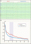
Figure 6:. EEG burst suppression in late recovery of independent function following post-cardiac arrest coma.
A. (A) Example of EEG burst suppression pattern on day 10 in a post-cardiac arrest patient with prolonged coma (from Becker et al.). B. Example of a 7 Hz peak in the power spectrum of intra-burst activity from EEG in (A) (from Forgacs et al.).
Figure 7:
Figure 7:. Isolation syndrome in incomplete rescue of neuronal cell types following post-cardiac arrest coma.
A. Time evolution of burst structure in representative comatose post–cardiac arrest patients with BSP on acute electroencephalography (EEG) in the prospective cohort studied by Forgacs et al.. Each plot represents a single patient with intra-burst spectral features over days (orange, light red, and dark red spectra). In all patients with BSP and favorable outcome, bursting frequency increased with time (right side of the inverted U shape). In all patients with BSP and unfavorable outcomes, there were either no features present during bursting, or, if any feature was present (including theta), it was present briefly and/or the frequency of bursting activity decreased with time (examples of all of these patterns are shown over the left side of the inverted U shape). At the top of the inverted U a “tipping point” is proposed over which neurons either live or die. B. Co-registration of CT scan and fluorodeoxyglucose imaging in a post-cardiac coma patient with sharp boundary of preserved neuronal tissue in the anterior forebrain (from). Red arrows indicate preservation of cerebral metabolism across anterior forebrain structures which survived treatment of cardiac arrest with therapeutic hypothermia, and posterior cortical regions which underwent widespread apoptotic cell death. The sharp separation of outcome suggests that within this patient a tipping point might have separated these two fates allowing for a future method of induced PDS to rescue more of the neurons. C. Demonstration of successful command following to spoken command to “keep opening and closing your right hand” from.

Similar articles

Cited by

References

    1. Posner JB, Saper CB, Schiff ND & Claassen J. (2019) Plum and Posner’s diagnosis and treatment of stupor and coma. 5 edn, (Oxford University Press; ).
    1. Laureys S, Schiff ND. (2012) Coma and consciousness: paradigms (re)framed by neuroimaging. Neuroimage. 61, 478–91. - PubMed
    1. Laureys S, Celesia GG, Cohadon F, Lavrijsen J, León-Carrión J, Sannita WG, Sazbon L, Schmutzhard E, von Wild KR, Zeman A, Dolce G; European Task Force on Disorders of Consciousness. (2010) Unresponsive wakefulness syndrome: a new name for the vegetative state or apallic syndrome. BMC Med. 8, 68. - PMC - PubMed
    1. Giacino JT, Katz DI, Schiff ND, Whyte J, Ashman EJ, Ashwal S, Barbano R, Hammond FM, Laureys S, Ling GSF, Nakase-Richardson R, Seel RT, Yablon S, Getchius TSD, Gronseth GS, Armstrong MJ. (2018) Practice guideline update recommendations summary: Disorders of consciousness: Report of the Guideline Development, Dissemination, and Implementation Subcommittee of the American Academy of Neurology; the American Congress of Rehabilitation Medicine; and the National Institute on Disability, Independent Living, and Rehabilitation Research. Neurology 91, 450–460. - PMC - PubMed
    1. Giacino JT, Ashwal S, Childs N, Cranford R, Jennett B, Katz DI, Kelly JP, Rosenberg JH, Whyte J, Zafonte RD, Zasler ND. (2002) The minimally conscious state: definition and diagnostic criteria. Neurology. 58, 349–53. - PubMed

MeSH terms

LinkOut - more resources