. 2024 Jun;300(6):107372.
doi: 10.1016/j.jbc.2024.107372. Epub 2024 May 15.

Cardioprotective properties of OMT-28, a synthetic analog of omega-3 epoxyeicosanoids

Affiliations

Cardioprotective properties of OMT-28, a synthetic analog of omega-3 epoxyeicosanoids

Joshua Kranrod et al. J Biol Chem. 2024 Jun.

Abstract

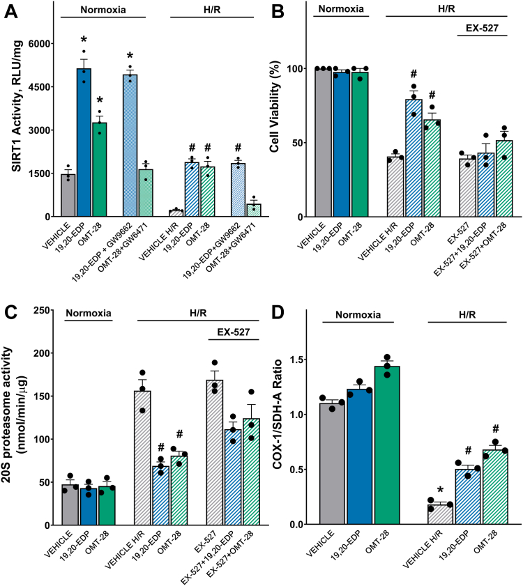
OMT-28 is a metabolically robust small molecule developed to mimic the structure and function of omega-3 epoxyeicosanoids. However, it remained unknown to what extent OMT-28 also shares the cardioprotective and anti-inflammatory properties of its natural counterparts. To address this question, we analyzed the ability of OMT-28 to ameliorate hypoxia/reoxygenation (HR)-injury and lipopolysaccharide (LPS)-induced endotoxemia in cultured cardiomyocytes. Moreover, we investigated the potential of OMT-28 to limit functional damage and inflammasome activation in isolated perfused mouse hearts subjected to ischemia/reperfusion (IR) injury. In the HR model, OMT-28 (1 μM) treatment largely preserved cell viability (about 75 versus 40% with the vehicle) and mitochondrial function as indicated by the maintenance of NAD+/NADH-, ADP/ATP-, and respiratory control ratios. Moreover, OMT-28 blocked the HR-induced production of mitochondrial reactive oxygen species. Pharmacological inhibition experiments suggested that Gαi, PI3K, PPARα, and Sirt1 are essential components of the OMT-28-mediated pro-survival pathway. Counteracting inflammatory injury of cardiomyocytes, OMT-28 (1 μM) reduced LPS-induced increases in TNFα protein (by about 85% versus vehicle) and NF-κB DNA binding (by about 70% versus vehicle). In the ex vivo model, OMT-28 improved post-IR myocardial function recovery to reach about 40% of the baseline value compared to less than 20% with the vehicle. Furthermore, OMT-28 (1 μM) limited IR-induced NLRP3 inflammasome activation similarly to a direct NLRP3 inhibitor (MCC950). Overall, this study demonstrates that OMT-28 possesses potent cardio-protective and anti-inflammatory properties supporting the hypothesis that extending the bioavailability of omega-3 epoxyeicosanoids may improve their prospects as therapeutic agents.

Keywords: 17,18-EEQ; OMT-28; analog; cardioprotection; eicosanoid; oxylipin.


Conflict of interest statement

Conflict of interest The authors declare the following financial interests/personal relationships which may be considered as potential competing interests. A. K. is an employee, R. F. and W.-H. S. are co-founders of OMEICOS Therapeutics GmbH. J. M. S. received a collaborative research grant from OMEICOS Therapeutics GmbH. All other authors declared no competing interests for this work.

Figures

Figure 1
Figure 1
OMT-28 protects HL-1 cells against HR injury.A, structure of 19,20-EDP, 17,18-EEQ and OMT-28. HL-1 cells treated with 1 μM either 17(R),18(S)-EEQ, 17(S),18(R)-EEQ, racemic 17,18-EEQ, 19,20-EDP, or OMT-28 with or without tAUCB (1 μM) and subjected to normoxia or hypoxia-reoxygenation. Colorimetric analysis of EpFA-induced changes on cell viability and mitochondrial oxidative metabolism following HR injury. B, WST-8 (CCK-8) cell viability assay. C, cell proliferation (MTT). Values represent mean ± SEM, data were obtained by analyzing responses of three independent cell preparations and using at least three technical replicas. p < 0.05 statistically significant, one-way ANOVA, Bonferroni post hoc test, ∗ versus vehicle control normoxia; # versus vehicle control HR.
Figure 2
Figure 2
OMT-28 protects NRCMs against HR injury through pleiotropic mechanisms. The cytoprotective effect of OMT-28 was reduced by various inhibitors following HR injury in neonatal rat cardiomyocytes. NRCM subjected to normoxia or hypoxia-reoxygenation were treated with vehicle, 19,20-EDP (1 μM), or OMT-28 (1 μM) with or without inhibitors. A, pan-PI3K inhibition with Wortmannin (WM, 100 nM). B, PI3Kα-selective inhibition with PI-103 (100 nM). C, Gαi inhibition with pertussis toxin (PTX, 200 ng/ml). D, PPAR inhibition with GSK3787 (1 μM), GW6471 (1 μM), or GW9662 (1 μM) to selectively inhibit isoforms β/δ, α, and γ, respectively. E, ELISA quantification of OMT-28-induced PPARα DNA-binding. F, ELISA quantification of 19,20-EDP-induced PPARγ DNA-binding. G, sKATP inhibition with HMR-1098 (10 μM). Values represent mean ± SEM, data were obtained by analyzing responses of three independent cell preparations and using at least three technical replicas, p < 0.05 statistically significant, one-way ANOVA, Bonferroni post hoc test, ∗ versus vehicle control normoxia; # versus vehicle control HR.
Figure 3
Figure 3
Cytoprotection induced by OMT-28 is dependent upon PPARα-mediated preservation of SIRT1 activity.A, SIRT1 activity in NRCMs subjected to normoxia or HR injury and incubated with vehicle, 19,20-EDP (1 μM), OMT-28 (1 μM) with and without PPAR-inhibitors: GW9662 (PPARγ antagonist, 1 μM) and GW6471 (PPARα antagonist, 1 μM). HL-1 cells subjected to normoxia or HR injury and incubated with vehicle, 19,20-EDP (1 μM), OMT-28 (1 μM) or SIRT1 inhibitor (EX-527, 10 μM) were assessed for, B, cell viability in HL-1 cells, C, 20S proteasome activity in HL-1 cells. D, ELISA quantification of the SDH-A/COX-1 ratio as a marker of mitobiogenesis in NRCMs. Values represent mean ± SEM, data were obtained by analyzing responses of three independent cell preparations and using at least three technical replicas, p < 0.05 statistically significant, one-way ANOVA, Bonferroni post hoc test, ∗ versus vehicle control normoxia; # versus vehicle control HR.
Figure 4
Figure 4
OMT-28 improves mitochondrial function, ADP/ATP-, and NAD+/NADH-ratios. Mitochondrial function was assessed in NRCMs subjected to normoxia or HR injury and incubated with vehicle, 19,20-EDP (1 μM), or OMT-28 (1 μM). A, schematic representing potential points of interaction between OMT-28, the TCA cycle and mitochondrial respiratory chain. B, complex I respiratory control ratio (RCR). C, complex II RCR. D, NAD+/NADH-ratios. E, ADP/ATP- ratio. F, NADH dehydrogenase activity. G, SDH enzymatic activity. H, COX-1 enzymatic activity. I, aconitase enzymatic activity. J, citrate synthase enzymatic activity. Values represent mean ± SEM, data were obtained by analyzing responses of three independent cell preparations and using at least three technical replicas, p < 0.05 statistically significant, one-way ANOVA, Bonferroni post hoc test, ∗ versus vehicle control normoxia; # versus vehicle control HR.
Figure 5
Figure 5
OMT-28 limits HR-induced mitochondrial ROS production in NRCMs. Representative images and quantification of mitochondrial ROS produced in NRCMs treated with vehicle, 19,20-EDP (1 μM), or OMT-28 (1 μM) before hypoxia or at the beginning of reoxygenation. A, pre-hypoxia treatment representative images. B, pre-hypoxia treatment ROS quantification. C, post-hypoxia treatment representative images. D, post-hypoxia treatment ROS quantification. Values represent mean ± SEM, data were obtained by analyzing responses of three independent cell preparations and using at least three technical replicas, p < 0.05 statistically significant, one-way ANOVA, Bonferroni post hoc test, ∗ versus vehicle control normoxia; # versus vehicle control HR. Image created with Biorender.com and published with permission.
Figure 6
Figure 6
OMT-28 limits LPS-induced cell death and injury in HL-1 cells. Assessment of the cytoprotective effect of OMT-28 following LPS injury. A, cell viability (CCK-8) was assessed in cells exposed to 24 h LPS (1 μg/ml) and treated with either vehicle, 19,20-EDP or OMT-28 (10 nM, 100 nM, and 1 μM) with or without a SIRT1 inhibitor (EX-527,1 μM). B, cell viability (CCK-8) was assessed in cells exposed for 1, 6, 12 or 24 h LPS (1 μg/ml) and treated with either vehicle, 19,20-EDP or OMT-28 (1 μM). C, cell proliferation (MTT), and D, 20S proteasome activity in cells exposed to 24 h LPS (1 μg/ml) and treated with either vehicle, 19,20-EDP (1 μM) or OMT-28 (1 μM). Values represent mean ± SEM, data were obtained by analyzing responses of three independent cell preparations and using at least three technical replicas, p < 0.05 statistically significant, one-way ANOVA, Bonferroni post hoc test, ∗ versus control conditions; # versus LPS control.
Figure 7
Figure 7
OMT-28 limits LPS-induced inflammation in cardiac cells. Analysis of several inflammatory markers expressed by cardiac cells exposed to 24 h LPS (1 μg/ml) and treated with either vehicle, 19,20-EDP or OMT-28. A, ELISA quantification of TNFα expression in HL-1 treated with either vehicle, 19,20-EDP or OMT-28 (10 nM, 100 nM, and 1 μM) following LPS insult. Analysis of inflammatory markers in NRCM. B, TNFα expression, C, MCP-1 expression, D, TGF-β expression, and E, NF-κβ DNA-binding activity. Values represent mean ± SEM, data were obtained by analyzing responses of three independent cell preparations and using at least three technical replicas, p < 0.05 statistically significant, one-way ANOVA, Bonferroni post hoc test, ∗ versus vehicle control in PBS treated cells: # versus vehicle control in LPS-treated cells.
Figure 8
Figure 8
OMT-28 enhances postischemic-reperfusion myocardial function recovery. Perfusion of hearts with OMT-28 (1 μM) or MCC950 (1 μM) resulted in improved postischemic functional recovery. A, left ventricular developed pressure (LVDP) at baseline before drug treatment (B20), during ischemia, and at 10, 20, 30, and 40 min following reperfusion (R10, R20, R30, and R40). B, rate of contraction (dP/dt max). C, rate of relaxation (dP/dt min). D, LVDP recovery at 40 min reperfusion as a percentage of baseline. E, rate of contraction at 40 min reperfusion as a percentage of baseline. F, rate of relaxation at 40 min reperfusion as a percentage of baseline. G, heart rate assessed as beats per minute (BPM) at the end of reperfusion (R40). Values represent mean ± SEM, n = 6, p < 0.05 statistically significant, one-way ANOVA, Bonferroni post hoc test, # versus IR vehicle control.
Figure 9
Figure 9
OMT-28 limits IR-induced inflammasome and autophagy responses. Perfusion of hearts with OMT-28 (1 μM) or MCC950 (1 μM) inhibited IR-induced activation of mitochondrial autophagy and the NLRP3 inflammasome. Representative immunoblots and densiometric quantification of mitochondrial autophagy-associated markers and NLRP3 inflammasome were assessed by immunoblotting. Protein expression was normalized to either VDAC (mitochondria) or GAPDH (cytosolic) loading controls. IR-induced mitochondrial expression of A, dynamin-related protein-1 (DRP-1), B, PTEN-induced kinase 1 (PINK1), C, Parkin, D, p62 and, E, microtubule-associated proteins 1A/1B light chain 3B (LC3B-II). Immunoblots were re-probed for multiple markers. The representative images for A, B, and G share the same tissue source. The representative images for C, D, and E share the same tissue source. IR-induced cytosolic expression of F, Nucleotide NLR family pyrin domain containing 3 (NLRP3) and G, thioredoxin interacting protein (TXNIP). H, cardiac caspase-1 enzymatic activity assessed in the cytosolic fraction following IR injury. I, cardiac quantification of interleukin-1β levels (IL-1B) expression following IR injury. Values represent mean ± SEM, n = 3 to 6, p < 0.05 statistically significant, one-way ANOVA, Bonferroni post hoc test, ∗ versus aerobic control (AERO); # versus IR vehicle control.
Figure 10
Figure 10
Schematic of OMT-28 protective mechanisms. Conceptual illustration demonstrating the potential cardioprotective role OMT-28 has toward inflammatory and ischemic injury. Hearts subjected to stressors such as excessive inflammation or ischemic injury have decreased viability, decrease mitochondrial quality, and elevated inflammatory responses leading to reduced function. OMT-28 protective effects are mediated via an unknown GPCR leading to activation of an intracellular signaling pathway involving PPARα and SIRT1 activation that preserves mitochondrial quality and limits inflammation resulting in a robust cardioprotective response. Image created with Biorender.com and published with permission.

References

    1. Arnold C., Markovic M., Blossey K., Wallukat G., Fischer R., Dechend R., et al. Arachidonic acid-metabolizing cytochrome P450 enzymes are targets of omega-3 fatty acids. J. Biol. Chem. 2010;285:32720–32733. - PMC - PubMed
    1. Schunck W.H., Konkel A., Fischer R., Weylandt K.H. Therapeutic potential of omega-3 fatty acid-derived epoxyeicosanoids in cardiovascular and inflammatory diseases. Pharmacol. Ther. 2018;183:177–204. - PubMed
    1. Fischer R., Konkel A., Mehling H., Blossey K., Gapelyuk A., Wessel N., et al. Dietary omega-3 fatty acids modulate the eicosanoid profile in man primarily via the CYP-epoxygenase pathway. J. Lipid Res. 2014;55:1150–1164. - PMC - PubMed
    1. Ostermann A.I., West A.L., Schoenfeld K., Browning L.M., Walker C.G., Jebb S.A., et al. Plasma oxylipins respond in a linear dose-response manner with increased intake of EPA and DHA: results from a randomized controlled trial in healthy humans. Am. J. Clin. Nutr. 2019;109:1251–1263. - PubMed
    1. Jamieson K.L., Endo T., Darwesh A.M., Samokhvalov V., Seubert J.M. Cytochrome P450-derived eicosanoids and heart function. Pharmacol. Ther. 2017;179:47–83. - PubMed

MeSH terms

LinkOut - more resources