Clinical Trial
. 2024 May 15;12(5):e008659.
doi: 10.1136/jitc-2023-008659.

Safety and biological outcomes following a phase 1 trial of GD2-specific CAR-T cells in patients with GD2-positive metastatic melanoma and other solid cancers

Affiliations
Clinical Trial

Safety and biological outcomes following a phase 1 trial of GD2-specific CAR-T cells in patients with GD2-positive metastatic melanoma and other solid cancers

Tessa Gargett et al. J Immunother Cancer. .

Abstract

Background: Chimeric antigen receptor (CAR) T cell therapies specific for the CD19 and B-cell maturation antigen have become an approved standard of care worldwide for relapsed and refractory B-cell malignancies. If CAR-T cell therapy for non-hematological malignancies is to achieve the same stage of clinical development, then iterative early-phase clinical testing can add value to the clinical development process for evaluating CAR-T cell products containing different CAR designs and manufactured under differing conditions.

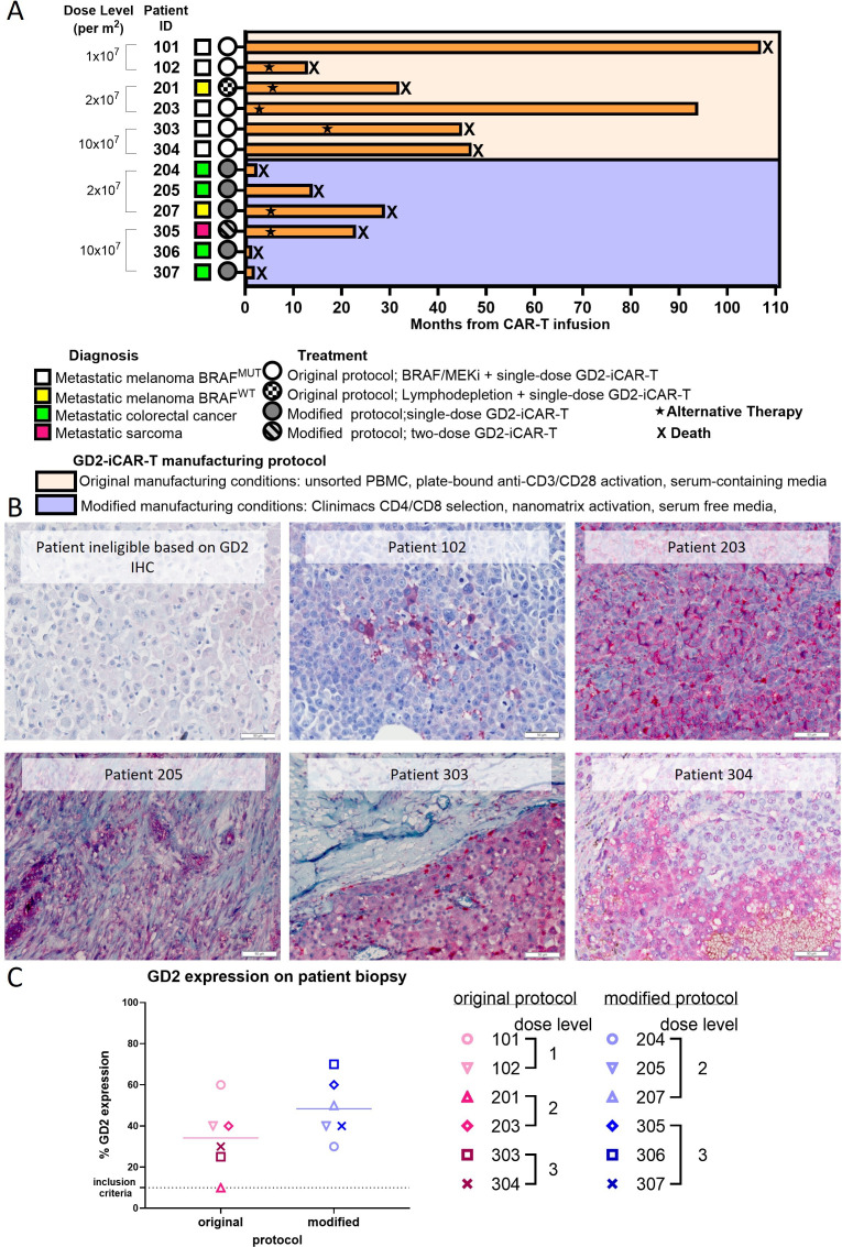
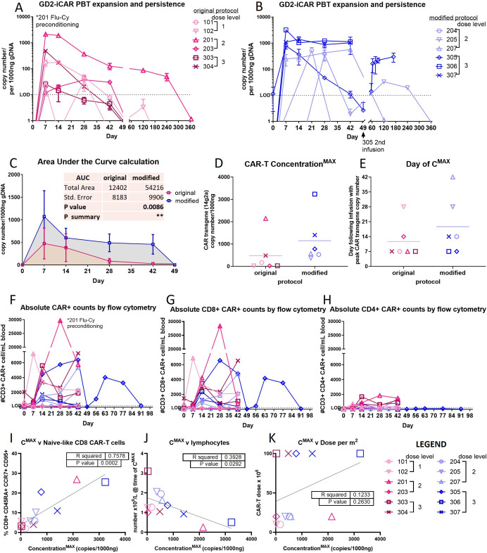
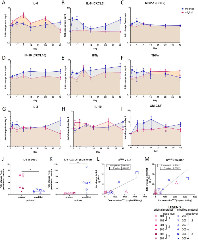
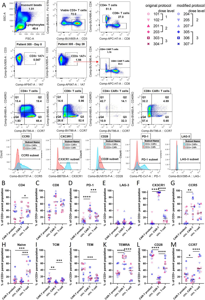
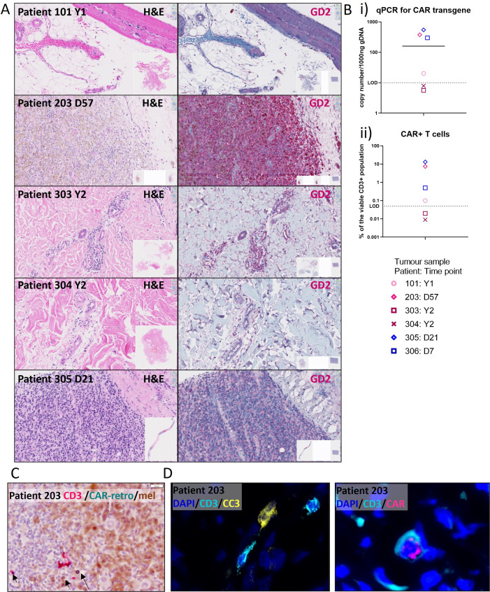
Methods: We conducted a phase 1 trial of third-generation GD2-specific CAR-T cell therapy, which has previously been tested in neuroblastoma patients. In this study, the GD2-CAR-T therapy was evaluated for the first time in metastatic melanoma patients in combination with BRAF/MEK inhibitor therapy, and as a monotherapy in patients with colorectal cancer and a patient with fibromyxoid sarcoma. Feasibility and safety were determined and persistence studies, multiplex cytokine arrays on sera and detailed immune phenotyping of the original CAR-T products, the circulating CAR-T cells, and, in select patients, the tumor-infiltrating CAR-T cells were performed.

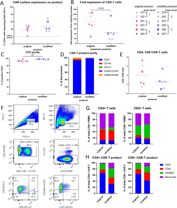
Results: We demonstrate the feasibility of manufacturing CAR-T products at point of care for patients with solid cancer and show that a single intravenous infusion was well tolerated with no dose-limiting toxicities or severe adverse events. In addition, we note significant improvements in CAR-T cell immune phenotype, and expansion when a modified manufacturing procedure was adopted for the latter 6 patients recruited to this 12-patient trial. We also show evidence of CAR-T cell-mediated immune activity and in some patients expanded subsets of circulating myeloid cells after CAR-T cell therapy.

Conclusions: This is the first report of third-generation GD2-targeting CAR-T cells in patients with metastatic melanoma and other solid cancers such as colorectal cancer, showing feasibility, safety and immune activity, but limited clinical effect.

Trial registration number: ACTRN12613000198729.

Keywords: Chimeric antigen receptor - CAR; Combination therapy; Solid tumor.


Conflict of interest statement

Competing interests: None declared.

Figures

Figure 1
Figure 1
Tumor expression of GD2 in CARPETS study patients. Archived patient tumor tissues (formalin-fixed, paraffin embedded; FFPE) collected for diagnostic pathology were screened for GD2 expression, with an inclusion criterion of ≥10% tumor-cell GD2 expression. (A) Swimmer plot showing outcomes after GD2-CAR-T cell infusions in patients with metastatic melanoma (with or without BRAF/MEK inhibitor therapy), metastatic colorectal cancer and metastatic sarcoma. (B) Examples of GD2 expression detected by immunohistochemistry on FFPE archival tumor tissues at diagnosis. (C) Summary of tumor GD2 expression levels in patients as reported by an independent pathologist. Metastatic melanoma patients treated using the original manufacturing protocol (pink symbols), metastatic melanoma and other solid cancer patients treated using the modified protocol (blue symbols). Significance was assessed by unpaired t test, GraphPad Prism V.10.1.1. CAR, chimeric antigen receptor; CARPETS, Phase 1 study of GD2 Chimeric Antigen Receptor-Expressing PEripheral Blood T CellS.
Figure 2
Figure 2
Autologous GD2-specific chimeric antigen receptor (CAR) T cell products manufactured using the original protocol (pink symbols) or modified protocol (blue symbols). Quality control assessments for CAR-T cell products: (A) transduction efficiency as determined by surface CAR expression at day 7 of culture, detected using the 1A7 anti-idiotypic antibody. (B) Fold-expansion during product culture period of 8–12 days. (C) CD3 purity at the end of the expansion period. (D) Purity of the CAR-T cell product at end of the expansion period. (E) Ratio of CD4+to CD8+ CAR T cells at end of the expansion period. (F) Flow cytometry gating strategy and representative plots for patient 304 (original manufacturing protocol) showing effector memory (TEM); central memory (TCM); TEMRA; T-naïve-like phenotype. (G) Phenotype of CD4+ and CD8+T cells from patient peripheral blood used for manufacturing and (H) the final GD2-CAR-T cell product. CAR-T cell product from the original manufacturing protocol had significantly more TEM T cells (see online supplemental figure S2). Significance was assessed by unpaired t-test, *p<0.05, **p<0.01, ***p<0.001, GraphPad Prism V.10.1.1.
Figure 3
Figure 3
Biokinetics of GD2-iCAR-PBT according to CAR-T cell manufacturing method. Peripheral expansion and persistence were determined by quantitative PCR for CAR transgene with a Taqman probe designed for the 14g2a scFv DNA sequence, using genomic DNA isolated from whole blood: (A) Original manufacturing protocol; (B) Modified manufacturing protocol. (C) Area under the curve (AUC) calculation. (D) Peak expansion (CMAX) as determined by copy number per 1000 ng genomic DNA. (E) Day of peak expansion following infusion of CAR-T cell product. Significance was assessed by unpaired t-test, or Pearson correlation analysis on GraphPad Prism V.10.1.1. Flow cytometry for CAR transgene was performed using the 1A7 anti-idiotypic antibody staining of isolated peripheral blood mononuclear cells in TruCount tubes. Absolute counts of (F) CD3+CAR+ cells (G) CD3+CD4+ CAR+ cells, and (H) CD3+CD8+ CAR+ cells per mL blood. Correlation analysis between maximum expansion of CAR-T cells and (I) Naïve-like T cells in the product; (J) Absolute lymphocyte number at the time of maximum expansion; (K) Dose level. Original manufacturing protocol (pink symbols); Modified manufacturing protocol (blue symbols). CAR, chimeric antigen receptor. AUC calculation *p <0.05, **p<0.01, ***p<0.001, GraphPad Prism V.10.1.1.
Figure 4
Figure 4
Summary of fold-change from day 0 baseline serum samples in serum cytokines after individual GD2-iCAR-PBT infusions according to CAR-T cell manufacturing method. (A–I) Fold-change in serum cytokines IL-6, IL-8, MCP-1, IP-10, IFNγ, TNFα, IL-2, IL-10, GM-CSF by cytometric bead array from the. (J) IL-6 on day 7 postinfusion and (K) IL-8 at 24 hours postinfusion; unpaired t-test, p=0.019 and p=0.015, respectively. Pearson correlation analysis between peak expansion (CMAX) and maximum fold-change in (L) IL-8 (M) GM-CSF. Original manufacturing protocol (pink symbols); modified manufacturing protocol (blue symbols). The following cytokines were also assessed TGFβ, IL-4, IL-12p70, IL-17A but did not show consistent changes from baseline. Patient’s individual cytokine profiles are provided in online supplemental figure S5 and absolute quantification (pg/mL) with statistical analysis is provided in online supplemental figure S6. Significance was assessed by unpaired t-test p*<0.05, **p<0.01, ***p<0.001, GraphPad Prism V.10.1.1.CAR, chimeric antigen receptor;
Figure 5
Figure 5
Phenotype of circulating CAR-positive T cells compared with the preinfusion GD2-iCAR-PBT product, and endogenous circulating T cells (CAR-negative) (A) Gating strategy and representative plots for patient 305 at day 28 postinfusion. A representative plot of CD3 vs CAR (1A7 anti-idiotypic antibody) from day 0 is provided to show background staining of the 1A7 antibody. Percentage of viable CD3+CAR+ T cells (for the infusion product and circulating CAR-T cell populations) or CD3+CAR T cells (for the circulating endogenous T cell population) that are (B) CD4+, (C) CD8+, (D) PD-1+, (E) LAG-3+, (F) CX3CR1+, (G) CCR5+,H) Naïve (CCR7+CD45RO−), (I) TCM (CCR7+, CD45RO+), (J) TEM (CCR7−, CD45RO+), (K) TEMRA (CCR7−, CD45RO−),L) CD28+, (M) CCR7+. Significance was determined by one-way ANOVA with Dunnett’s multiple comparisons test, *p<0.05, **p<0.01, ***p<0.001, ****p<0.0001, GraphPad Prism V.10.1.1. Further representative plots for patient 201 are also provided in online supplemental figure S8. ANOVA, analysis of variance; CAR, chimeric antigen receptor.
Figure 6
Figure 6
Evidence of tumor CAR-T cell infiltration. Biopsies were collected at various time points after each CAR-T cell infusion depending on patient consent and clinical circumstances (range day 21 to year 3; for full details see online supplemental table S3). (A) Tissue pieces from post-treatment biopsies were preserved in formalin with paraffin embedding (FFPE) and stained with H&E (left column) and using GD2 IHC (right column, dark red GD2 with hematoxylin counter stain). Insets show the whole scanned tissue section and region of interest in red. (B) CAR-T cells infiltrating tumor as assessed by (i) quantitative PCR for CAR transgene with a Taqman probe designed for the 14g2a scFv DNA sequence on genomic DNA derived from tumor biopsies from five patients and from an ascites sample from one patient (306); (ii) Flow cytometry using the 1A7 anti-idiotypic antibody for three patients with sufficient fresh single cell suspensions. (C) In situ hybridisation (ISC; RNAScope) was used to detect CD3 (magenta) and retroviral mRNA (CARretro; blue/green) in FFPE tissue, with melanoma cells showing brown melanin pigment. (D) Immunofluorescence analysis was performed on available fresh-frozen tissue to detect CD3, CAR (magenta), and cleaved caspase 3 (CC3; yellow) expression (representative images from the patient 203; for full tissue sections and patient 101 images see online supplemental figure S10). LOD, limit of detection; CAR, chimeric antigen receptor.

Similar articles

Cited by

References

    1. Pule MA, Savoldo B, Myers GD, et al. . Virus-specific T cells engineered to coexpress tumor-specific receptors: persistence and antitumor activity in individuals with neuroblastoma. Nat Med 2008;14:1264–70. 10.1038/nm.1882 - DOI - PMC - PubMed
    1. Wikstrand CJ, Fredman P, Svennerholm L, et al. . Detection of glioma-associated Gangliosides GM2, GD2, GD3, 3'-IsoLM1 3',6'-IsoLD1 in central nervous system tumors in vitro and in vivo using EPITOPE-defined Monoclonal antibodies. Prog Brain Res 1994;101:213–23. 10.1016/s0079-6123(08)61951-2 - DOI - PubMed
    1. Gargett T, Ebert LM, Truong NTH, et al. . Gd2-targeting CAR-T cells enhanced by transgenic IL-15 expression are an effective and clinically feasible therapy for glioblastoma. J Immunother Cancer 2022;10:e005187. 10.1136/jitc-2022-005187 - DOI - PMC - PubMed
    1. Grant SC, Kostakoglu L, Kris MG, et al. . Targeting of small-cell lung cancer using the anti-GD2 ganglioside monoclonal antibody 3F8: a pilot trial. Eur J Nucl Med 1996;23:145–9. 10.1007/BF01731837 - DOI - PubMed
    1. Fisher JPH, Flutter B, Wesemann F, et al. . Effective combination treatment of GD2-expressing neuroblastoma and Ewing’s sarcoma using anti-Gd2 Ch14.18/CHO antibody with Vgamma9Vdelta2+ gammadeltaT cells. Oncoimmunology 2016;5:e1025194. 10.1080/2162402X.2015.1025194 - DOI - PMC - PubMed

Publication types

Substances