

MR Imaging Features of Critical Spinal Demyelinating Lesions Associated with Progressive Motor Impairment
- PMID: 38754997
- PMCID: PMC11286007
- DOI: 10.3174/ajnr.A8304
MR Imaging Features of Critical Spinal Demyelinating Lesions Associated with Progressive Motor Impairment
Abstract
Background and purpose: Progressive MS is typically heralded by a myelopathic pattern of asymmetric progressive motor weakness. Focal individual "critical" demyelinating spinal cord lesions anatomically associated with progressive motor impairment may be a compelling explanation for this clinical presentation as described in progressive solitary sclerosis (single CNS demyelinating lesion), progressive demyelination with highly restricted MR imaging lesion burden (2-5 total CNS demyelinating lesions; progressive paucisclerotic MS), and progressive, exclusively unilateral hemi- or monoparetic MS (>5 CNS demyelinating progressive unilateral hemi- or monoparetic MS [PUHMS] lesions). Critical demyelinating lesions appear strikingly similar across these cohorts, and we describe their specific spinal cord MR imaging characteristics.
Materials and methods: We performed a retrospective, observational MR imaging study comparing spinal cord critical demyelinating lesions anatomically associated with progressive motor impairment with any additional "noncritical" (not anatomically associated with progressive motor impairment) spinal cord demyelinating lesions. All spinal cord MR images (302 cervical and 91 thoracic) were reviewed by an experienced neuroradiologist with final radiologic assessment on the most recent MR imaging. Anatomic association with clinical progressive motor impairment was confirmed independently by MS subspecialists.
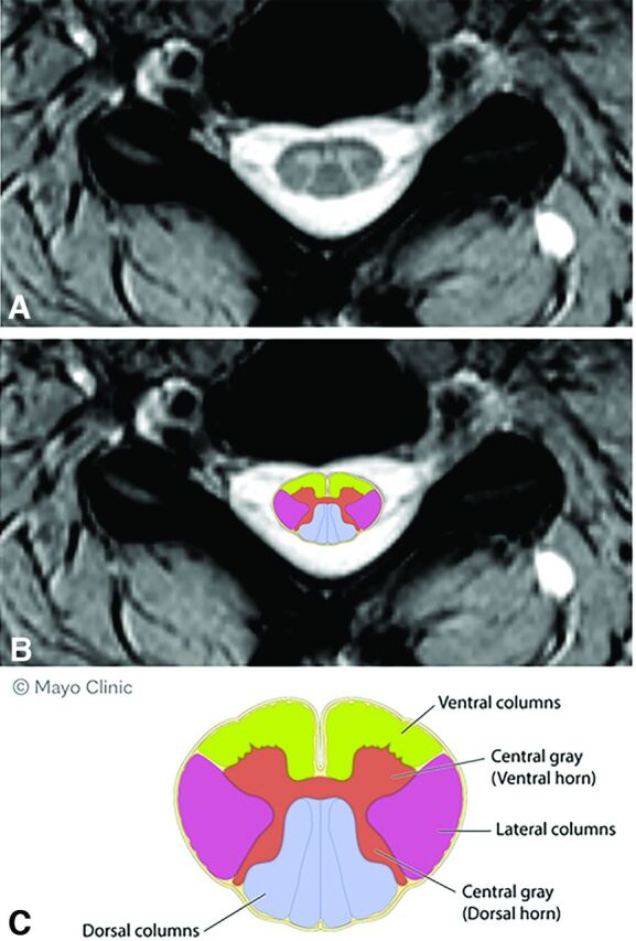
Results: Ninety-one individuals (PUHMS, 37 [41%], progressive paucisclerosis 35 [38%], progressive solitary sclerosis 19 [21%]) with 91 critical and 98 noncritical spinal cord MR imaging demyelinating lesions were evaluated. MR imaging characteristics that favored critical spinal cord demyelinating lesions over noncritical lesions included moderate-to-severe, focal, lesion-associated spinal cord atrophy: 41/91 (45%) versus 0/98 (0%) (OR, 161.91; 9.43 to >999.9); lateral column axial location (OR, 10.43; 3.88-28.07); central region (OR, 3.23; 1.78-5.88); ventral column (OR, 2.98; 1.55-5.72); and larger lesion size of the axial width (OR, 2.01;1.49-2.72), transverse axial size (OR, 1.66; 1.36-2.01), or lesion area (OR, 1.14; 1.08-1.2). Multiple regression analysis revealed focal atrophy and lateral axial location as having the strongest association with critical demyelinating lesions.
Conclusions: Focal, lesion-associated atrophy, lateral column axial location, and larger lesion size are spinal cord MR imaging characteristics of critical demyelinating lesions. The presence of critical demyelinating lesions should be sought as these features may be associated with the development of progressive motor impairment in MS.
© 2024 by American Journal of Neuroradiology.
Figures


Similar articles
-
Uncommon Non-MS Demyelinating Disorders of the Central Nervous System.Curr Neurol Neurosci Rep. 2025 Jul 1;25(1):45. doi: 10.1007/s11910-025-01432-8. Curr Neurol Neurosci Rep. 2025. PMID: 40591029 Review.
-
Siponimod for multiple sclerosis.Cochrane Database Syst Rev. 2021 Nov 16;11(11):CD013647. doi: 10.1002/14651858.CD013647.pub2. Cochrane Database Syst Rev. 2021. PMID: 34783010 Free PMC article.
-
Translating state-of-the-art spinal cord MRI techniques to clinical use: A systematic review of clinical studies utilizing DTI, MT, MWF, MRS, and fMRI.Neuroimage Clin. 2015 Dec 4;10:192-238. doi: 10.1016/j.nicl.2015.11.019. eCollection 2016. Neuroimage Clin. 2015. PMID: 26862478 Free PMC article.
-
Neurodegeneration and Demyelination in the Multiple Sclerosis Spinal Cord: Clinical, Pathological, and 7T MRI Perspectives.Neurology. 2025 Apr 8;104(7):e210259. doi: 10.1212/WNL.0000000000210259. Epub 2025 Mar 13. Neurology. 2025. PMID: 40080735 Free PMC article.
-
WITHDRAWN: Interventions for fatigue and weight loss in adults with advanced progressive illness.Cochrane Database Syst Rev. 2017 Apr 7;4(4):CD008427. doi: 10.1002/14651858.CD008427.pub3. Cochrane Database Syst Rev. 2017. PMID: 28387447 Free PMC article.
References
Publication types
MeSH terms
LinkOut - more resources
Full Text Sources
Medical