. 2024 May 16;15(1):4148.
doi: 10.1038/s41467-024-48632-3.

CEBPA restricts alveolar type 2 cell plasticity during development and injury-repair

Affiliations

CEBPA restricts alveolar type 2 cell plasticity during development and injury-repair

Dalia Hassan et al. Nat Commun. .

Abstract

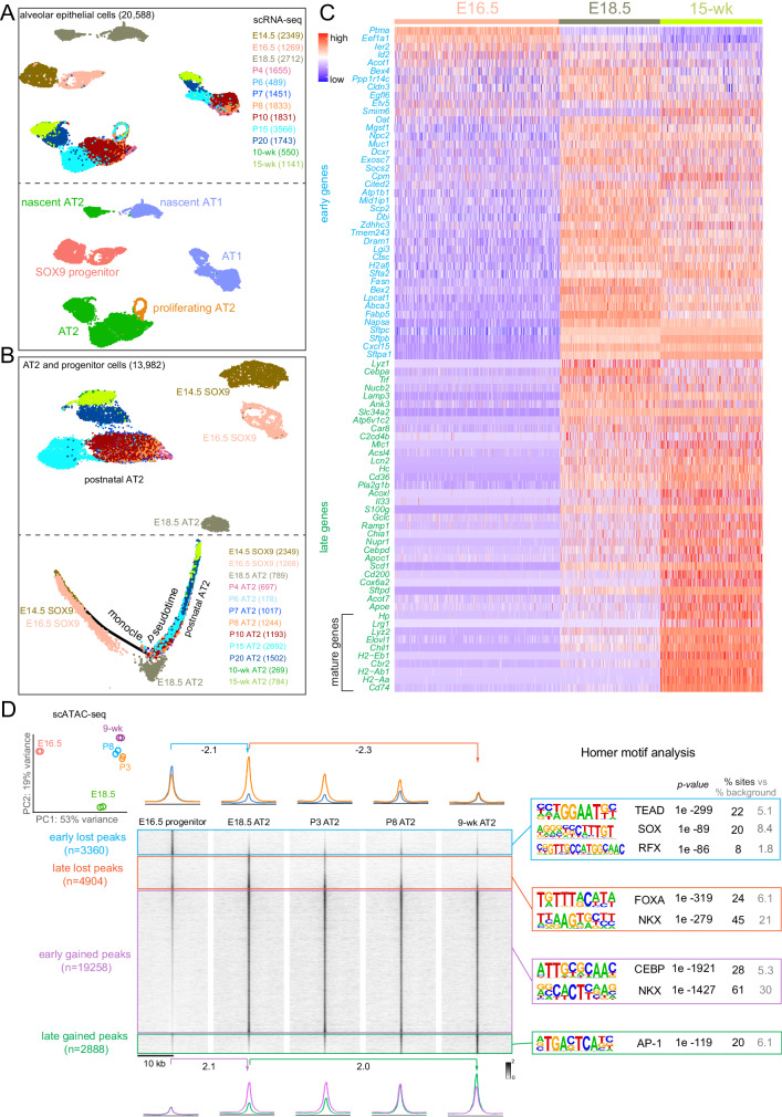
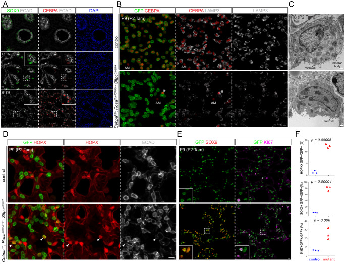
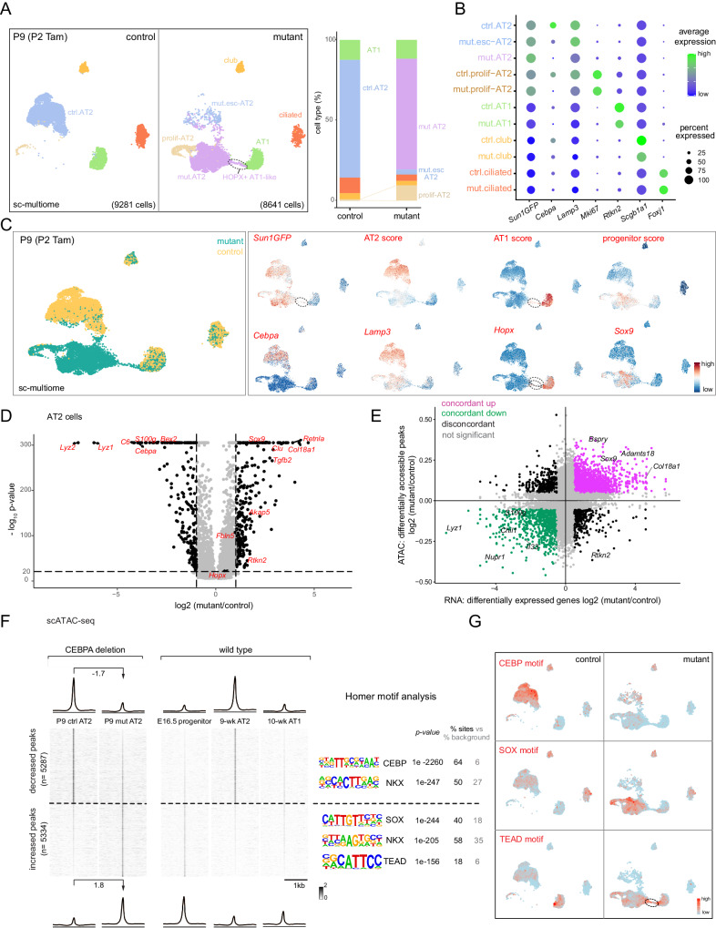
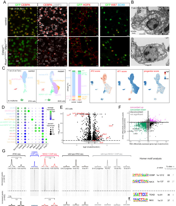
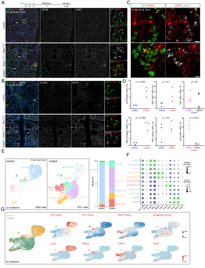
Cell plasticity theoretically extends to all possible cell types, but naturally decreases as cells differentiate, whereas injury-repair re-engages the developmental plasticity. Here we show that the lung alveolar type 2 (AT2)-specific transcription factor (TF), CEBPA, restricts AT2 cell plasticity in the mouse lung. AT2 cells undergo transcriptional and epigenetic maturation postnatally. Without CEBPA, both neonatal and mature AT2 cells reduce the AT2 program, but only the former reactivate the SOX9 progenitor program. Sendai virus infection bestows mature AT2 cells with neonatal plasticity where Cebpa mutant, but not wild type, AT2 cells express SOX9, as well as more readily proliferate and form KRT8/CLDN4+ transitional cells. CEBPA promotes the AT2 program by recruiting the lung lineage TF NKX2-1. The temporal change in CEBPA-dependent plasticity reflects AT2 cell developmental history. The ontogeny of AT2 cell plasticity and its transcriptional and epigenetic mechanisms have implications in lung regeneration and cancer.


Conflict of interest statement

The authors declare no competing interests.

Figures

Fig. 1
Fig. 1. AT2 cells undergo transcriptomic and epigenomic maturation postnatally, separate from their embryonic specification.
A Aggregated scRNA-seq UMAPs of alveolar epithelial cells of wild type lungs from 12-time points color coded by time (top) or cell type (bottom). Cell numbers are in parenthesis. Each time point consists of at least 2 mice profiled as one sample. B Top: UMAP of AT2 and SOX9 progenitor cells subset from in (A), color-coded by time, and their monocle pseudotime analysis (bottom) showing molecular progression from E14.5 SOX9 progenitors through E18.5 nascent AT2 to 15-wk mature AT2 cells. Cell numbers are in parenthesis. C Expression heatmap of 82 AT2-specific genes, classified as early if present in at least 25% of cells at E16.5. The remaining late genes are considered mature if the fold increase from E18.5 to 15-wk is more than 4. D Principal component analysis (PCA) of scATAC-seq pseudobulk duplicates showing the distinct epigenome of E18.5 nascent AT2 cells. ScATAC-seq heatmaps and profile plots were categorized and color-coded as early lost (E16.5 vs E18.5), late lost (E18.5 vs 9-wk), early gained (E16.5 vs E18.5) and late gained (E18.5 vs 9-wk) and their Homer motif analysis. Peak numbers are in parenthesis. Each time point consists of at least 2 mice profiled as one sample.
Fig. 2
Fig. 2. CEBPA promotes AT2 and suppresses progenitor programs in neonatal AT2 cells.
A Confocal images of immunostained wild type lungs show little CEBPA expression at E14.5 and E16.5 when branch tips are dominated by SOX9 progenitors but CEBPA expression in cuboidal cells outlined with E-Cadherin (ECAD) at E18.5 (n = 3 mice each). B Confocal images of immunostained neonatal AT2-specific Cebpa mutant and littermate control lungs showing loss of CEBPA in GFP+ recombined cells (asterisk: escaper), without affecting its expression in alveolar macrophages (AM) in the airspace, and reduced LAMP3. Tam, 250 μg tamoxifen. Images are representative of at least three lungs (same for subsequent immunostainings). C Transmission electron microscopy (TEM) images show a reduction in lamellar bodies in mutant AT2 cells without affecting their apical microvilli (n = 2 mice each). Tam, 250 μg tamoxifen. See Supplementary Fig. 4B for more examples and quantification. D Confocal images showing lineage labeled mutant AT2 cells expressing an AT1 marker HOPX and no longer cuboidal (ECAD outline) (arrowhead). E Confocal images showing lineage labeled mutant AT2 cells ectopically expressing a progenitor marker SOX9 and a proliferation marker KI67. F Quantification of (D) and (E). Each symbol represents one mouse from littermate pairs. P values were calculated using two-tailed Student’s t test. Scale: 10 μm for all except for (C) 1 μm.
Fig. 3
Fig. 3. Single-cell multiome defines CEBPA-dependent neonatal AT2 cell program and plasticity.
A Sc-multiome UMAPs of purified epithelial cells from Cebpa mutant and littermate control lungs color-coded by cell type (left) and the corresponding percentages (right). Esc, escaper; prolif, proliferating; Tam, 250 μg tamoxifen. See Supplementary Fig. 5A for the sorting strategy. Each sample includes 1 male and 1 female mouse profiled as one sample (same for subsequent sc-multiome experiments). B Dot plot showing the lineage marker (Sun1GFP), Cebpa, and cell type markers. See also feature plots in (C) and Supplementary Fig. 5B. C Sc-multiome UMAP color-coded for genotype (left) and feature plots of metagene scores (top) and representative genes (bottom). The circled population is specific to the mutant, GFP + , and expresses AT1 genes, thus labeled as HOPX + AT1-like cells in (A). See Source Data for metagene lists. D Volcano plot (two-tailed, non-parametric Wilcoxon rank sum test) showing downregulation of AT2 genes and upregulation of progenitor genes in mutant AT2 cells (right) compared to control AT2 cells (left) defined in (A). E Scatter plot correlating changes in the accessibility of scATAC-seq peaks (y-axis) and scRNA-seq expression of their nearest genes (x-axis), color-coded as concordant or discordant as well as the directionality of change. F ScATAC-seq heatmaps and profile plots of decreased and increased peak sets in the mutant and associated log2 fold changes, as well as the corresponding scATAC-seq data in wild type cells and associated Homer motifs. G Feature plots of motif activity scores showing that the mutant has lower CEBP, and higher SOX and TEAD (circle) activities.
Fig. 4
Fig. 4. CEBPA recruits NKX2-1 to promote the AT2 program and indirectly restricts the progenitor program.
A Heatmaps and profile plots of CEBPA and NKX2-1 binding for decreased and increased peak sets from Fig. 3F, as well as associated frequency distributions of CEBP, NKX, SOX motifs. CEBPA binds to decreased peaks but not increased peaks. NKX2-1 binding decreases (log2 fold change) for decreased peaks and increases (log2 fold change) for increased peaks in the mutant, corresponding to AT2 and progenitor/AT1-specific binding in wild type lungs. Diagram: a recruitment model, in which CEBPA normally recruits NKX2-1 to activate AT2 genes, whereas without CEBPA, NKX2-1 is released from AT2 genes and possibly relocates to progenitor and AT1 genes. Loss of NKX2-1 binding due to Cebpa deletion is associated with chromatin closure (open vs closed). See Supplementary Fig. 6A for nuclei sorting strategy. B Representative coverage plots of (A) showing a decreased peak near an AT2 gene Il33, and an increased peak near a progenitor gene Acaca. C Venn diagram showing NKX2-1 and CEBPA co-bound and single-bound peak sets in purified P2 AT2 cells (left) and frequency distributions of NKX and CEBP motifs for the co-bound peak set (right). D Heatmaps and profile plots for the 3 peak sets in (C) and associated log2 fold changes showing the largest decreases for the co-bound peak set.
Fig. 5
Fig. 5. CEBPA maintains the AT2 program without affecting the progenitor program in mature AT2 cells.
A Confocal images of immunostained adult AT2-specific Cebpa mutant and littermate control lungs showing loss of CEBPA in GFP+ recombined cells (asterisk: escaper), without affecting its expression in alveolar macrophages in the airspace (AM), and reduced LAMP3, but no extra HOPX, SOX9, or KI67. Tam, two doses of 3 mg each tamoxifen at 48 h interval (same for the rest of Fig. 5) (n = 3 mice each). Scale: 10 μm. B TEM images showing a reduction in lamellar bodies in mutant AT2 cells without affecting their apical microvilli. Large granules in mutant AT2 cells lack characteristic lamellae (n = 2 mice each). Scale: 1 μm. See Supplementary Fig. 6C for quantification. C Sc-multiome UMAPs of purified epithelial cells from Cebpa mutant and littermate control lungs color-coded by cell type (left), the corresponding percentages (middle), and metagene scores. Esc, escaper. See Source Data for metagene lists. D Dot plot showing the lineage marker (Sun1GFP), Cebpa, and cell type markers. Rtkn2, but not Spock2, is expressed in HOPXlow AT1-like cells. E Volcano plot (two-tailed, non-parametric Wilcoxon rank sum test) showing downregulation of AT2 genes but minimal upregulation of progenitor/AT1 genes in mutant AT2 cells (left) compared to control AT2 cells(right) defined in (C). Compare with Fig. 3D. F Scatter plot correlating changes in the accessibility of scATAC-seq peaks (y-axis) and scRNA-seq expression of their nearest genes (x-axis), color-coded as concordant or discordant as well as the directionality of change. Compared to Fig. 3E, few concordant pairs are upregulated. See Source Data for the complete list. G Heatmaps and profile plots of decreased and increased scATAC-seq peak sets in the adult mutant and associated log2 fold changes, as well as the corresponding CEBPA and NKX2-1 binding and scATAC-seq data in wild type cells and associated Homer motifs. Decreased peaks have CEBPA binding and decreased NKX2-1 binding, corresponding to ATAC accessibility and NKX2-1 binding in wild type AT2 cells. Increased peaks are many fewer and have no CEBPA binding and increased NKX2-1 binding, corresponding to NKX2-1 binding in wild type AT1 cells.
Fig. 6
Fig. 6. Viral infection expands CEBPA-dependent plasticity in mature AT2 cells. Figure 6. Viral infection expands CEBPA-dependent plasticity in mature AT2 cells.
A Experimental timeline of tamoxifen injection (Tam, 3 mg), Sendai virus (SeV) or saline (PBS) administration, and lung harvest at 14 dpi (day post-infection). Confocal images of immunostained infected Cebpa mutant and littermate control lungs, showing mutant-specific activation of SOX9 and increase in KI67 near airways (aw) and lobe edges (inset; scale: 10 μm). Scale: 100 μm. B Confocal images of lungs in (A) showing increased KRT8 and CLDN4. Scale: 100 μm (inset: 10 μm). C Confocal images of lungs in (A) showing lineage-labeled HOPX+ cells with little LAMP3 (arrowhead). Scale: 10 μm. D Quantification of (A, B, C). KI67+ cells in the control and Cebpa mutant are stratified by CEBPA and SOX9 expression, respectively. Each symbol represents one mouse from littermate pairs. P values were calculated using two-tailed Student’s t test. E Sc-multiome UMAPs of purified epithelial cells from infected Cebpa mutant and littermate control lungs color-coded by cell type (left) and the corresponding percentages (right). Esc, escaper; prolif, proliferating. F Dot plot showing the lineage marker (Sun1GFP), Cebpa, and cell type markers. G Sc-multiome UMAP color-coded for genotype (left) and feature plots of metagene scores (top) and representative genes (bottom). A published damage-associated transient progenitor (DATP) score marks KRT8/CLDN4+ cells.
Fig. 7
Fig. 7. Diagram of CEBPA restricting AT2 cell plasticity during development and injury-repair.
A In a wild type lung, SOX9 progenitors undergo specification and maturation to become neonatal/nascent and mature AT2 cells, sequentially, expressing CEBPA. A small fraction of AT2 cells at both stages become AT1-like cells with varying levels of HOPX. Sendai virus (SeV) infection induces KRT8/CLDN4 transitional cells as well as AT1-like cells, as a sequential or parallel response of injury-repair. Cebpa deletion in neonatal, but not mature, AT2 cells leads to reversion to the progenitor program and proliferation, in addition to an increase of AT1-like cells upon either deletion. Sendai virus infection bestows mature AT2 cells with the plasticity to revert to progenitors as well as to more readily transition to the KRT8/CLDN4 state. B In an aerial view of the cell plasticity landscape, the progenitor program, although still present in neonatal AT2 cells, is submerged under the AT2 program promoted by CEBPA. Although CEBPA does not bind to genomic regions of plasticity, without CEBPA, the AT2 program subcedes to expose the progenitor program. As AT2 cells mature, the progenitor program disappears so that, even without the AT2 program, no progenitor program is visible. After infection, mature AT2 cells reshape their plasticity landscape, but only the KRT8/CLDN4 transitional program rises enough to manifest over the AT2 program. Without CEBPA and the AT2 program, the progenitor program is visible and the transitional program more evident.

Update of

References

    1. Takahashi K, Yamanaka S. Induction of pluripotent stem cells from mouse embryonic and adult fibroblast cultures by defined factors. Cell. 2006;126:663–676. doi: 10.1016/j.cell.2006.07.024. - DOI - PubMed
    1. Perez-Gonzalez A, Bevant K, Blanpain C. Cancer cell plasticity during tumor progression, metastasis and response to therapy. Nat. Cancer. 2023;4:1063–1082. doi: 10.1038/s43018-023-00595-y. - DOI - PMC - PubMed
    1. Ring NAR, Valdivieso K, Grillari J, Redl H, Ogrodnik M. The role of senescence in cellular plasticity: lessons from regeneration and development and implications for age-related diseases. Dev. Cell. 2022;57:1083–1101. doi: 10.1016/j.devcel.2022.04.005. - DOI - PubMed
    1. Lam NT, Sadek HA. Neonatal heart regeneration: comprehensive literature review. Circulation. 2018;138:412–423. doi: 10.1161/CIRCULATIONAHA.118.033648. - DOI - PMC - PubMed
    1. Desai, T. J., Brownfield, D. G. & Krasnow, M. A. Alveolar progenitor and stem cells in lung development, renewal and cancer. Nature 507:190–194 (2014). - PMC - PubMed

MeSH terms