. 2024 Jun 1;17(6):dmm050561.
doi: 10.1242/dmm.050561. Epub 2024 Jun 28.

Female Alms1-deficient mice develop echocardiographic features of adult but not infantile Alström syndrome cardiomyopathy

Affiliations

Female Alms1-deficient mice develop echocardiographic features of adult but not infantile Alström syndrome cardiomyopathy

Eleanor J McKay et al. Dis Model Mech. .

Abstract

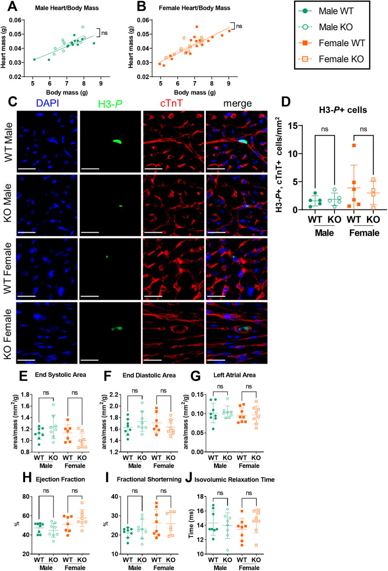
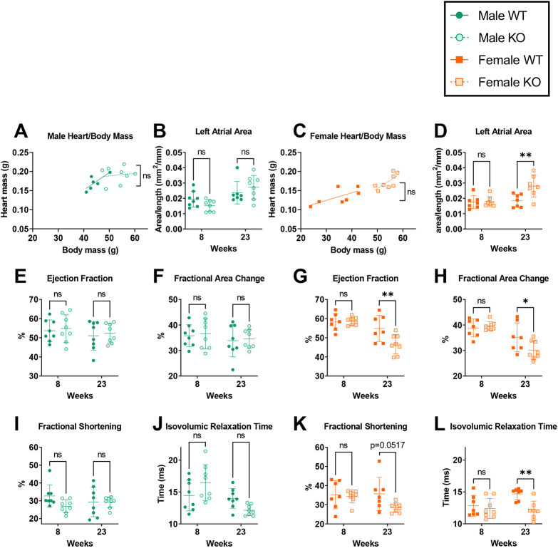
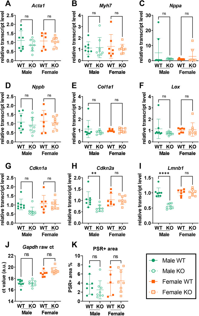
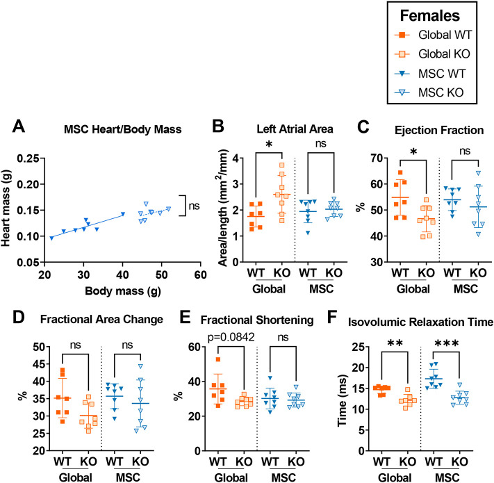
Alström syndrome (AS), a multisystem disorder caused by biallelic ALMS1 mutations, features major early morbidity and mortality due to cardiac complications. The latter are biphasic, including infantile dilated cardiomyopathy and distinct adult-onset cardiomyopathy, and poorly understood. We assessed cardiac function of Alms1 knockout (KO) mice by echocardiography. Cardiac function was unaltered in Alms1 global KO mice of both sexes at postnatal day 15 (P15) and 8 weeks. At 23 weeks, female - but not male - KO mice showed increased left atrial area and decreased isovolumic relaxation time, consistent with early restrictive cardiomyopathy, as well as reduced ejection fraction. No histological or transcriptional changes were seen in myocardium of 23-week-old female Alms1 global KO mice. Female mice with Pdgfra-Cre-driven Alms1 deletion in cardiac fibroblasts and in a small proportion of cardiomyocytes did not recapitulate the phenotype of global KO at 23 weeks. In conclusion, only female Alms1-deficient adult mice show echocardiographic evidence of cardiac dysfunction, consistent with the cardiomyopathy of AS. The explanation for sexual dimorphism remains unclear but might involve metabolic or endocrine differences between sexes.

Keywords: ALMS1; Alstrom syndrome; Alström syndrome; Cardiomyopathy; Ciliopathy; Heart; Primary cilia.


Conflict of interest statement

Competing interests R.K.S. has received consulting fees from Novartis, Astra Zeneca and Alnylam, research contribution in kind from Pfizer, and speaking fees from Novo Nordisk, Eli Lilly and Amryt.

Figures

Fig. 1.
Fig. 1.
Neither male nor female global Alms1 knockout mice exhibit a cardiac phenotype at postnatal day 15. (A,B) Linear regression of heart and body mass in wild-type (WT) and knockout (KO) male (A) and female (B) mice. Lines in linear regression graphs (A,B) represent lines of best fit. Comparisons between lines of best fit were undertaken by simple linear regression, with square brackets showing comparison of y intercepts. No significant change was seen between gradients. (C) Representative immunofluorescence images showing cardiac left ventricles of mice as indicated, stained for proliferative marker phosphorylated histone H3 (H3-P, green) and co-stained for cardiac muscle troponin T (cTnT, red). Nuclei were stained with DAPI (blue). Scale bars: 20 μm. (D) Quantification of images shown in C, showing percentage of cTnT-positive cells that also showed staining for H3-P. (E-J) Echocardiography data. Area values calculated from echocardiography (E-G) are normalised to total body mass, showing no changes in end-systolic or end-diastolic areas of the left ventricle, the left atrial area, and ejection fraction, fractional shortening or isovolumic relaxation times. Each data point represents an individual animal, with error bars in D-J representing the mean±s.d. Comparison between groups in panels D-J was undertaken using two-way ANOVA with Tukey's multiple comparisons test. Animals used per experiment were n=13, 11, 17 and 12 (A,B); n=5, 5, 6 and 4 (D); n=8, 8, 7 and 8 (E-J) for WT males, KO males, WT females and KO females, respectively. ns, not significant.
Fig. 2.
Fig. 2.
Systolic and diastolic dysfunction develops with age in female but not male global Alms1 knockout mice. (A,C) Linear regression of heart to body mass in wild-type (WT) and knockout (KO) male (A) and female (C) mice at 24 weeks of age, showing that increased raw heart mass in Alms1 KO mice is proportionate to total body mass. Lines in linear regression graphs (A,C) represent the of best fit. Comparisons between lines of best fit (A,C) were undertaken by simple linear regression, with square brackets showing comparison of y intercepts. No significant change was seen between gradients. (B, D-L) Echocardiography data of male and female WT and KO mice aged 8 and 23 weeks, showing no changes in male KO mice, but several structural and functional changes in female KO mice at 23 weeks of age. Each data point represents an individual animal. Left atrial area values (B,D) are normalised to nose–anus length. Error bars in B, D-L represent the mean±s.d., comparison between groups was undertaken using two-way ANOVA with Šídák's multiple comparisons test. Animals used per experiment were n=8, 8, 7 and 8 for WT males, KO males, WT females and KO females, respectively. Statistical significance: *P<0.05, **P<0.01, ****P<0.0001. ns, not significant.
Fig. 3.
Fig. 3.
Neither transcriptional nor histological correlates were found for the echocardiography phenotype of global Alms1 knockout mice. (A-I) Transcriptional analysis of hearts obtained at 24 weeks from wild-type (WT) and knockout (KO) male and female mice as indicated, showing minimal transcriptional changes in the cardiac tissue of Alms1 KO males and females. Data present crossing point (Cp) values of qPCR data (indicating the number of cycles which, in turn, indicate a significant increase in signal intensity) normalised to Gapdh values run in duplex. (J) An example of raw Gapdh Cp values from duplexed reactions. Data show that Gapdh transcript levels do not significantly differ between WT and KO animals of each sex. (K) Quantification of Picrosirius Red (PSR) staining by pixel thresholding, showing that Gapdh transcript levels do not significantly differ between WT and KO animals of each sex. Samples analysed from 24-week-old male and female Alms1 knockout mice. Each data point represents an individual animal, with error bars representing the mean±sd. Comparisons between groups were performed using two-way ANOVA with Tukey's multiple comparisons test. Animals used per experiment were n=8, 8, 7 and 8 for WT males, KO males, WT females and KO females, respectively. Statistical significance: **P<0.01. ns, not significant.
Fig. 4.
Fig. 4.
Mesenchymal stem cell-specific Alms1 knockout in female mice does not recapitulate the phenotype of global Alms1 knockout. All global knockout (KO) data were repeated according to those shown in Fig. 2 for comparison to mesenchymal stem cell (MSC)-specific Alms1 KO. (A) Linear regression of heart to body mass in wild-type (WT) and knockout (KO) female mice at 24 weeks of age as indicated, showing that the increase in raw heart mass in Alms1 MSC KO female mice is proportionate to total body mass. Lines in linear regression graphs represent lines of best fit. Comparisons between lines of best fit (A) was undertaken using simple linear regression, with square brackets showing comparison of y intercepts. No significant change was seen between gradients. (B-F) Data obtained from analysis of echocardiography in mice as indicated performed at 23 weeks of age, showing a failure of female Alms1 MSC KO mice to recapitulate the cardiac phenotype seen in female Alms1 global mice. Left atrial area (B) was normalised to nose–anus length. Each data point represents an individual animal, with error bars in B-F representing the mean±s.d. Global WT/KO and MSC WT and MSC KO experiments were performed with identical design at different times; this is reflected by the dotted line separating the two cohorts. Comparison between WT and KO in B-F was undertaken using unpaired two-tailed Student's t-test followed by Bonferroni correction for multiple testing. Animals used per experiment were n=7, 8, 8 and 8 for global WT, global KO, MSC WT and MSC KO, respectively. *P<0.05, **P<0.01 and ***P<0.001. ns, not significant.

Similar articles

Cited by

  • Translating multiscale research in rare disease.
    Hooper KM, Justice MJ, Lek M, Liu KJ, Rauen KA. Hooper KM, et al. Dis Model Mech. 2024 Jun 1;17(6):dmm052009. doi: 10.1242/dmm.052009. Epub 2024 Jul 10. Dis Model Mech. 2024. PMID: 38982973 Free PMC article.

References

    1. Arsov, T., Larter, C. Z., Nolan, C. J., Petrovsky, N., Goodnow, C. C., Teoh, N. C., Yeh, M. M. and Farrell, G. C. (2006a). Adaptive failure to high-fat diet characterizes steatohepatitis in Alms1 mutant mice. Biochem. Biophys. Res. Commun. 342, 1152-1159. 10.1016/j.bbrc.2006.02.032 - DOI - PubMed
    1. Arsov, T., Silva, D. G., O'Bryan, M. K., Sainsbury, A., Lee, N. J., Kennedy, C., Manji, S. S. M., Nelms, K., Liu, C., Vinuesa, C. G.et al. (2006b). Fat aussie - a new Alström syndrome mouse showing a critical role for ALMS1 in obesity, diabetes, and spermatogenesis. Mol. Endocrinol. 20, 1610-1622. 10.1210/me.2005-0494 - DOI - PubMed
    1. Baig, S., Veeranna, V., Bolton, S., Edwards, N., Tomlinson, J. W., Manolopoulos, K., Moran, J., Steeds, R. P. and Geberhiwot, T. (2018). Treatment with PBI-4050 in patients with Alström syndrome: study protocol for a phase 2, single-Centre, single-arm, open-label trial. BMC Endocr. Disord. 18, 88. 10.1186/s12902-018-0315-6 - DOI - PMC - PubMed
    1. Baig, S., Paisey, R., Dawson, C., Barrett, T., Maffei, P., Hodson, J., Rambhatla, S. B., Chauhan, P., Bolton, S., Dassie, F.et al. (2020a). Defining renal phenotype in Alström syndrome. Nephrol. Dial. Transplant. 35, 994-1001. 10.1093/ndt/gfy293 - DOI - PubMed
    1. Baig, S., Dowd, R., Edwards, N. C., Hodson, J., Fabritz, L., Vijapurapu, R., Liu, B., Geberhiwot, T. and Steeds, R. P. (2020b). Prospective cardiovascular magnetic resonance imaging in adults with Alström syndrome: silent progression of diffuse interstitial fibrosis. Orphanet J. Rare Dis. 15, 139. 10.1186/s13023-020-01426-4 - DOI - PMC - PubMed

MeSH terms

Substances