

Maximizing microcalcification detectability in low-dose dedicated cone-beam breast CT: parallel cascades-based theoretical analysis
- PMID: 38756437
- PMCID: PMC11095120
- DOI: 10.1117/1.JMI.11.3.033501
Maximizing microcalcification detectability in low-dose dedicated cone-beam breast CT: parallel cascades-based theoretical analysis
Abstract
Purpose: We aim to determine the combination of X-ray spectrum and detector scintillator thickness that maximizes the detectability of microcalcification clusters in dedicated cone-beam breast CT.
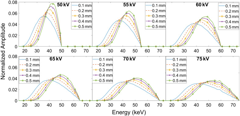
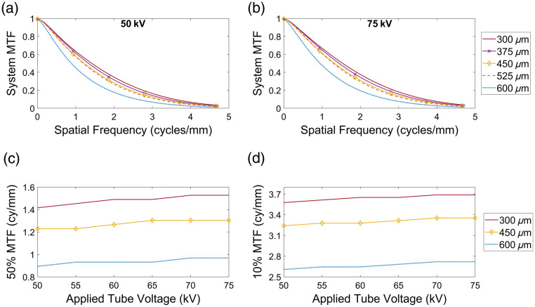
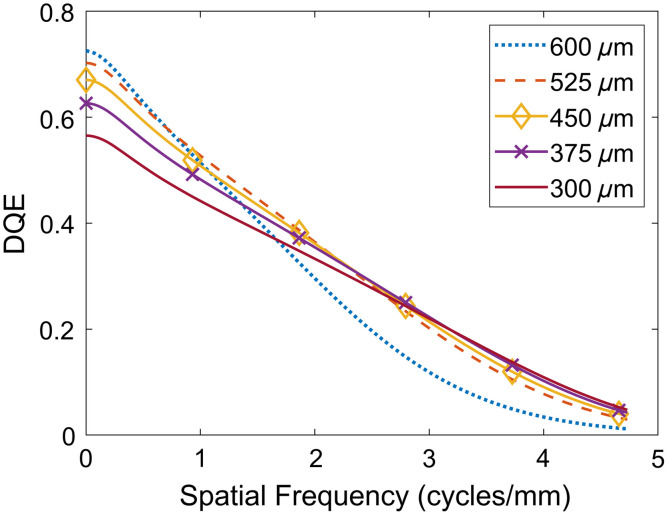
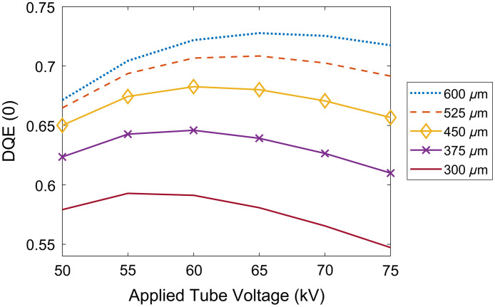
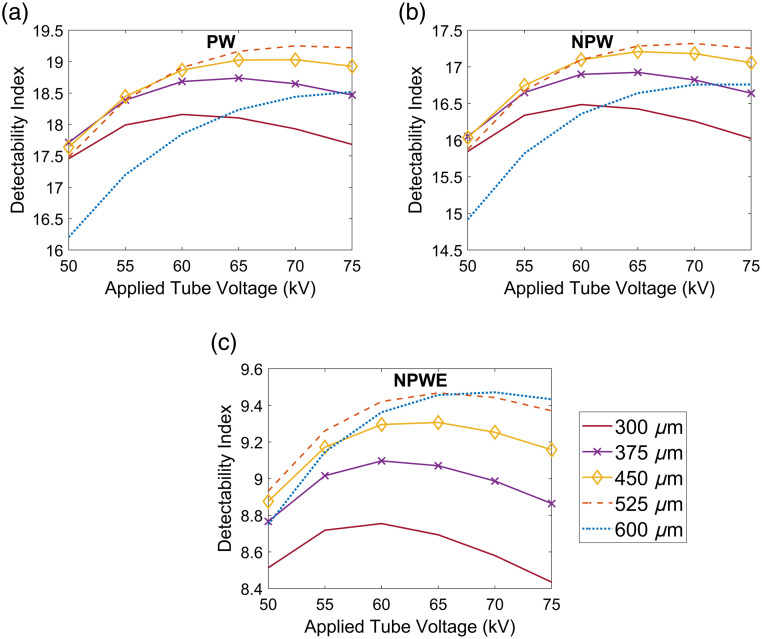
Approach: A cascaded linear system analysis was implemented in the spatial frequency domain and was used to determine the detectability index using numerical observers for the imaging task of detecting a microcalcification cluster with 0.17 mm diameter calcium carbonate spheres. The analysis considered a thallium-doped cesium iodide scintillator coupled to a complementary metal-oxide semiconductor detector and an analytical filtered-back-projection reconstruction algorithm. Independent system parameters considered were the scintillator thickness, applied X-ray tube voltage, and X-ray beam filtration. The combination of these parameters that maximized the detectability index was considered optimal.
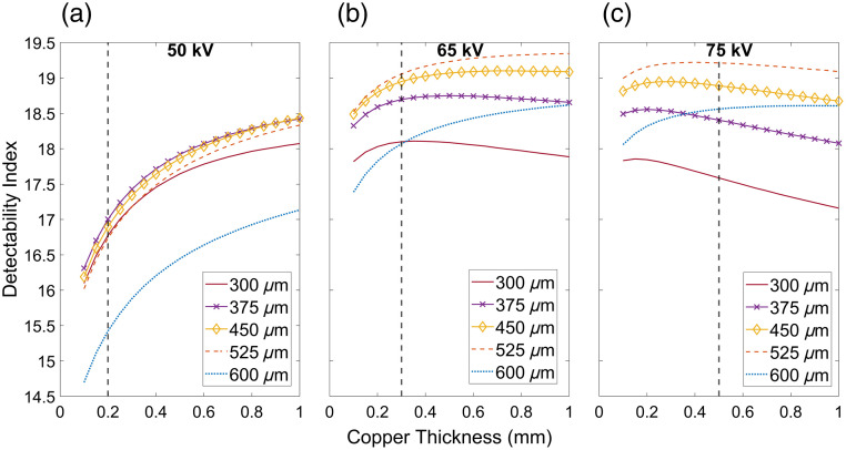
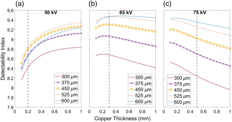
Results: Prewhitening, nonprewhitening, and nonprewhitening with eye filter numerical observers indicate that the combination of 0.525 to 0.6 mm thick scintillator, 70 kV, and 0.25 to 0.4 mm added copper filtration maximized the detectability index at a mean glandular dose (MGD) of 4.5 mGy.
Conclusion: Using parallel cascade systems' analysis, the combination of parameters that could maximize the detection of microcalcifications was identified. The analysis indicates that a harder beam than that used in current practice may be beneficial for the task of detecting microcalcifications at an MGD suitable for breast cancer screening.
Keywords: breast CT; breast cancer; cascaded systems; microcalcifications; numerical observers.
© 2024 Society of Photo-Optical Instrumentation Engineers (SPIE).
Figures

Similar articles
-
The Generalized NEQ and Detectability Index for Tomosynthesis and Cone-Beam CT: From Cascaded Systems Analysis to Human Observers.Proc SPIE Int Soc Opt Eng. 2010 Mar 22;7622:10.1117/12.845462. doi: 10.1117/12.845462. Proc SPIE Int Soc Opt Eng. 2010. PMID: 24307930 Free PMC article.
-
Investigation of statistical iterative reconstruction for dedicated breast CT.Med Phys. 2013 Aug;40(8):081904. doi: 10.1118/1.4811328. Med Phys. 2013. PMID: 23927318 Free PMC article.
-
Modeling and evaluation of a high-resolution CMOS detector for cone-beam CT of the extremities.Med Phys. 2018 Jan;45(1):114-130. doi: 10.1002/mp.12654. Epub 2017 Nov 27. Med Phys. 2018. PMID: 29095489 Free PMC article.
-
Microcalcification detectability in breast CT images using CNN observers.Med Phys. 2024 Feb;51(2):933-945. doi: 10.1002/mp.16922. Epub 2023 Dec 28. Med Phys. 2024. PMID: 38154070 Free PMC article.
-
Cone beam breast CT with a high pitch (75 μm), thick (500 μm) scintillator CMOS flat panel detector: visibility of simulated microcalcifications.Med Phys. 2013 Oct;40(10):101915. doi: 10.1118/1.4820440. Med Phys. 2013. PMID: 24089917 Free PMC article.
References
-
- Hendrick R. E., et al. , “Comparison of acquisition parameters and breast dose in digital mammography and screen-film mammography in the American College of Radiology Imaging Network digital mammographic imaging screening trial,” Am. J. Roentgenol. 194(2), 362–369 (2010).AJROAM10.2214/AJR.08.2114 - DOI - PMC - PubMed
Grants and funding
LinkOut - more resources
Full Text Sources
