

Ossifying fibromyxoid tumor of the soft tissue in the left upper arm and a review of the literature: A case report
- PMID: 38756906
- PMCID: PMC11097289
- DOI: 10.3892/etm.2024.12549
Ossifying fibromyxoid tumor of the soft tissue in the left upper arm and a review of the literature: A case report
Abstract
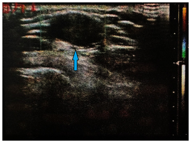
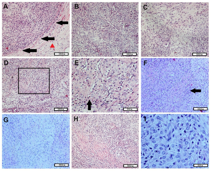
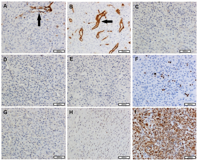
Ossifying fibromyxoid tumor (OFMT) of the soft parts is a mesenchymal neoplasm of uncertain lineage. Fibromyxoid matrix and peripheral metaplastic bone are common histological features of this type of tumor. In the present study, a case of OFMT in a 33-year-old female was reported. The patient was referred to the First Affiliated Hospital of China Medical University (Shenyan, China) in January 2018. The patient had developed a mass in the left upper arm 6 months prior to presentation, which was slowly enlarging. The tumor was 1.5 cm in diameter, with hard texture. Histologically, the tumor showed a clear boundary with no invasion into the adjacent tissue. The majority of tumor cells were round and medium-sized, with abundant pale cytoplasm, without obvious atypia and densely arranged in sheets. The tumor tissue was characterized by cartilage-like morphology and fibromyxoid and hyalinization matrix. Mitotic index was <1/10 high-power fields. Additionally, tumor cells were positive for S-100 and vimentin expression, but negative for smooth muscle actin, CD34, cytokeratin, desmin, human melanoma black 45 and melanoma A. Ki67 index was ~1%. The patient underwent surgery and the tumor was totally removed. No recurrence was observed at the final 6-year follow-up. Based on the aforementioned findings, the patient was diagnosed as typical OFMT. Slow growth and clear boundaries often suggest an indolent nature to this type of tumor. However, close follow-up should be performed due to its malignant potential.
Keywords: case report; ossifying fibromyxoid tumor; upper arm.
Copyright © 2024, Spandidos Publications.
Conflict of interest statement
The authors declare that they have no competing interests.
Figures
Similar articles
-
Ossifying fibromyxoid tumor of soft parts: a clinicopathologic study of 70 cases with emphasis on atypical and malignant variants.Am J Surg Pathol. 2003 Apr;27(4):421-31. doi: 10.1097/00000478-200304000-00001. Am J Surg Pathol. 2003. PMID: 12657926 Review.
-
Ossifying Fibromyxoid tumor of soft parts in head and neck: case report and literature review.Diagn Pathol. 2018 Mar 27;13(1):21. doi: 10.1186/s13000-018-0699-7. Diagn Pathol. 2018. PMID: 29587781 Free PMC article. Review.
-
Ossifying fibromyxoid tumor: report of a case with cytomorphologic description.Diagn Cytopathol. 2015 Aug;43(8):646-9. doi: 10.1002/dc.23287. Epub 2015 Apr 23. Diagn Cytopathol. 2015. PMID: 25914177
-
Malignant ossifying fibromyxoid tumor of the tongue: case report and review of the literature.Head Face Med. 2013 Jun 24;9:16. doi: 10.1186/1746-160X-9-16. Head Face Med. 2013. PMID: 23800162 Free PMC article. Review.
-
Ossifying fibromyxoid tumor: modified myoepithelial cell tumor? Report of three cases with immunohistochemical and electron microscopic studies.Ultrastruct Pathol. 2005 Nov-Dec;29(6):535-48. doi: 10.1080/01913120500323605. Ultrastruct Pathol. 2005. PMID: 16316954
Cited by
-
A rare case of atypical ossifying fibromyxoid tumor: multiple recurrences and metastasis.Skeletal Radiol. 2025 Oct;54(10):2193-2198. doi: 10.1007/s00256-025-04929-y. Epub 2025 Apr 13. Skeletal Radiol. 2025. PMID: 40221618
References
-
- Fletcher CDM, Bridge JA, Hogendoorn PCW, Mertens F. World Health Organization Classifiction of Tumors of Soft Tissue and Bone. IARC Press, Lyon, 2013.
-
- Sharif MA, Mushtaq S, Mamoon N, Khadim MT. Ossifying fibromyxoid tumor of oral cavity. J Coll Physicians Surg Pak. 2008;18:181–182. - PubMed
Publication types
LinkOut - more resources
Full Text Sources