. 2024 May 16:7:0374.
doi: 10.34133/research.0374. eCollection 2024.

Coumarin Glycosides Reverse Enterococci-Facilitated Enteric Infections

Affiliations

Coumarin Glycosides Reverse Enterococci-Facilitated Enteric Infections

Wenjiao Xu et al. Research (Wash D C). .

Abstract

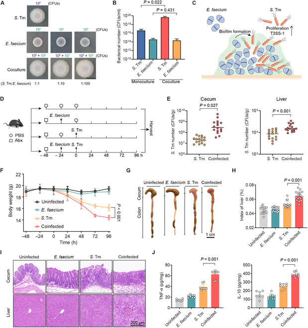
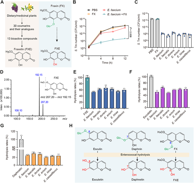
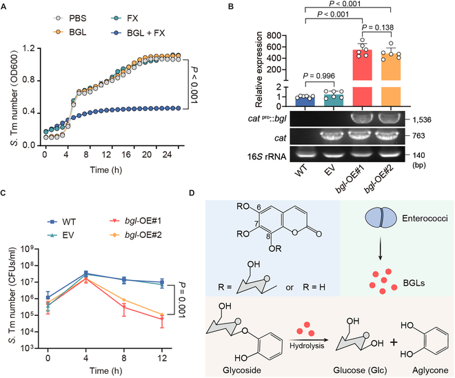
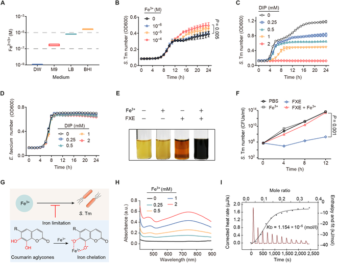
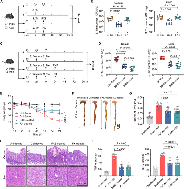
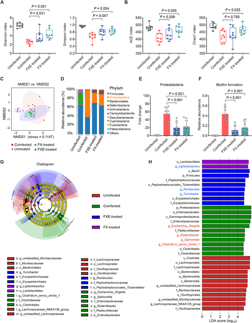
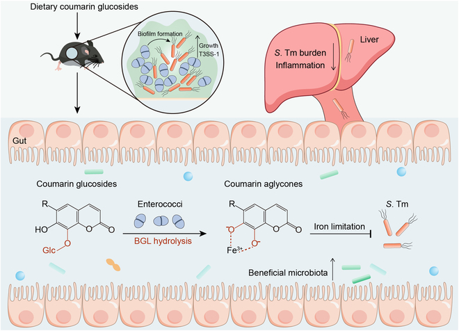
Commensal enterococci with pathogenic potential often facilitate the growth of diverse pathogens, thereby exacerbating infections. However, there are few effective therapeutic strategies to prevent and intervene in enterococci-mediated polymicrobial infections. Here, we find that enterococci at high density drive the expansion and pathogenicity of enteric Salmonella enterica serotype Typhimurium (S. Tm). Subsequently, we show that the driving role of enterococci in such infections is counteracted by dietary coumarin glycosides in vivo. Enterococci, which are tolerant of iron-deficient environments, produce β-glucosidases to hydrolyze coumarin glycosides into bioactive aglycones, inhibiting S. Tm growth and ameliorating the severity of S. Tm-induced symptoms by inducing iron limitation. Overall, we demonstrate that coumarin glycosides as a common diet effectively reverse enterococci-facilitated enteric infections, providing an alternative intervention to combat polymicrobial infections.


Conflict of interest statement

Competing interests: The authors declare that they have no competing interests.

Figures

Fig. 1.
Fig. 1.
Enterococci promote the expansion and pathogenicity of S. Tm. (A) Representative macrocolony images of S. Tm 15E475, E. faecium CAU369, and cocultures (1:1, 1:10, and 1:100) on agar plates. The starting inoculum of S. Tm is 106 CFUs in all macrocolonies. Scale bar, 1 cm. (B) Bacterial counts of S. Tm 15E475 and E. faecium CAU369 in mono- or cocultured (1:100) macrocolonies. n = 3 biological replicates with 3 technical replicates. (C) Schematic diagram of E. faecium enhanced growth and virulence of S. Tm within biofilms. (D) Experimental scheme. In the coinfected group, antibiotic-treated (Abx) mice were orally precolonized with E. faecium CAU369 before challenge with S. Tm 15E475. Mice were colonized with E. faecium CAU369 or S. Tm 15E475 alone as the mono-infected control. n = 16 per group from 2 independent experiments. (E) Enumeration of S. Tm in cecal contents and livers at 96 h after S. Tm infection. (F) Body weight of uninfected, mono-infected, and coinfected mice. (G) Macroscopic phenotype of cecum and colon from corresponding treatments. (H) Organ indices of the liver. (I) Hematoxylin and eosin (H&E) staining images of the corresponding samples. (J) Levels of TNF-α and IL-10 in the cecum. Results represent the means ± SE. P values were calculated using one-way ANOVA with the LSD post hoc test (B) or independent-samples t test (E, F, H, and J).
Fig. 2.
Fig. 2.
Coumarin glycosides inhibit S. Tm growth in the presence of enterococci. (A) Scheme representation of screening bioactive coumarin compounds. Glc, glucose. (B) Population dynamics of S. Tm 15E475 in the cocultures with E. faecium CAU369, FX, or E. faecium CAU369 plus FX for 12 h. (C) Enumeration of S. Tm 15E475 in cocultures with FX or diverse enterococcal species plus FX at 12 h. (D) The MS/MS spectrum and chemical structure of FXE. m/z, mass/charge ratio. (E to G) Hydrolytic rates of diverse enterococcal species to FX (E), esculin (F), and daphnetin (G) based on LC-MS/MS analysis. (H) Schematic diagram illustrating the hydrolysis of glycosidic bonds in dietary coumarin glycosides by enterococci. n = 3 biological replicates with 3 technical replicates. Results represent the means ± SE. P values were calculated using the independent-samples t test.
Fig. 3.
Fig. 3.
Enterococci-derived BGLs hydrolyze coumarin glycosides into antibacterial aglycones. (A) Growth curves of S. Tm 15E475 in cocultures with FX, BGL, or BGL plus FX for 24 h. OD600, optical density at 600 nm. (B) Relative expression of bgl in wild-type (WT; E. faecalis JH2-2), EV, and overexpression strains (bgl-OE#1 and bgl-OE#2). (C) Growth dynamics of S. Tm 15E475 in cocultures with FX plus wild-type, EV, bgl-OE#1, or bgl-OE#2, respectively. (D) Schematic representation of enterococci hydrolyzing glycosides to aglycones via secreting BGLs. n = 3 biological replicates with 3 technical replicates. Results represent the means ± SE. P values were calculated using one-way ANOVA with the LSD post hoc test (B) or independent-samples t test (C).
Fig. 4.
Fig. 4.
Coumarin aglycones inhibit S. Tm growth by chelating iron(III). (A) The concentration of iron in different media including distilled water (DW), M9, Lilly–Barnett (Lilly–Barnett), and BHI. (B) Growth curves of S. Tm 15E475 in M9 broth with the addition of iron(III) for 24 h. (C) Growth curves of S. Tm 15E475 with the addition of DIP for 24 h. (D) Growth curves of E. faecium CAU369 with the addition of DIP for 24 h. (E and F) The addition of excess iron(III) offsets the antibacterial activity of FXE. (E) The color change reaction of S. Tm 15E475 liquid cocultured with FXE, excess iron(III), or FXE plus excess iron(III), respectively. (F) The number of S. Tm 15E475 in the presence of FXE with excess iron(III). (G) Strategy scheme. Iron limitation caused by coumarin aglycones inhibits the growth of S. Tm. (H) The UV-Vis spectra of gradient concentrations of iron(III) with FXE (1 mM) in 5% DMSO buffer. a.u., arbitrary units. (I) Affinity of the chelation between iron(III) and FXE based on the ITC analysis. Iron(III) (2 mM) was dropped into 1 mM FXE in 5% DMSO buffer at 25 °C. Thermodynamic parameters were calculated, including the equilibrium dissociation constant (KD = 1.154 × 10⁻⁵ mol/l), molar binding enthalpy (ΔH = −38.60 kJ/mol), number of binding sites (n = 0.098), and molar binding entropy (ΔS = −34.92 J/mol−1 K−1). n = 3 biological replicates with 3 technical replicates. Results represent the means ± SE. P values were calculated using the independent samples t test.
Fig. 5.
Fig. 5.
Coumarin glycosides reverse the driving role of enterococci in vivo. (A) Experimental scheme. In the S. Tm group, Abx-pretreated mice were orally infected with 107 CFUs of S. Tm 15E475. In the FXET or FXT groups, mice were treated with FXE (100 mg/kg) or FX (100 mg/kg) at 24 h after the single S. Tm infection. n = 12 per group from 3 independent experiments. (B) S. Tm 15E475 counts in cecal contents and livers. (C) Experimental scheme. In the coinfected group, Abx-pretreated mice were orally precolonized with 109 CFUs of E. faecium CAU369 for 24 h before being challenged with 107 CFUs of S. Tm 15E475. In the FXE or FX-treated groups, mice were treated with FXE (100 mg/kg) or FX (100 mg/kg) at 24 h after the coinfection of E. faecium CAU369 and S. Tm 15E475. n = 16 per group from 2 independent experiments. (D) S. Tm 15E475 counts in cecal contents and livers. (E) Body weight of differentially treated mice. (F) Phenotype of the cecum and colon from the corresponding treatments. (G) Organ indices of the liver. (H) Hematoxylin and eosin staining images from the corresponding treatments. (I) Levels of TNF-α and IL-10 in the cecum. Results represent the means ± SE. P values were calculated using one-way ANOVA with the LSD post hoc test.
Fig. 6.
Fig. 6.
Coumarin glycosides ameliorate microbiota dysbiosis induced by the coinfection. (A and B) α-Diversity evaluation of the cecal microbiota. (A) Shannon and Simpson indices. (B) ACE and Chao1 indices. (C) Nonmetric multidimensional scaling (NMDS) score plot of the cecal microbiota based on the binary Jaccard distance metrics. (D and E) Relative abundance of the top 10 phyla (D) and the phylum Proteobacteria (E) in the cecal microbiota. (F) Relative abundance of biofilm formation in the cecal microbiota based on Bugbase analysis. (G) Taxonomic cladogram obtained from the LEfSe analysis of the cecal microbiota. Biomarker taxa are highlighted by colored circles and shaded areas. The diameter of each circle reflects the abundance of those taxa in the community. (H) Taxa with different abundance in the cecal microbiota. A cutoff value of ≥4.5 was used for the linear discriminant analysis (LDA). Results represent the means ± SE. P values were calculated using one-way ANOVA with the LSD post hoc test.
Fig. 7.
Fig. 7.
Scheme of coumarin glucosides reversing enterococci facilitated S. Tm infection. The intestinal domination of enterococci promotes the expansion and pathogenicity of S. Tm. After the oral administration of coumarin glucosides, enterococci inhibit the growth and invasion of S. Tm by hydrolyzing dietary coumarin glycosides to antibacterial aglycones. These catechol aglycones suppress the growth of pathogenic bacteria via causing iron limitation. Meanwhile, coumarin glycosides and their aglycones ameliorate the microbiota dysbiosis of enterococci-mediated polymicrobial infections.

Similar articles

Cited by

References

    1. Zitvogel L, Kroemer G. Immunostimulatory gut bacteria. Science. 2019;366(6469):1077–1078. - PubMed
    1. Freedberg DE, Zhou MJ, Cohen ME, Annavajhala MK, Khan S, Moscoso DI, Brooks C, Whittier S, Chong DH, Uhlemann AC, et al. . Pathogen colonization of the gastrointestinal microbiome at intensive care unit admission and risk for subsequent death or infection. Intensive Care Med. 2018;44(8):1203–1211. - PMC - PubMed
    1. Xu W, Fang Y, Zhu K. Enterococci facilitate polymicrobial infections. Trends Microbiol. 2024;32(2):162–177. - PubMed
    1. Berkell M, Mysara M, Xavier BB, Werkhoven CH, Monsieurs P, Lammens C, Ducher A, Vehreschild MJGT, Goossens H, Gunzburg J, et al. . Microbiota-based markers predictive of development of Clostridioides difficile infection. Nat Commun. 2021;12(1):2241. - PMC - PubMed
    1. Ch’ng JH, Muthu M, Chong KKL, Wong JJ, Tan CAZ, Koh ZJS, Lopez D, Matysik A, Nair ZJ, Barkham T, et al. . Heme cross-feeding can augment Staphylococcus aureus and Enterococcus faecalis dual species biofilms. ISME J. 2022;16(8):2015–2026. - PMC - PubMed

LinkOut - more resources