. 2020 Feb 24;20(2):582-601.
doi: 10.5114/aoms.2020.93216. eCollection 2024.

TLR4 knockdown by miRNA-140-5p improves tendinopathy: an in vitro study

Affiliations

TLR4 knockdown by miRNA-140-5p improves tendinopathy: an in vitro study

Tu Jun et al. Arch Med Sci. .

Abstract

Introduction: The purpose of this study was to determine whether TLR4 knockdown induced by miRNA-140-5p improves tendinopathy in an in vitro experiment.

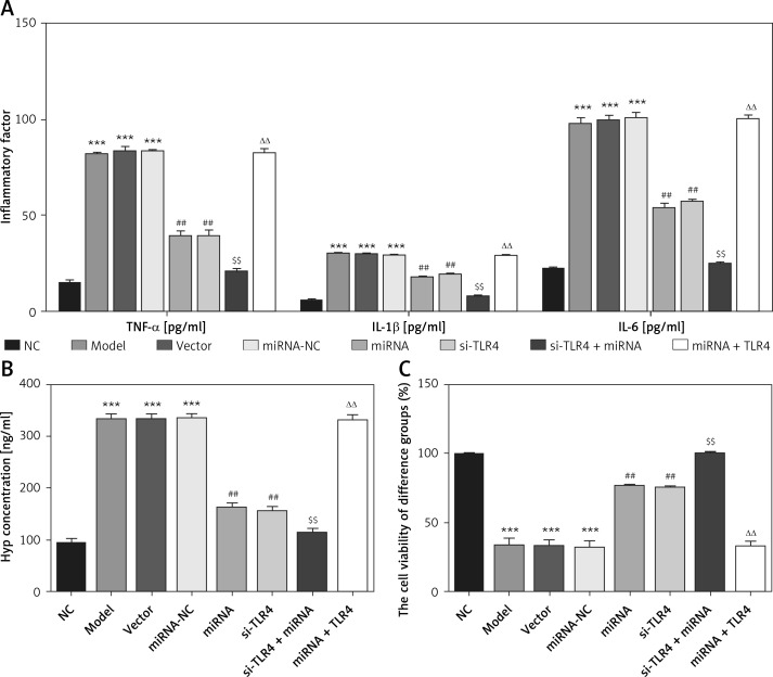
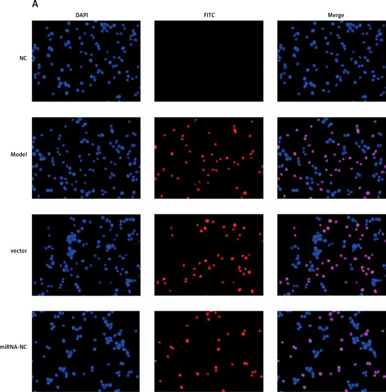
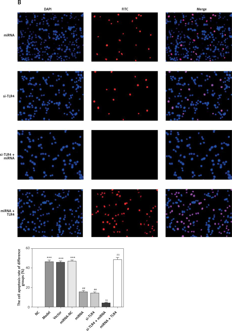
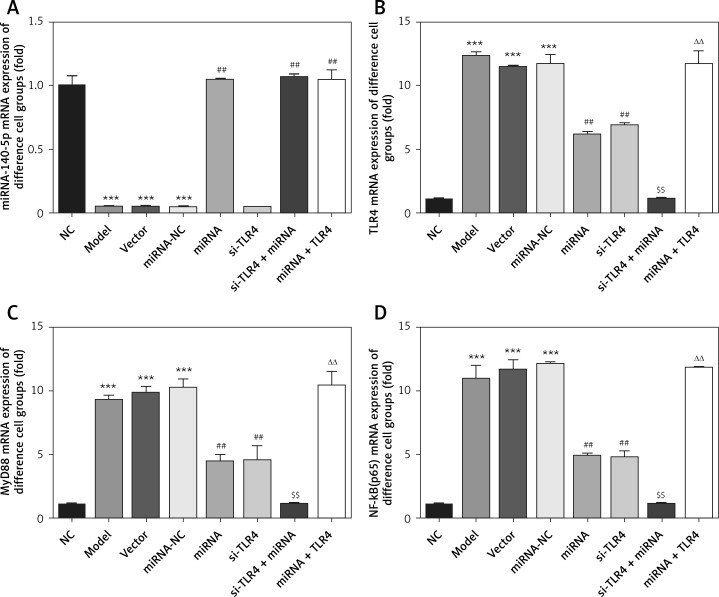
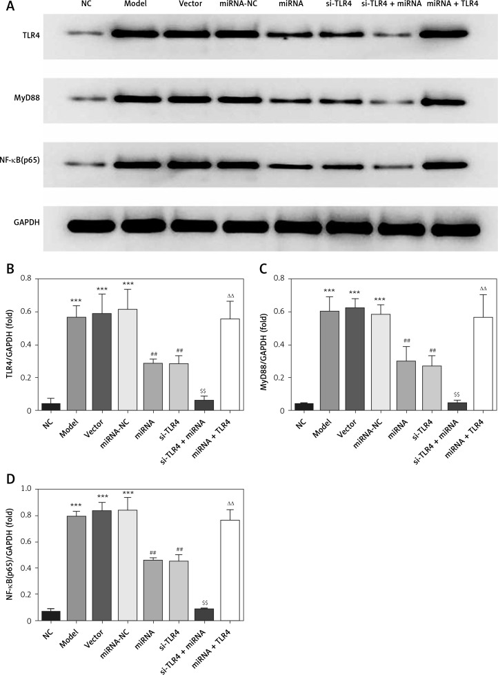
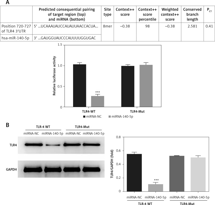
Material and methods: Extraction of tendon-derived stem cells (TDSCs) from SD rats was performed using TGF-β1 to develop a tendinopathy cell model. In the first step, we knocked down TLR4 by si-TLR4 to investigate TLR4 in tendinopathy development, and the next we used miRNA-140-5p to investigate miRNA-140-5p in tendinopathy development. The inflammatory factors and Hyp concentration were evaluated by ELISA assay; the cell viability was measured by MTT assay; the cell apoptosis was evaluated by TUNEL and/or flow cytometry. The relative mRNA was measured by RT-qPCR assay; the relative proteins expression was evaluated by cellular immunofluorescence and/or WB assay. The correlation between miRNA-140-5p and TLR4 was analyzed by Luciferase reporter assay.

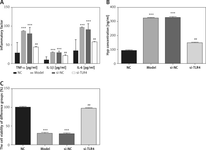
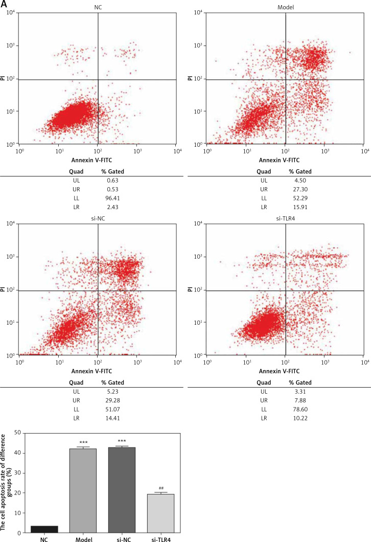
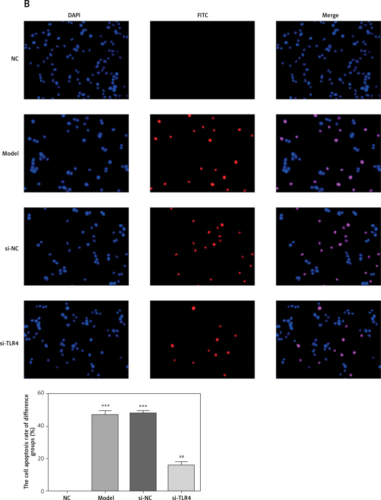
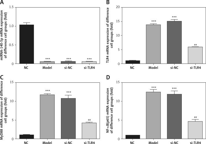
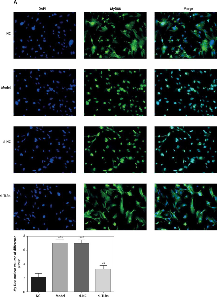
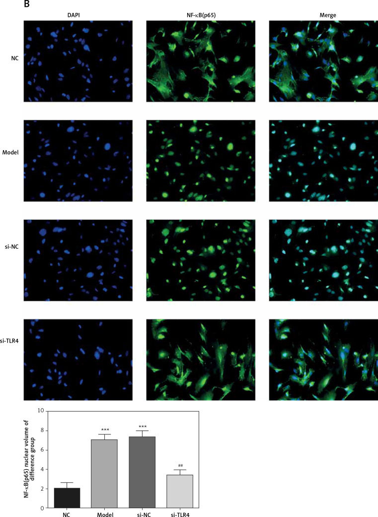
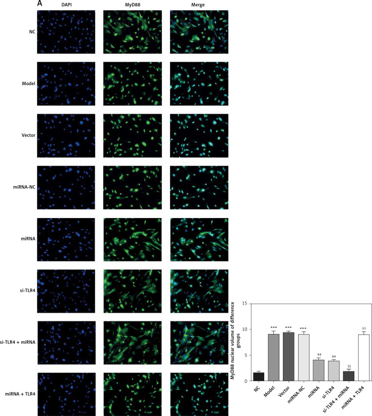
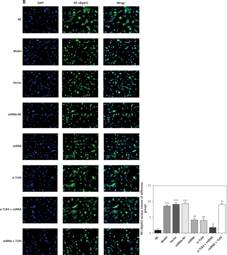
Results: With miRNA-140-5p overexpression or TLR4 knockdown, the cell viability was significantly increased with cell apoptosis depressing compared with the Model group (p < 0.05, respectively). Meanwhile, the inflammatory factors TNF-α, IL-1β and IL-6 and Hyp concentration were significantly improved (p < 0.05, respectively), whereas the TLR4, MyD88 and NF-κB(p65) protein expression levels were significantly depressed with TLR4 knockdown by si-TLR4 or miRNA-140-5p which target TLR4.

Conclusions: The present results showed that TLR4 knockdown induced by miRNA-140-5p or si-TLR4 improved tendinopathy in an in vitro cell experiment.

Keywords: MyD88; NF-κB(p65); TLR4; miRNA-140-5p; tendinopathy.


Conflict of interest statement

The authors declare no conflict of interest.

Figures

Figure 1
Figure 1
TDSC identification
Figure 2
Figure 2
Influence of TLR4 knock-down on inflammatory factors, Hyp and TDSCs proliferation. NC – The TDSCs were treated with normal treatment; Model – the TDSCs treated with 50 ng/ml TGF-β1; si-NC – the cells were transfected with si-NC (negative control) based on the model; si-TLR4 – the cells were transfected with si-TLR4 (knockdown TLR4) based on the model. A – TLR4 knock-down affects inflammatory factors. ***P < 0.001, compared with NC group; ##p < 0.01, compared with Model group. B – TLR4 knock-down affects Hyp concentration by ELISA assay. ***P < 0.001, compared with NC group; ##p < 0.01, compared with Model group. C – TLR4 knock-down affects cell viability by MTT assay. ***P < 0.001, compared with NC group; ##p < 0.01, compared with Model group
Figure 3
Figure 3
Influence of TLR4 knock-down on TDSC apoptosis. NC – the TDSCs were treated with normal treatment; Model – the TDSCs were treated with 50 ng/ml TGF-β1; si-NC – the cells were transfected with si-NC (negative control) based on the model; si-TLR4 – the cells were transfected with si-TLR4 (knockdown TLR4) based on the model. A – TLR4 knockdown affect cell apoptosis by flow cytometry. ***P < 0.001, compared with NC group; ##p < 0.01, compared with Model group. B – TLR4 knockdown affect cell apoptosis by TUNEL assay. ***P < 0.001, compared with NC group; ##p < 0.01, compared with Model group
Figure 3
Figure 3
Influence of TLR4 knock-down on TDSC apoptosis. NC – the TDSCs were treated with normal treatment; Model – the TDSCs were treated with 50 ng/ml TGF-β1; si-NC – the cells were transfected with si-NC (negative control) based on the model; si-TLR4 – the cells were transfected with si-TLR4 (knockdown TLR4) based on the model. A – TLR4 knockdown affect cell apoptosis by flow cytometry. ***P < 0.001, compared with NC group; ##p < 0.01, compared with Model group. B – TLR4 knockdown affect cell apoptosis by TUNEL assay. ***P < 0.001, compared with NC group; ##p < 0.01, compared with Model group
Figure 4
Figure 4
Influence of TLR4 knock-down on related genes by RT-qPCR assay. NC – the TDSCs were treated with normal treatment; Model – the TDSCs were treated with 50 ng/ml TGF-β1; si-NC – the cells were transfected with si-NC (negative control) based on the model; si-TLR4 – the cells were transfected with si-TLR4 (knockdown TLR4) based on the model ***P < 0.001, compared with NC group; ##p < 0.01, compared with Model group.
Figure 5
Figure 5
Influence of TLR4 knock-down on amounts of MyD88 and NF-κB(p65) that are transported to nucleus. NC – the TDSCs were treated with normal treatment; Model – the TDSCs were treated with 50 ng/ml TGF-β1; si-NC – the cells were transfected with si-NC (negative control) based on the model; si-TLR4 – the cells were transfected with si-TLR4 (knockdown TLR4) based on the model. A – TLR4 knock-down affect MyD88 nuclear volume. ***P < 0.001, compared with NC group; ##p < 0.01, compared with Model group. B – TLR4 knock-down affect NF-κB(p65) nuclear volume. ***P < 0.001, compared with NC group; ##p < 0.01, compared with Model group
Figure 5
Figure 5
Influence of TLR4 knock-down on amounts of MyD88 and NF-κB(p65) that are transported to nucleus. NC – the TDSCs were treated with normal treatment; Model – the TDSCs were treated with 50 ng/ml TGF-β1; si-NC – the cells were transfected with si-NC (negative control) based on the model; si-TLR4 – the cells were transfected with si-TLR4 (knockdown TLR4) based on the model. A – TLR4 knock-down affect MyD88 nuclear volume. ***P < 0.001, compared with NC group; ##p < 0.01, compared with Model group. B – TLR4 knock-down affect NF-κB(p65) nuclear volume. ***P < 0.001, compared with NC group; ##p < 0.01, compared with Model group
Figure 6
Figure 6
Influence of miRNA-140-5p on inflammatory factors, Hyp and TDSCs proliferation. NC – The TDSCs were treated with normal treatment; Model – The TDSCs were treated with 50 ng/ml TGF-β1; Vector – The cells were transfected with empty vector based on the model; miRNA-NC – The cells were transfected with miRNA-NC (negative control) based on the model; miRNA – The cells were transfected with miRNA-140-5p based on the model; si-TLR4 – The cells were transfected with si-TLR4 (knockdown TLR4) based on the model; si-TLR4 + miRNA – The cells were transfected with miRNA-140-5p and si-TLR4 (knockdown TLR4) based on the model; miRNA + TLR4 – The cells were transfected with miRNA-140-5p and TLR4 (TLR4 overexpression) based on the model. A – miRNA-140-5p affect inflammatory factors by ELISA assay. ***P < 0.001, compared with NC group; ##p < 0.01, compared with miRNA-NC group; $$p < 0.01, compared with miRNA group; ΔΔp < 0.01, compared with si-TLR4 + miRNA group. B – miRNA-140-5p affect Hyp concentration by ELISA assay. ***P < 0.001, compared with NC group; ##p < 0.01, compared with miRNA-NC group; $$p < 0.01, compared with miRNA group; ΔΔp < 0.01, compared with si-TLR4 + miRNA group. C – miRNA-140-5p affects cell proliferation by MTT assay. ***P < 0.001, compared with NC group; ##p < 0.01, compared with miRNA-NC group; $$p < 0.01, compared with miRNA group; ΔΔp < 0.01, compared with si-TLR4 + miRNA group
Figure 7
Figure 7
miRNA-140-5p affects cell apoptosis by TUNEL assay. NC – the TDSCs were treated with normal treatment; Model – the TDSCs were treated with 50 ng/ml TGF-β1; Vector – the cells were transfected with empty vector based on the model; miRNA-NC – the cells were transfected with miRNA-NC (negative control) based on the model; miRNA – the cells were transfected with miRNA-140-5p based on the model; si-TLR4 – the cells were transfected with si-TLR4 (knockdown TLR4) based on the model; si-TLR4 + miRNA – the cells were transfected with miRNA-140-5p and si-TLR4 (knockdown TLR4) based on the model; miRNA + TLR4 – the cells were transfected with miRNA-140-5p and TLR4 (TLR4 overexpression) based on the model. ***P < 0.001, compared with NC group; ##p < 0.01, compared with miRNA-NC group; $$p < 0.01, compared with miRNA group; ΔΔp < 0.01, compared with si-TLR4 + miRNA group
Figure 7
Figure 7
miRNA-140-5p affects cell apoptosis by TUNEL assay. NC – the TDSCs were treated with normal treatment; Model – the TDSCs were treated with 50 ng/ml TGF-β1; Vector – the cells were transfected with empty vector based on the model; miRNA-NC – the cells were transfected with miRNA-NC (negative control) based on the model; miRNA – the cells were transfected with miRNA-140-5p based on the model; si-TLR4 – the cells were transfected with si-TLR4 (knockdown TLR4) based on the model; si-TLR4 + miRNA – the cells were transfected with miRNA-140-5p and si-TLR4 (knockdown TLR4) based on the model; miRNA + TLR4 – the cells were transfected with miRNA-140-5p and TLR4 (TLR4 overexpression) based on the model. ***P < 0.001, compared with NC group; ##p < 0.01, compared with miRNA-NC group; $$p < 0.01, compared with miRNA group; ΔΔp < 0.01, compared with si-TLR4 + miRNA group
Figure 8
Figure 8
miRNA-140-5p affect relative mRNA expression by RT-qPCR assay. NC – the TDSCs were treated with normal treatment; Model – the TDSCs were treated with 50 ng/ml TGF-β1; Vector – the cells were transfected with empty vector based on the model; miRNA-NC – The cells were transfected with miRNA-NC (negative control) based on the model; miRNA – the cells were transfected with miRNA-140-5p based on the model; si-TLR4 – the cells were transfected with si-TLR4 (knockdown TLR4) based on the model; si-TLR4 + miRNA – the cells were transfected with miRNA-140-5p and si-TLR4 (knockdown TLR4) based on the model; miRNA + TLR4 – the cells were transfected with miRNA-140-5p and TLR4 (TLR4 overexpression) based on the model. ***P < 0.001, compared with NC group; ##p < 0.01, compared with miRNA-NC group; $$p < 0.01, compared with miRNA group; ΔΔp < 0.01, compared with si-TLR4 + miRNA group
Figure 9
Figure 9
miRNA-140-5p affects relative protein expression by WB assay. NC – the TDSCs cells were treated with normal treatment; Model – the TDSCs were treated with 50 ng/ml TGF-β1; Vector – the cells were transfected with empty vector based on the model; miRNA-NC – the cells were transfected with miRNA-NC (negative control) based on the model; miRNA – the cells were transfected with miRNA-140-5p based on the model; si-TLR4 – the cells were transfected with si-TLR4 (knockdown TLR4) based on the model; si-TLR4 + miRNA – the cells were transfected with miRNA-140-5p and si-TLR4 (knockdown TLR4) based on the model; miRNA + TLR4 – the cells were transfected with miRNA-140-5p and TLR4 (TLR4 overexpression) based on the model. ***P < 0.001, compared with NC group; ##p < 0.01, compared with miRNA-NC group; $$p < 0.01, compared with miRNA group; ΔΔp < 0.01, compared with si-TLR4 + miRNA group
Figure 10
Figure 10
miRNA-140-5p affect MyD88 and NF-κB(p65) protein nuclear volume. NC – the TDSCs cells were treated with normal treatment; Model – the TDSCs were treated with 50 ng/ml TGF-β1; Vector – the cells were transfected with empty vector based on the model; miRNA-NC – the cells were transfected with miRNA-NC (negative control) based on the model; miRNA – the cells were transfected with miRNA-140-5p based on the model; si-TLR4 – the cells were transfected with si-TLR4 (knockdown TLR4) based on the model; si-TLR4 + miRNA – the cells were transfected with miRNA-140-5p and si-TLR4 (knockdown TLR4) based on the model; miRNA + TLR4 – the cells were transfected with miRNA-140-5p and TLR4 (TLR4 overexpression) based on the model. A – miRNA-140-5p affects MyD88 nuclear volume. ***P < 0.001, compared with NC group; ##p < 0.01, compared with miRNA-NC group; $$p < 0.01, compared with miRNA group; ΔΔp < 0.01, compared with si-TLR4 + miRNA group. B – miRNA-140-5p affects NF-κB(p65) nuclear volume. ***P < 0.001, compared with NC group; ##p < 0.01, compared with miRNA-NC group; $$p < 0.01, compared with miRNA group; ΔΔp < 0.01, compared with si-TLR4 + miRNA group
Figure 10
Figure 10
miRNA-140-5p affect MyD88 and NF-κB(p65) protein nuclear volume. NC – the TDSCs cells were treated with normal treatment; Model – the TDSCs were treated with 50 ng/ml TGF-β1; Vector – the cells were transfected with empty vector based on the model; miRNA-NC – the cells were transfected with miRNA-NC (negative control) based on the model; miRNA – the cells were transfected with miRNA-140-5p based on the model; si-TLR4 – the cells were transfected with si-TLR4 (knockdown TLR4) based on the model; si-TLR4 + miRNA – the cells were transfected with miRNA-140-5p and si-TLR4 (knockdown TLR4) based on the model; miRNA + TLR4 – the cells were transfected with miRNA-140-5p and TLR4 (TLR4 overexpression) based on the model. A – miRNA-140-5p affects MyD88 nuclear volume. ***P < 0.001, compared with NC group; ##p < 0.01, compared with miRNA-NC group; $$p < 0.01, compared with miRNA group; ΔΔp < 0.01, compared with si-TLR4 + miRNA group. B – miRNA-140-5p affects NF-κB(p65) nuclear volume. ***P < 0.001, compared with NC group; ##p < 0.01, compared with miRNA-NC group; $$p < 0.01, compared with miRNA group; ΔΔp < 0.01, compared with si-TLR4 + miRNA group
Figure 11
Figure 11
Analysis of correlation between miRNA-140-5p and TLR4. miRNA-140-5p – the cell containing TLR4-WT or TLR4-Mul were transfected with miRNA-140-5p mimics; miRNA-NC – the cell contained TLR4-WT or TLR4-Mul were transfected with miRNA-NC (negative control); TLR4-WT – TLR4-wild type; TLR4-Mut – TLR4-mutation. A – the correlation between miRNA-140-5p and TLR4 by Double Luciferase Report assay. ***P < 0.001, compared with miRNA-NC. B – TLR4 protein expression in different groups by WB assay. ***P < 0.001, compared with miRNA-NC

Similar articles

Cited by

References

    1. Koca TT, Arslan A, Özdemir F, Acet G. Clinical features of patients diagnosed with degenerative rotator cuff tendon disease: a 6-month prospective-definitive clinical study from turkey. J Phys Ther Sci 2017; 29: 1433-7. - PMC - PubMed
    1. Snedeker JG, Foolen J. Tendon injury and repair – a perspective on the basic mechanisms of tendon disease and future clinical therapy. Acta Biomater 2017; 63: 18-36. - PubMed
    1. Morita W, Snelling SJ, Dakin SG, Carr AJ. Profibrotic mediators in tendon disease: a systematic review. Arthritis Res Ther 2016; 18: 269. - PMC - PubMed
    1. Ackermann PW, Hart DA. General overview and summary of concepts regarding tendon disease topics addressed related to metabolic disorders. Adv Exp Med Biol 2016; 920: 293-8. - PubMed
    1. Schwellnus MP, Swanevelder S, Jordaan E, Derman W, Van Rensburg DCJ. Underlying chronic disease, medication use, history of running injuries and being a more experienced runner are independent factors associated with exercise-associated muscle cramping: a cross-sectional study in 15778 distance runners. Clin J Sport Med 2018; 28: 289-98. - PubMed