

Mapping Thyroid Hormone Action in the Human Brain
- PMID: 38757586
- PMCID: PMC11295854
- DOI: 10.1089/thy.2024.0120
Mapping Thyroid Hormone Action in the Human Brain
Abstract
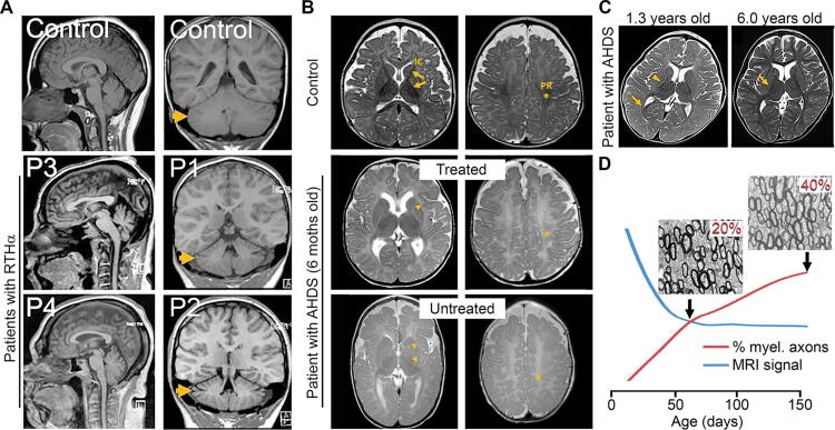
Background: Normal brain development, mood, and cognitive functions depend on thyroid hormone (TH) action. However, little is known about how TH mediates its actions in the human brain. This is due to limited access to human brains deprived of TH during fetal and early postnatal life, as well as from adults with altered thyroid status. One way to partially bypass these limitations is by using magnetic resonance imaging and spectroscopy, two neuroimaging techniques that provide detailed, noninvasive information on human brain structure and function. Another way is using human-induced pluripotent stem cell (hiPSCs)-derived three-dimensional in vitro systems, known as brain organoids, which allow for the study of fundamental aspects of the early stages of human brain development. Summary: This narrative review focuses on neuroimaging and brain organoid studies. Neuroimaging of human brains performed in individuals with different thyroid conditions provides information on the volume, myelination, blood flow, neural activity, and connectivity of different areas. Such studies show that suboptimal thyroid status can impact human brain development and its normal function throughout life. This is true not only for patients with sporadic congenital hypothyroidism, during pregnancy or early after birth, but also for adult patients with hypo- or hyperthyroidism, patients carrying mutations that manifest as impaired sensitivity to TH, and even for normal individuals during aging. Studies using brain organoids generated from hiPSCs of healthy individuals or patients with thyroid genetic conditions provide insights into how TH can impact the early development of the human cerebral cortex. Conclusions: The developmental alterations in children born to mothers with different degrees of gestational hypothyroidism or who developed hypothyroidism early in life are remarkable, affecting multiple brain regions and pathways, including the cerebral cortex, hippocampus, cerebellum, interhemispheric and corticospinal tracts, and associative nuclei. The data connecting such changes to poor neurological outcomes in adult patients with hypothyroidism represent an objective link between thyroid-specific functional brain alterations and behavior. Growing brain organoids require TH, which is critical for human neurogenesis and oligodendrogenesis. These models have proven useful in screening drugs with potential therapeutic effects for patients with genetic thyroid diseases.
Keywords: MRI; brain development; brain organoids; congenital hypothyroidism; resistance to thyroid hormone.
Figures
References
Publication types
MeSH terms
Substances
Grants and funding
LinkOut - more resources
Full Text Sources
