Multicenter Study
. 2024 Jul-Aug;38(4):2138-2150.
doi: 10.1111/jvim.17103. Epub 2024 May 17.

Short colon syndrome in cats

Affiliations
Multicenter Study

Short colon syndrome in cats

Audrey P King et al. J Vet Intern Med. 2024 Jul-Aug.

Abstract

Background: Shortening of the colon has been described in cats, but its imaging and clinicopathological features remain poorly understood.

Objectives: Description of the signalment, clinical presentation, imaging, endoscopic and histological features of short colon syndrome in cats.

Animals: Ninety-three cats diagnosed with short colon.

Methods: Multi-institutional, descriptive, retrospective case series study. Medical records were searched for a diagnosis of short colon on abdominal ultrasonography, computed tomography, endoscopy, autopsy, or a combination of these modalities.

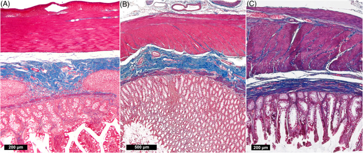
Results: The median age of included cats was 12 years at the time of diagnosis. Diarrhea was the most common clinical sign (60/92; 65%), followed by vomiting (36/92; 39%), weight loss (36/92; 39%), and inappetence (24/92; 26%). Thirteen percent of cats (12/92) had no signs of gastrointestinal disease at the time of diagnosis. In addition to a shortened colonic length, 79% (66/84) of cats had concomitant colonic thickening on ultrasonographic examination. On colonoscopy, mucosal ulcerations of the colonic wall were seen in 39% (9/23) of cats. Histopathologically, all cats but 1 (diagnosed simultaneously with colonic small cell lymphoma) had lymphoplasmacytic colitis, and when small intestinal biopsies were performed, concurrent lymphoplasmacytic enteritis or small cell lymphoma of the small intestine.

Conclusions and clinical importance: Lymphoplasmacytic colitis is seen commonly in cats with short colon, suggesting a potential link between these entities.

Keywords: colonic shortening; decreased colonic length; lack of colonic flexure; lymphoplasmacytic colitis.


Conflict of interest statement

Authors declare no conflict of interest.

Figures

FIGURE 1
FIGURE 1
Sonogram of a cat with short colon. (A) Composite sagittal image of the colon. The colon is severely decreased in length, measuring 5‐10 cm maximum. The ileocecocolic junction and distal portion of the ileum (arrow) are located within the left hemiabdomen, with absence of the transverse and ascending segments of the colon. There is mural thickening (up to 4 mm, yellow double‐sided arrow), characterized by thickening of the submucosa and muscularis layers. The urinary bladder (asterisk) is seen ventral to the most caudal portion of the colon. (B) Transverse image of the colon. A mildly enlarged caudal mesenteric (formerly “left colic”) lymph node (arrowheads) is dorsal to the colon. The mesentery surrounding the colon is hyperechoic.
FIGURE 2
FIGURE 2
Post‐contrast CT dorsal multiplanar reconstruction images displayed in a soft tissue algorithm of a normal cat vs cats with short colon. (A) Normal cat. The ileocecocolic junction is located within the right hemiabdomen. The colonic length measures 29 cm from the rectum to the ileocecocolic junction. (B, C) Cats with short colon. The colon terminates abruptly within the left hemiabdomen and a transverse and ascending colonic segment is not identified. In (B), the cecum is visualized and there is a normal 90° orientation of the ileum in relation to the colon. In (C), the cecum is absent, and the ileum is parallel to the colon at the level of the ileocecocolic junction. The colonic length measures approximately 13‐14 cm in both cases.
FIGURE 3
FIGURE 3
Post‐contrast CT multiplanar reconstruction (A: sagittal MIP, B: transverse, C: parasagittal MIP) in a soft‐tissue algorithm of a cat with short colon, correlated with an endoscopic image from the same cat. (A‐C) On the arterial phase, there is increased vascularization of the colonic wall, with numerous tortuous vessels associated with the mildly thickened colonic wall and associated tortuosity of the colic artery. (D) Endoscopic image taken at the level of the ileocecocolic junction, showing erythema and erosion/ulceration of the colonic mucosa.
FIGURE 4
FIGURE 4
Autopsy image from a normal cat (A), compared to a cat with short colon (B), and biopsy images from a cat with short colon (C, D). (B) A developed cecum is absent, and the small intestine is continuous with the colon. There is no overt ileocecal junction. (C) Ileum of a cat with short colon, diagnosed as moderate, multifocal lymphoplasmacytic enteritis. There are moderately increased numbers of lymphocytes and plasma cells in the lamina propria. The villi are shortened and blunted with a reduced villous: crypt ratio. (D) Colon of the same cat (C), with moderately increased numbers of plasma cells and lymphocytes in the lamina propria. Scattered neutrophils are also observed.
FIGURE 5
FIGURE 5
Masson's trichrome stains, normal cat (A) compared to cats with short colon syndrome (B, C). Fibrous connective tissue (blue staining) is present in the submucosa (just above the mucosa) in the image in the normal cat, as well as the 2 cats with short colon syndrome. The tunica muscularis layers (top of image) in the normal cat has minimal blue staining. The 2 cats with short colon syndrome have mild (B) and moderately (C) increased amounts of fibrous connective tissue in the tunica muscularis layers, more pronounced in the inner layer.
FIGURE 6
FIGURE 6
Kaplan‐Meier curve of cats diagnosed with short colon. The blue solid line represents the survival curve, and the dashed red lines represent the 95% confidence limits. The + symbols indicate censored data (time to last follow‐up).

References

    1. Fluke MH, Hawkins EC, Elliott GS, Blevins WE. Short colon in two cats and a dog. J Am Vet Med Assoc. 1989;195(1):87‐90. - PubMed
    1. Schlesinger DP, Philibert D, Breur GJ. Agenesis of the cecum and the ascending and transverse colon in a 12‐yer‐old cat. Can Vet J. 1992;33(8):544‐546. - PMC - PubMed
    1. Oishi A, Une R, Baba S, Fukushima K, Suzuki K, Miyoshi N. Short colon in a cat. J Jpn Vet Medical Assoc. 2006;59(6):403‐408.
    1. Koenig JB, Rodriguez A, Colquhoun JK, Stämpfli H. Congenital colonic malformation (“short colon”) in a 4‐month‐old standardbred foal. Can Vet J. 2007;48(4):420‐422. - PMC - PubMed
    1. Remmel P, Gros L, Mortier J, Freiche V. Congenital partial colonic agenesis in dogs and cats: clinical, biological, diagnostic imaging, endoscopic and histopathologic characterization, a retrospective study. Vet Sci. 2023;10(9):577. - PMC - PubMed

Publication types

MeSH terms

LinkOut - more resources