Dendritic cell-specific intercellular adhesion molecule-3-grabbing nonintegrin (DC-SIGN) is a cellular receptor for delta inulin adjuvant
- PMID: 38757764
- PMCID: PMC11296934
- DOI: 10.1111/imcb.12774
Dendritic cell-specific intercellular adhesion molecule-3-grabbing nonintegrin (DC-SIGN) is a cellular receptor for delta inulin adjuvant
Abstract
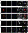
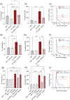
Delta inulin, or Advax, is a polysaccharide vaccine adjuvant that significantly enhances vaccine-mediated immune responses against multiple pathogens and was recently licensed for use in the coronavirus disease 2019 (COVID-19) vaccine SpikoGen. Although Advax has proven effective as an immune adjuvant, its specific binding targets have not been characterized. In this report, we identify a cellular receptor for Advax recognition. In vitro uptake of Advax particles by macrophage cell lines was substantially greater than that of latex beads of comparable size, suggesting an active uptake mechanism by phagocytic cells. Using a lectin array, Advax particles were recognized by lectins specific for various carbohydrate structures including mannosyl, N-acetylgalactosamine and galactose moieties. Expression in nonphagocytic cells of dendritic cell-specific intercellular adhesion molecule-3-grabbing nonintegrin (DC-SIGN), a C-type lectin receptor, resulted in enhanced uptake of fluorescent Advax particles compared with mock-transfected cells. Advax uptake was reduced with the addition of ethylenediaminetetraacetic acid and mannan to cells, which are known inhibitors of DC-SIGN function. Finally, a specific blockade of DC-SIGN using a neutralizing antibody abrogated Advax uptake in DC-SIGN-expressing cells. Together, these results identify DC-SIGN as a putative receptor for Advax. Given the known immunomodulatory role of DC-SIGN, the findings described here have implications for the use of Advax adjuvants in humans and inform future mechanistic studies.
Keywords: Adjuvant; C‐type lectin; DC‐SIGN; vaccine.
© 2024 The Authors. Immunology & Cell Biology published by John Wiley & Sons Australia, Ltd on behalf of the Australian and New Zealand Society for Immunology, Inc.
Conflict of interest statement
CONFLICT OF INTEREST
NP is the research director for Vaxine P/L. All authors attest they meet the criteria for authorship.
Figures


Similar articles
-
The S1 spike protein of SARS-CoV-2 upregulates the ERK/MAPK signaling pathway in DC-SIGN-expressing THP-1 cells.Cell Stress Chaperones. 2024 Apr;29(2):227-234. doi: 10.1016/j.cstres.2024.03.002. Epub 2024 Mar 5. Cell Stress Chaperones. 2024. PMID: 38453000 Free PMC article.
-
Evaluation of the safety and efficacy of AdvaxTM as an adjuvant: A systematic review and meta-analysis.Adv Med Sci. 2022 Mar;67(1):10-17. doi: 10.1016/j.advms.2021.09.002. Epub 2021 Sep 22. Adv Med Sci. 2022. PMID: 34562856
-
Delta inulin alone or combined with CpG oligonucleotide enhances antibody-dependent influenza vaccine protection in mice and nonhuman primate newborns.Immunol Cell Biol. 2025 Jun 29:10.1111/imcb.70045. doi: 10.1111/imcb.70045. Online ahead of print. Immunol Cell Biol. 2025. PMID: 40582987
-
Cost-effectiveness of using prognostic information to select women with breast cancer for adjuvant systemic therapy.Health Technol Assess. 2006 Sep;10(34):iii-iv, ix-xi, 1-204. doi: 10.3310/hta10340. Health Technol Assess. 2006. PMID: 16959170
-
Investigation of the biodistribution, breakdown and excretion of delta inulin adjuvant.Vaccine. 2017 Aug 3;35(34):4382-4388. doi: 10.1016/j.vaccine.2017.06.045. Epub 2017 Jul 1. Vaccine. 2017. PMID: 28676380
Cited by
-
HeberNasvac: Development and Application in the Context of Chronic Hepatitis B.Euroasian J Hepatogastroenterol. 2024 Jul-Dec;14(2):221-237. doi: 10.5005/jp-journals-10018-1457. Epub 2024 Dec 27. Euroasian J Hepatogastroenterol. 2024. PMID: 39802853 Free PMC article. Review.
-
One Health adjuvant selection for vaccines against zoonotic infections.Explor Med. 2025;6:1001316. doi: 10.37349/emed.2025.1001316. Epub 2025 May 7. Explor Med. 2025. PMID: 40444176 Free PMC article.
-
Progress and prospect of polysaccharides as adjuvants in vaccine development.Virulence. 2024 Dec;15(1):2435373. doi: 10.1080/21505594.2024.2435373. Epub 2024 Dec 5. Virulence. 2024. PMID: 39601191 Free PMC article. Review.
-
Chimerolectins: Classification, structural architecture, and functional perspectives.Protein Sci. 2025 Sep;34(9):e70261. doi: 10.1002/pro.70261. Protein Sci. 2025. PMID: 40815298 Free PMC article. Review.
-
Post-Hoc Analysis of Potential Correlates of Protection of a Recombinant SARS-CoV-2 Spike Protein Extracellular Domain Vaccine Formulated with Advax-CpG55.2-Adjuvant.Int J Mol Sci. 2024 Aug 30;25(17):9459. doi: 10.3390/ijms25179459. Int J Mol Sci. 2024. PMID: 39273405 Free PMC article. Clinical Trial.
References
-
- Barclay T, Ginic-Markovic M, Cooper P, Petrovsky N. Inulin - A versatile polysaccharide with multiple pharmaceutical and food chemical uses. Journal of Excipients and Food Chemicals 2010; 1: 27–50.
Publication types
MeSH terms
Substances
Grants and funding
LinkOut - more resources
Full Text Sources
Molecular Biology Databases

