. 2024 Jun 13;98(6):e0049424.
doi: 10.1128/jvi.00494-24. Epub 2024 May 17.

Classical swine fever virus non-structural protein 4A recruits dihydroorotate dehydrogenase to facilitate viral replication

Affiliations

Classical swine fever virus non-structural protein 4A recruits dihydroorotate dehydrogenase to facilitate viral replication

Bing-Qian Zhao et al. J Virol. .

Abstract

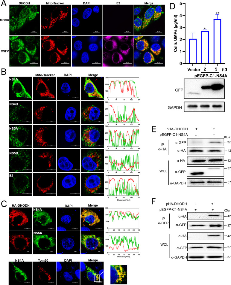
Mitochondria are energy producers in cells, which can affect viral replication by regulating the host innate immune signaling pathways, and the changes in their biological functions are inextricably linked the viral life cycle. In this study, we screened a library of 382 mitochondria-targeted compounds and identified the antiviral inhibitors of dihydroorotate dehydrogenase (DHODH), the rate-limiting enzyme in the de novo synthesis pathway of pyrimidine ribonucleotides, against classical swine fever virus (CSFV). Our data showed that the inhibitors interfered with viral RNA synthesis in a dose-dependent manner, with half-maximal effective concentrations (EC50) ranging from 0.975 to 26.635 nM. Remarkably, DHODH inhibitors obstructed CSFV replication by enhancing the innate immune response including the TBK1-IRF3-STAT1 and NF-κB signaling pathways. Furthermore, the data from a series of compound addition and supplementation trials indicated that DHODH inhibitors also inhibited CSFV replication by blocking the de novo pyrimidine synthesis. Remarkably, DHODH knockdown demonstrated that it was essential for CSFV replication. Mechanistically, confocal microscopy and immunoprecipitation assays showed that the non-structural protein 4A (NS4A) recruited and interacted with DHODH in the perinuclear. Notably, NS4A enhanced the DHODH activity and promoted the generation of UMP for efficient viral replication. Structurally, the amino acids 65-229 of DHODH and the amino acids 25-40 of NS4A were pivotal for this interaction. Taken together, our findings highlight the critical role of DHODH in the CSFV life cycle and offer a potential antiviral target for the development of novel therapeutics against CSF.

Importance: Classical swine fever remains one of the most economically important viral diseases of domestic pigs and wild boar worldwide. dihydroorotate dehydrogenase (DHODH) inhibitors have been shown to suppress the replication of several viruses in vitro and in vivo, but the effects on Pestivirus remain unknown. In this study, three specific DHODH inhibitors, including DHODH-IN-16, BAY-2402234, and Brequinar were found to strongly suppress classical swine fever virus (CSFV) replication. These inhibitors target the host DHODH, depleting the pyrimidine nucleotide pool to exert their antiviral effects. Intriguingly, we observed that the non-structural protein 4A of CSFV induced DHODH to accumulate around the nucleus in conjunction with mitochondria. Moreover, NS4A exhibited a strong interaction with DHODH, enhancing its activity to promote efficient CSFV replication. In conclusion, our findings enhance the understanding of the pyrimidine synthesis in CSFV infection and expand the novel functions of CSFV NS4A in viral replication, providing a reference for further exploration of antiviral targets against CSFV.

Keywords: DHODH; classical swine fever virus (CSFV); interaction; non-structural protein 4A (NS4A); pyrimidine; viral replication.


Conflict of interest statement

The authors declare no conflict of interest.

Figures

Fig 1
Fig 1
The inhibitors are screened from the mitochondrial-targeted compound library. (A) Screening process for the compounds that inhibit CSFV replication. (B) Each dot represents the inhibition rate of the compound (1 µM) against CSFV (MOI of 1). (C) The fluorescence intensity of CSFV E2 for the 13 compounds screened against CSFV was expressed as relative fluorescence intensity. The red columns indicated the DHODH inhibitor. (D) CC50, EC50, and selectivity index (SI = CC50/EC50) values of six DHODH inhibitors against CSFV. Data are presented as the mean ± SD from three independent experiments. **P < 0.01.
Fig 2
Fig 2
DHODH-IN-16, BAY-2402234 and Brequinar inhibit CSFV replication. (A) Cells were treated with either DMSO or different concentrations of DHODH-IN-16, BAY-2402234, and Brequinar for 24 h after infection with CSFV (MOI of 1). Viral replication was determined by RT-qPCR and Western blotting. The effect of DHODH-IN-16, BAY-2402234, and Brequinar on cell cytotoxicity was quantified using the CCK-8 assay. (B) Different concentrations of DHODH-IN-16, BAY-2402234, and Brequinar were added to cells after infected with CSFV (MOI of 1). At 24 hpi, cells were fixed and stained with mouse anti-CSFV E2 antibody (green) and observed by fluorescence microscopy. Bars = 50 µm. (C and D) IPEC or ST cells were treated with either DMSO or different concentrations of DHODH-IN-16, BAY-2402234, and Brequinar for 24 h after infection with CSFV (MOI of 1). Viral replication was determined by Western blotting. Data are presented as the mean ± SD from three independent experiments. **P < 0.01.
Fig 3
Fig 3
Broad-spectrum antiviral activity of DHODH inhibitors against RNA viruses. Cells were treated with either DMSO or different concentrations of DHODH-IN-16, BAY-2402234, and Brequinar for 24 h after infection with (A) JEV (MOI of 0.1), (B) BVDV (MOI of 0.5), (C) PRV (MOI of 0.1) and harvested for RT-qPCR and Western blotting. The effect of DHODH-IN-16, BAY-2402234, and Brequinar on cell cytotoxicity was quantified using CCK-8 assay. Data are presented as the mean ± SD from three independent experiments. **P < 0.01.
Fig 4
Fig 4
DHODH inhibitors affect the initial stage of CSFV replication. (A) Cells were treated with different concentrations of DHODH-IN-16, BAY-2402234, and Brequinar for 2 h and infected with CSFV (MOI of 10) for 1 h at 4°C and then at 37°C at 0 hpi (binding) or 1 hpi (entry). Cells were harvested for RT-qPCR for detecting viral RNA levels. (B) Time points of addition of DHODH inhibitors in CSFV infection. Cells were treated with 10 nM DHODH-IN-16, 10 nM BAY-2402234, or 500 nM Brequinar at various time points during the infection by CSFV (MOI of 1). Viral replication was determined by RT-qPCR and Western blotting. 12 h pretreatment, cells were pretreated with DHODH inhibitors for 12 h; 1 h entry, cells were treated with DHODH inhibitors and CSFV for 1 h before the mixture was washed and treated with 2% DMEM; after entry, cells were treated with DHODH inhibitors 1 h after CSFV entry; throughout, cells were treated with DHODH inhibitors for the whole time period, including the 12 h pretreatment. (C) The infected cells treated with 10 nM DHODH-IN-16, 10 nM BAY-2402234, or 500 nM Brequinar for 4 h were harvested and subjected to RT-qPCR for detecting the production of CSFV (−)strand RNA. (D) Cells were treated with different concentrations of DHODH inhibitors for 24 and 48 h after infection by CSFV (MOI of 1), and the supernatant was collected and used to infect fresh cells for 24 h. The viral genome was determined by RT-qPCR. Data are presented as the mean ± SD from three independent experiments. **P < 0.01.
Fig 5
Fig 5
DHODH inhibitors repress CSFV replication through enchancing the innate immune response. (A) Cells were infected with CSFV (MOI of 1) and treated with DMSO or 500 nm Brequinar. The treated cells were harvested at indicated time points and subjected to Western blotting using indicated antibodies against TBK1, pTBK1, IRF3, pIRF3, STAT1, pSTAT1, p65, p-p65, Npro, and β-actin. (B and C) The treated cells described above were lysed and subjected to RT-qPCR for detecting the mRNA levels of ISG15, IFIT1, MX1, IL-6, IL-8, and TNF-α. Data are presented as the mean ± SD from three independent experiments. *P < 0.05; **P < 0.01.
Fig 6
Fig 6
DHODH inhibitors block viral replication by interfering with host pyrimidine biosynthesis. (A) Schematic overview of de novo biosynthesis of pyrimidine nucleotide. (B) Cells infected with CSFV (MOI of 1) were treated with DMSO or different concentrations of pyrazofuran for 24 h, viral replication was determined by RT-qPCR and Western blotting. (C and D) Cells were treated with DMSO, different concentrations of DHO (C) or ORO (D) for 24 h after infection with CSFV (MOI of 0.1), viral replication was detected by RT-qPCR and Western blotting. (E–G) Cells infected with CSFV (MOI of 1) were treated with 500 nM Brequinar in medium with or without DHO or ORO and harvested at 24 h for (E) HPLC, (F and G) RT-qPCR, and Western blotting. (H) The infected cells pre-treated with Brequinar were treated with or without 100 µM uridine. After 24 h, cells were harvested and subjected to RT-qPCR and Western blotting for detecting viral replication. (I) Cells infected with BVDV were pre-treated with Brequinar and treated with or without 100 µM uridine. After 24 h, cells were harvested and subjected to Western blotting for detecting viral replication. Data are presented as the mean ± SD from three independent experiments. *P < 0.05; **P < 0.01.
Fig 7
Fig 7
DHODH is essential for CSFV infection. (A) Cells were infected with CSFV (MOI of 1), harvested at indicated time points, and then subjected to Western blotting and HPLC assay. (B) Cells were infected with CSFV (MOI of 1, 2, 10) for 24 h and then harvested for Western blotting and HPLC assay. (C) Cells transfected with siDHODH or siCtrl were inoculated with CSFV (MOI of 1) and harvested for Western blotting and HPLC. (D) Cells transfected with the plasmid pHA-DHODH or vector (pcDNA3.1-HA) were infected with CSFV (MOI of 1) for 24 h, harvested, and then subjected to Western blotting and HPLC. (E) The infected DHODH-knockdown cells were treated with or without 100 µM uridine and harvested at 24 h for Western blotting. Data are presented as the mean ± SD from three independent experiments. *P < 0.05; **P < 0.01.
Fig 8
Fig 8
CSFV NS4A interacts with DHODH. (A) Cells were infected with CSFV (MOI of 1) and fixed at 24 hpi. Then cells were stained with rabbit anti-DHODH antibody (green), mouse anti-E2 antibody (purple), and Mito-Tracker (red) and observed by confocal microscopy. The nuclei were stained with DAPI. Bars = 10 µm. (B) Cells transfected with indicated plasmids (pEGFP-C1-NS4A, pFlag-NS4B, -NS5A, -NS5B, or -E2) for 24 h were fixed and subjected to confocal microscopy by using mouse anti-Flag antibody (green) and mito-Tracker (red). The nuclei were stained with DAPI. Bars = 10 µm. (C) Cells transfected with indicated plasmids (pFlag-NS5A, or pEGFP-C1-NS4A) for 24 h were fixed and subjected to confocal microscopy using rabbit anti-DHODH antibody (red) and mouse anti-Flag antibody (green). The nuclei were stained with DAPI. Bars = 10 µm. (D) Cells transfected with pEGFP-C1-NS4A or vector (pEGFP-C1) for 24 h were harvested and subjected to Western blotting and HPLC assay. (E and F) Cells cotransfected with pEGFP-C1-NS4A and pHA-DHODH or control vector for 24 h were harvested and subjected to Co-IP assay by using anti-GFP or anti-HA nanobody magarose beads. Then, the whole cell lysates were subjected to Western blotting using rabbit anti-DHODH or anti-GFP antibody, along with GAPDH as a loading control.
Fig 9
Fig 9
The key domains of NS4A and DHODH interaction. (A) Schematic of full-length NS4A and its three truncated mutants. (B) Cells transfected with pEGFP-C1-NS4A or its truncated mutant plasmids (pEGFP-C1-NS4A-F1, -F2, or -F3) or control vector for 24 h were harvested and subjected to Western blotting and HPLC assay. (C) Cells cotransfected with pHA-DHODH and indicated plasmids (pEGFP-C1-NS4A-FL, -F1, -F2, or -F3) for 24 h were fixed and subjected to confocal microscopy using rabbit anti-HA antibody (red). The nuclei were stained with DAPI. Bars = 10 µm. (D) Cells cotransfected with pHA-DHODH and indicated plasmids (pEGFP-C1-NS4A-FL, -F1, -F2, or -F3) for 24 h were harvested for Co-IP assay by using anti-GFP antibody margarose beads, and whole-cell lysates were subjected to Western blotting using rabbit anti-HA antibody, along with GAPDH as a loading control. (E) Schematic of full-length DHODH and its three truncated mutants. (F) Cells transfected with pHA-DHODH or its truncated mutant plasmids (pHA-DHODH-F1, -F2, or -F3) or control vector for 24 h were harvested and subjected to Western blotting. (G) Cells cotransfected with pEGFP-C1-NS4A and indicated plasmids (pHA-DHODH-FL, -F1, -F2, or -F3) for 24 h were fixed and subjected to confocal microscopy using rabbit anti-HA antibody (red). The nuclei were stained with DAPI. Bars = 10 µm. (H) Cells were cotransfected with p-EGFP-C1-NS4A and indicated plasmids (pHA-DHODH-FL, -F1, -F2, or -F3) for 24 h and harvested for Co-IP using anti-GFP nanobody-margarose beads, and whole cell lysates were subjected to Western blotting using rabbit anti-HA antibody, along with GAPDH as a loading control.

Similar articles

References

    1. Yuan M, Yang X, Zhang X, Zhao X, Abid M, Qiu HJ, Li Y. 2022. Different types of vaccines against pestiviral infections: "barriers" for "pestis" Viruses 15:2. doi:10.3390/v15010002 - DOI - PMC - PubMed
    1. Liu C-C, Liu Y-Y, Zhou J-F, Chen X, Chen H, Hu J-H, Chen J, Zhang J, Sun R-C, Wei J-C, Go YY, Morita E, Zhou B. 2022. Cellular ESCRT components are recruited to regulate the endocytic trafficking and RNA replication compartment assembly during classical swine fever virus infection. PLoS Pathog 18:e1010294. doi:10.1371/journal.ppat.1010294 - DOI - PMC - PubMed
    1. Blome S, Staubach C, Henke J, Carlson J, Beer M. 2017. Classical swine fever-an updated review. Viruses 9:86. doi:10.3390/v9040086 - DOI - PMC - PubMed
    1. Rossi S, Staubach C, Blome S, Guberti V, Thulke HH, Vos A, Koenen F, Le Potier MF. 2015. Controlling of CSFV in European wild boar using oral vaccination: a review. Front Microbiol 6:1141. doi:10.3389/fmicb.2015.01141 - DOI - PMC - PubMed
    1. Xu H, Han G, Lu Y, Liu Z, Tao L, He F. 2021. Broad neutralization of CSFV with novel monoclonal antibodies in vivo. Int J Biol Macromol 173:513–523. doi:10.1016/j.ijbiomac.2021.01.142 - DOI - PubMed

MeSH terms

LinkOut - more resources