

MUC16: clinical targets with great potential
- PMID: 38758220
- PMCID: PMC11101557
- DOI: 10.1007/s10238-024-01365-5
MUC16: clinical targets with great potential
Abstract
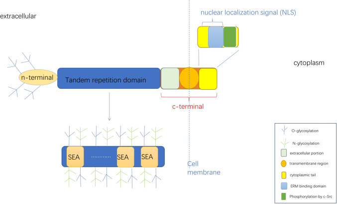
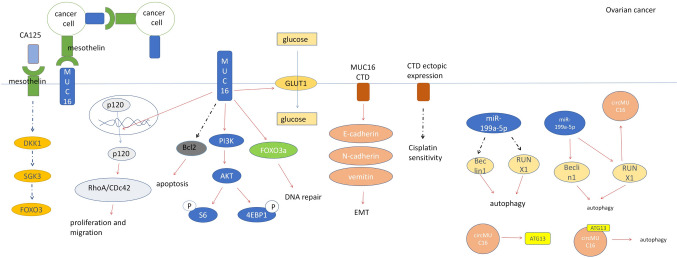
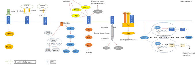
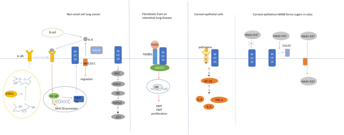
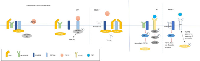
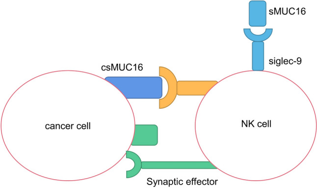
Mucin 16 (MUC16) is a membrane-bound mucin that is abnormally expressed or mutated in a variety of diseases, especially tumors, while being expressed in normal body epithelium. MUC16 and its extracellular components are often important cancer-related biomarkers. Abnormal expression of MUC16 promotes tumor progression through mesenchymal protein, PI3K/AKT pathway, JAK2/STAT3 pathway, ERK/FBW7/c-Myc, and other mechanisms, and plays an important role in the occurrence and development of tumors. In addition, MUC16 also helps tumor immune escape by inhibiting T cells and NK cells. Many drugs and trials targeting MUC16 have been developed, and MUC16 may be a new direction for future treatments. In this paper, the mechanism of action of MUC16 in the development of cancer, especially in the immune escape of tumor, is introduced in detail, indicating the potential of MUC16 in clinical treatment.
Keywords: Biomarker; CA125; Clinical trials; Immune escape; Mucin 16 (MUC16).
© 2024. The Author(s).
Conflict of interest statement
The authors declare that they have no competing interests.
Figures
Similar articles
-
MUC16/CA125 in cancer: new advances.Clin Chim Acta. 2025 Jan 15;565:119981. doi: 10.1016/j.cca.2024.119981. Epub 2024 Oct 4. Clin Chim Acta. 2025. PMID: 39368688 Review.
-
Ascites from ovarian cancer patients stimulates MUC16 mucin expression and secretion in human peritoneal mesothelial cells through an Akt-dependent pathway.BMC Cancer. 2019 Apr 30;19(1):406. doi: 10.1186/s12885-019-5611-7. BMC Cancer. 2019. PMID: 31039761 Free PMC article.
-
[MUC16: The Novel Target for Tumor Therapy].Zhongguo Fei Ai Za Zhi. 2022 Jul 20;25(7):452-459. doi: 10.3779/j.issn.1009-3419.2022.101.31. Zhongguo Fei Ai Za Zhi. 2022. PMID: 35899441 Free PMC article. Review. Chinese.
-
MUC16 facilitates cervical cancer progression via JAK2/STAT3 phosphorylation-mediated cyclooxygenase-2 expression.Genes Genomics. 2020 Feb;42(2):127-133. doi: 10.1007/s13258-019-00885-9. Epub 2019 Nov 17. Genes Genomics. 2020. PMID: 31736008
-
MUC16 (CA125): tumor biomarker to cancer therapy, a work in progress.Mol Cancer. 2014 May 29;13:129. doi: 10.1186/1476-4598-13-129. Mol Cancer. 2014. PMID: 24886523 Free PMC article. Review.
Cited by
-
Hypomethylation-associated ELF3 helps nasopharyngeal carcinoma to escape immune surveillance via MUC16-mediated glycolytic metabolic reprogramming.Am J Physiol Cell Physiol. 2024 Oct 1;327(4):C1125-C1142. doi: 10.1152/ajpcell.00438.2024. Epub 2024 Sep 2. Am J Physiol Cell Physiol. 2024. PMID: 39219440 Free PMC article.
-
Overlap of Genomic and Transcriptomic Genes Identified in Familial Eosinophilic Esophagitis.Gastroenterology. 2025 Jun;168(6):1101-1113.e18. doi: 10.1053/j.gastro.2025.01.235. Epub 2025 Feb 4. Gastroenterology. 2025. PMID: 39914776
-
Identification and Validation of Key Genes Involved in the Coupling of Mitochondria-Associated Endoplasmic Reticulum Membrane in Hemorrhoidal Disease.Int J Gen Med. 2025 May 31;18:2781-2798. doi: 10.2147/IJGM.S511281. eCollection 2025. Int J Gen Med. 2025. PMID: 40469970 Free PMC article.
-
CA125 as a Potential Biomarker in Non-Malignant Serous Effusions: Diagnostic and Prognostic Considerations.J Clin Med. 2025 Jun 11;14(12):4152. doi: 10.3390/jcm14124152. J Clin Med. 2025. PMID: 40565905 Free PMC article. Review.
-
The multifaceted roles of mucins family in lung cancer: from prognostic biomarkers to promising targets.Front Immunol. 2025 Jun 27;16:1608140. doi: 10.3389/fimmu.2025.1608140. eCollection 2025. Front Immunol. 2025. PMID: 40655139 Free PMC article. Review.
References
-
- Bast RC, Klug TL, St John E, et al. A radioimmunoassay using a monoclonal antibody to monitor the course of epithelial ovarian cancer. N Engl J Med. 1983;309:883–7. 10.1056/NEJM198310133091503. - PubMed
-
- Yin BW, Lloyd KO. Molecular cloning of the CA125 ovarian cancer antigen: identification as a new mucin, MUC16. J Biol Chem. 2001;276:27371–5. 10.1074/jbc.M103554200. - PubMed
Publication types
MeSH terms
Substances
Grants and funding
- 32271238/National Natural Science Foundation of China
- WKJ-ZJ-2117/National Health Commission Science Research Fund-Zhejiang Provincial Health Key Science and Technology Plan Project
- Zjwjw2021-40/Zhejiang Province Health Leader Talent
- LGD20H160003, LY20H160005 and LGF21H160010/Zhejiang Provincial Public Welfare Technology Research Plan Project
LinkOut - more resources
Full Text Sources
Medical
Research Materials
Miscellaneous
