

Repository of MRI-derived models of the breast with single and multiple benign and malignant tumors for microwave imaging research
- PMID: 38758760
- PMCID: PMC11101032
- DOI: 10.1371/journal.pone.0302974
Repository of MRI-derived models of the breast with single and multiple benign and malignant tumors for microwave imaging research
Abstract
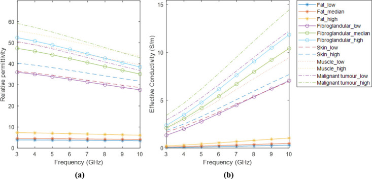
The diagnosis of breast cancer through MicroWave Imaging (MWI) technology has been extensively researched over the past few decades. However, continuous improvements to systems are needed to achieve clinical viability. To this end, the numerical models employed in simulation studies need to be diversified, anatomically accurate, and also representative of the cases in clinical settings. Hence, we have created the first open-access repository of 3D anatomically accurate numerical models of the breast, derived from 3.0T Magnetic Resonance Images (MRI) of benign breast disease and breast cancer patients. The models include normal breast tissues (fat, fibroglandular, skin, and muscle tissues), and benign and cancerous breast tumors. The repository contains easily reconfigurable models which can be tumor-free or contain single or multiple tumors, allowing complex and realistic test scenarios needed for feasibility and performance assessment of MWI devices prior to experimental and clinical testing. It also includes an executable file which enables researchers to generate models incorporating the dielectric properties of breast tissues at a chosen frequency ranging from 3 to 10 GHz, thereby ensuring compatibility with a wide spectrum of research requirements and stages of development for any breast MWI prototype system. Currently, our dataset comprises MRI scans of 55 patients, but new exams will be continuously added.
Copyright: © 2024 Pelicano et al. This is an open access article distributed under the terms of the Creative Commons Attribution License, which permits unrestricted use, distribution, and reproduction in any medium, provided the original author and source are credited.
Conflict of interest statement
The authors have declared that no competing interests exist.
Figures


References
-
- Solis-Nepote M, Reimer T, Pistorius S. An air-operated bistatic system for breast microwave radar imaging: pre-clinical validation. Annu Int Conf IEEE Eng Med Biol Soc. Berlin, Germany. 2019:1859–1862. - PubMed
-
- Meaney PM, Fanning MW, Li D, Poplack SP, Paulsen KD. A clinical prototype for active microwave imaging of the breast. IEEE Trans Microw Theory Tech. 2000;48(11):1841–1853. doi: 10.1109/22.883861 - DOI
MeSH terms
LinkOut - more resources
Full Text Sources
Medical
Miscellaneous
