. 2024 May 17;19(5):e0300644.
doi: 10.1371/journal.pone.0300644. eCollection 2024.

PAI-1 mediates acquired resistance to MET-targeted therapy in non-small cell lung cancer

Affiliations

PAI-1 mediates acquired resistance to MET-targeted therapy in non-small cell lung cancer

Yin Min Thu et al. PLoS One. .

Abstract

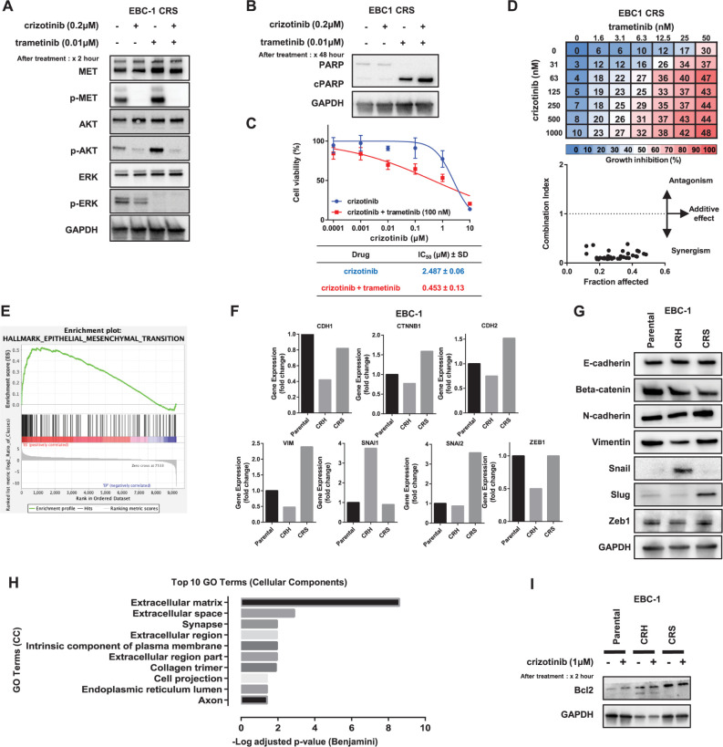
Mechanisms underlying primary and acquired resistance to MET tyrosine kinase inhibitors (TKIs) in managing non-small cell lung cancer remain unclear. In this study, we investigated the possible mechanisms acquired for crizotinib in MET-amplified lung carcinoma cell lines. Two MET-amplified lung cancer cell lines, EBC-1 and H1993, were established for acquired resistance to MET-TKI crizotinib and were functionally elucidated. Genomic and transcriptomic data were used to assess the factors contributing to the resistance mechanism, and the alterations hypothesized to confer resistance were validated. Multiple mechanisms underlie acquired resistance to crizotinib in MET-amplified lung cancer cell lines. In EBC-1-derived resistant cells, the overexpression of SERPINE1, the gene encoding plasminogen activator inhibitor-1 (PAI-1), mediated the drug resistance mechanism. Crizotinib resistance was addressed by combination therapy with a PAI-1 inhibitor and PAI-1 knockdown. Another mechanism of resistance in different subline cells of EBC-1 was evaluated as epithelial-to-mesenchymal transition with the upregulation of antiapoptotic proteins. In H1993-derived resistant cells, MEK inhibitors could be a potential therapeutic strategy for overcoming resistance with downstream mitogen-activated protein kinase pathway activation. In this study, we revealed the different mechanisms of acquired resistance to the MET inhibitor crizotinib with potential therapeutic application in patients with MET-amplified lung carcinoma.


Conflict of interest statement

I have read the journal’s policy and the authors of this manuscript have the following competing interests: Shinichi Toyooka received research funding from Eli Lilly Japan, Taiho (Japan) and Chugai (Japan), and lecture fees from Chugai. All other authors have declared that no competing interests exist.

Figures

Fig 1
Fig 1. Establishment of crizotinib-resistant clones.
(A) EBC-1, H1993 parental, and resistant cells were treated with crizotinib, tepotinib, and cabozantinib for 72 h, and cell viability was measured using an MTS assay. Each experiment was assayed in six replicates, and the average IC50 values (μM ± SD) were calculated from three independent experiments. (B) Morphological changes in parental and resistant cell lines were observed under a light microscope, and features were noted (40× magnification, light microscope). (C) Copy number assay on MET gene. The MET gene copy number was determined in human genomic DNA (HGD) and EBC-1 and H1993 parental and resistant cell lines. The copy number of each cell line with relative to the HGD copy number of 2 was presented, using the LINE-1 gene as endogenous control. (D) Protein expression analysis by Western blot in all parental and resistance cell lines. MET downstream signaling pathway markers were identified as any activation with or without crizotinib treatment. The cells were treated with crizotinib 1 μM for 2 h and subjected to immunoblotting. (E) Supervised hierarchical clustering analysis among the highly expressed significant 189 genes, which showed a significant difference with a p-value of less than 0.05 in parental cell lines compared with resistant cell lines. p- (Phosphorylated-), SD–standard deviation, NA–not applicable.
Fig 2
Fig 2. Contribution of PAI-1 upregulation in the EBC-1 CRH cell line.
(A) Expression of PAI-1 (SERPINE1 gene) in the EBC-1 cell line by RNA sequence analysis. (B) Protein expression analysis by Western blot of PAI-1 on EBC-1 cell lines showing consistent expression with RNA sequence analysis. (C) EBC-1 CRH cells were treated with crizotinib and the PAI-1 inhibitor tiplaxtinin (10μM) for 72 h, and cell viability was measured using the MTS assay. Each experiment was assayed in six-replicates, and the average IC50 value (μM ± SD) was calculated from three independent experiments. (D) Combination treatment with crizotinib and tiplaxtinin inhibited colony formation in EBC-1 CRH cells. The cells were treated with DMSO (control), crizotinib 0.2 μM, and tiplaxtinin 10 μM, individually or in combination. The relative number of colonies formed was compared to evaluate the effect of therapeutic agents. The average of three independent experiments was measured. (E) Knockdown of PAI-1 by transfection with shRNA. Control cell lines were treated with shNT (nontarget), and two knockdown cell lines were established and validated by assessing the expression of PAI-1 by Western blot. (F) Colony formation assay analysis of EBC-1 CRH PAI-1 knockdown and control cell lines treated with DMSO (control) or crizotinib (0.2 μM) in a similar manner. (G) Kaplan–Meier analysis of the overall survival (OS) of patient with high and low expression of SERPINE1 gene in lung cancer (HR = 1.15, longrank P = 0.019). *p < 0.05, **p < 0.01, ***p < 0.00.
Fig 3
Fig 3. MAPK pathway activation and EMT as a resistant mechanism in the EBC-1 CRS cell line.
(A) EBC-1 CRS cells were treated with crizotinib (0.2 μM) and trametinib (0.1 μM) for 2 h, individually and in combination, and the expression of MET downstream signaling markers was detected by immunoblotting. (B) The same cell line was treated with individual or a combination of crizotinib (0.2 μM) and trametinib (0.1 μM) for 48 h, and the lysate was subjected to immunoblotting on the apoptosis activity expression of PARP and cleaved PARP. (C) EBC-1 CRS cells were treated with crizotinib and trametinib (100nM) for 72 h, and the cell viability was measured using MTS assay. Each experiment was assayed in six-replicates, and the average IC50 value (μM ± SD) was calculated from three independent experiments. (D) EBC-1 CRS cells were treated with a combination of crizotinib and trametinib for 96 h, and cell viability was determined. The mean value of the growth inhibition percent at each concentration was presented. The combination index (CI) plot indicates the CI and fraction affected (inhibition ratio) among the different drug concentrations. (E) Gene set enrichment analysis (GSEA) of the EBC-1 CRS cell line compared with parental cells in which the enrichment plot of the hallmark EMT-related gene set was presented, with an enrichment score (ES) of 0.52 and a nominal p-value of 0.0. (F) Gene expression data of RNA sequence analysis on the EMT-related genes CDH1, CTNN2B, CDH2, VIM, SNAI1, SNAI2, and ZEB1 encoding E-cadherin, beta-catenin, N-cadherin, vimentin, Snail, Slug, and Zeb1, respectively. Fold changes in each resistant cell line were calculated and compared with those in the parental cell line. (G) Western blot analysis of EMT-related protein expression in EBC-1 cell lines, with upregulated mesenchymal markers as well as N-cadherin and slug in EBC-1 CRS cell lines compared with parental cells. (H) DAVID functional gene ontology (GO) analysis of gene enrichment in the EBC-1 CRS cell line. The top 10 terms on the cellular component (CC) gene set were presented, and enrichment of genes related to the extracellular matrix was observed. (I) The expression of the antiapoptotic protein Bcl-2 in the EBC-1 CRS cell line was determined after treatment with or without crizotinib (1 μM) for 2 h and subjected to immunoblotting.
Fig 4
Fig 4. MAPK pathway activation and EMT features are upregulated in the H1993 CRH cell line.
(A) H1993 CRH cells were treated with crizotinib (0.2 μM) and trametinib (0.1 μM) for 2 h, individually and in combination, and the lysates were subjected to immunoblotting. Expression of MET downstream signaling markers was detected. (B) The same cell line was treated with individual or a combination of crizotinib (0.2 μM) and trametinib (0.1 μM) for 48 h, and the lysate was subjected to immunoblotting. Apoptosis analysis of PARP and cleaved PARP expression in combination treatment with crizotinib and trametinib in H1993 CRH cells was observed by using Western blot data. (C) Combination treatment with crizotinib and trametinib inhibited colony formation in H1993 CRH cells. A colony formation assay was performed in H1993 CRH cells using a combination of crizotinib (0.2 μM) and trametinib (0.1 μM) individually or in combination treatment. Compared with DMSO (control), three independent assays were performed, and the average number of relative colonies formed was evaluated. (D) H1993 CRH was treated with a combination of crizotinib and trametinib for 96 h, and cell viability was determined. Data represented the mean value of the growth inhibition ratio at each concentration. The combination index (CI) plot indicates the CI and fraction affected (inhibition ratio) at different drug concentrations. (E) Gene set enrichment analysis (GSEA) in RNA sequence data of H1993 CRH cell lines compared with parental cell lines showing enrichment in the EMT-related gene set in RNA sequence data of H1993 CRH, with an enrichment score (ES) of 0.56 and a nominal p-value of 0.0. (F) Gene expression analysis of H1993 cells from RNA sequence data comparing genes encoding EMT markers in parental and resistant cell lines. The fold changes in each resistant cell line were calculated and compared with those in the parental cell line. (G) Western blot analysis of H1993 parental and resistant cells on EMT markers, which is consistent with the RNA sequence gene signature results. p- (Phosphorylated-), *p < 0.05, **p < 0.01, ***p < 0.001.

Similar articles

Cited by

References

    1. Sung H, Ferlay J, Siegel RL, Laversanne M, Soerjomataram I, Jemal A, et al.. Global Cancer Statistics 2020: GLOBOCAN Estimates of Incidence and Mortality Worldwide for 36 Cancers in 185 Countries. CA Cancer J Clin. 2021;71(3):209–49. Epub 2021/02/05. doi: 10.3322/caac.21660 . - DOI - PubMed
    1. Duma N, Santana-Davila R, Molina JR. Non-Small Cell Lung Cancer: Epidemiology, Screening, Diagnosis, and Treatment. Mayo Clin Proc. 2019;94(8):1623–40. Epub 2019/08/06. doi: 10.1016/j.mayocp.2019.01.013 . - DOI - PubMed
    1. Cancer Genome Atlas Research N. Comprehensive molecular profiling of lung adenocarcinoma. Nature. 2014;511(7511):543–50. Epub 2014/08/01. doi: 10.1038/nature13385 ; PubMed Central PMCID: PMC4231481. - DOI - PMC - PubMed
    1. Drilon A, Clark JW, Weiss J, Ou S-HI, Camidge DR, Solomon BJ, et al.. Antitumor activity of crizotinib in lung cancers harboring a MET exon 14 alteration. Nature Medicine. 2020;26(1):47–51. doi: 10.1038/s41591-019-0716-8 - DOI - PMC - PubMed
    1. Wolf J, Seto T, Han J-Y, Reguart N, Garon EB, Groen HJM, et al.. Capmatinib in MET Exon 14–Mutated or MET-Amplified Non–Small-Cell Lung Cancer. New England Journal of Medicine. 2020;383(10):944–57. doi: 10.1056/nejmoa2002787 - DOI - PubMed

MeSH terms