

Numerical study of pedicle screw construction and locking compression plate fixation in posterior pelvic ring injuries: Analyzed by finite element method
- PMID: 38758846
- PMCID: PMC11098222
- DOI: 10.1097/MD.0000000000038258
Numerical study of pedicle screw construction and locking compression plate fixation in posterior pelvic ring injuries: Analyzed by finite element method
Abstract
Background: The aim of this study was to compare the biomechanical performance of pedicle screw construction and locking compression plate fixation in posterior pelvic ring injuries analyzed by finite element method.
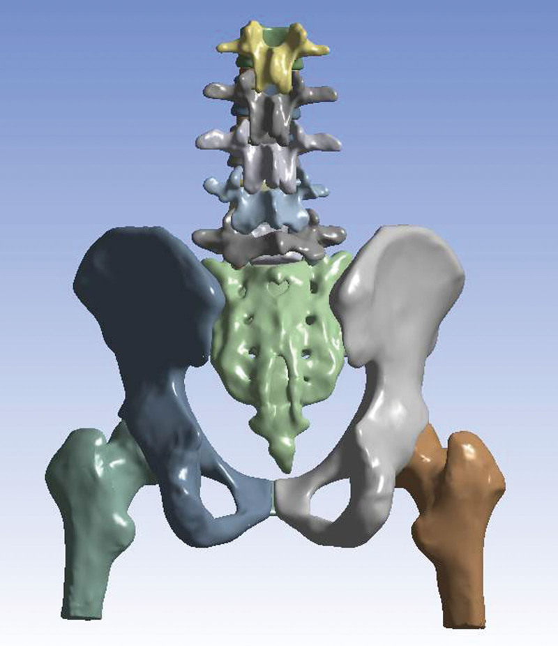
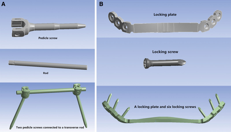
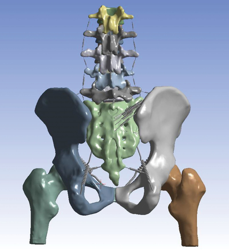
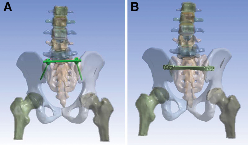
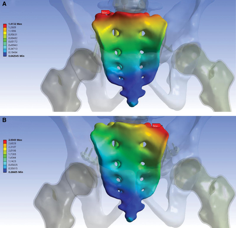
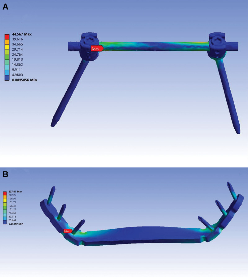
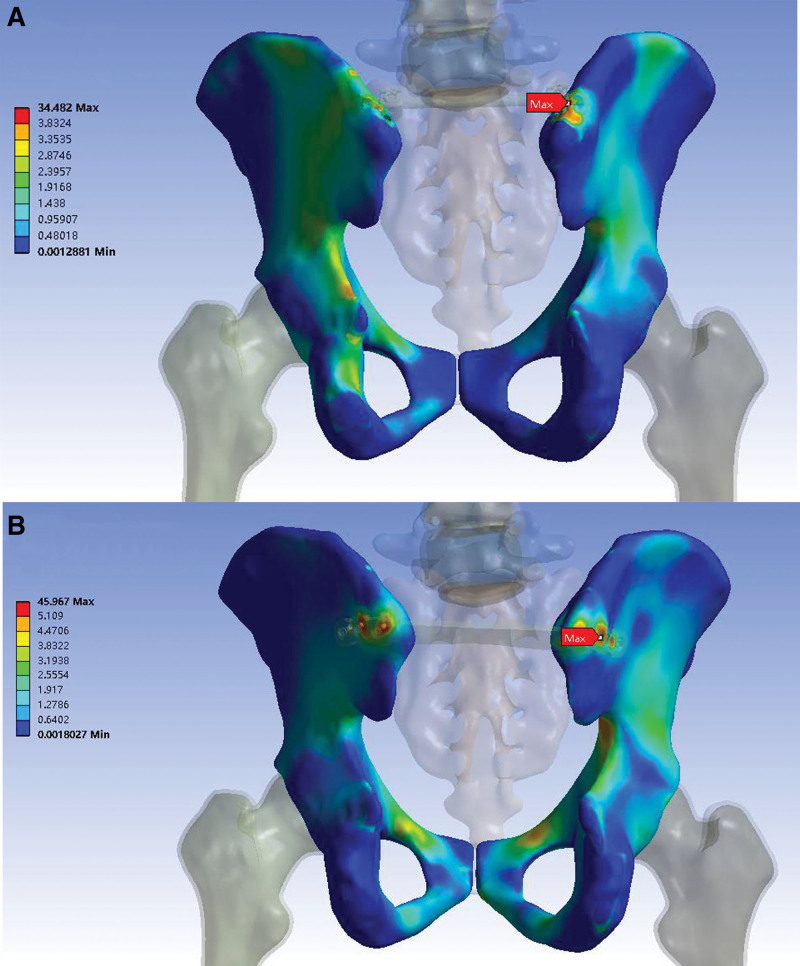
Methods: A 3-dimensional finite element model of the spine-pelvis-femur complex with ligaments was reconstructed from computed tomography images. An unstable posterior pelvic ring injury was created, which was fixed with a pedicle screw construction or locking compression plate. A follower load of 400 N was applied to the upper surface of the vertebrae to simulate the upper body weight, while the ends of the proximal femurs were fixed. The construct stiffness, the maximum vertical displacement, the maximum posterior displacement, the maximum right displacement, and the overall maximum displacement of the sacrum, and stress distributions of the implants and pelvises were assessed.
Results: The construct stiffness of the pedicle screw model (435.14 N/mm) was 2 times that of the plate model (217.01 N/mm). The maximum vertical displacement, the maximum posterior displacement, the maximum right displacement, and the overall maximum displacement of the sacrum in the pedicle screw model were smaller than those in the plate model (0.919, 1.299, 0.259, and 1.413 mm in the pedicle screw model, and 1.843, 2.300, 1.053, and 2.895 mm in the plate model, respectively). The peak stresses of the implant and pelvis in the pedicle screw model decreased by 80.4% and 25% when compared with the plate model (44.57 and 34.48 MPa in the pedicle screw model, and 227.47 and 45.97 MPa in the plate model, respectively).
Conclusion: The study suggested that the pedicle screw construction could provide better fixation stability than the locking compression plate and serves as the recommended fixation method for the treatment of posterior pelvic ring injuries.
Copyright © 2024 the Author(s). Published by Wolters Kluwer Health, Inc.
Conflict of interest statement
The authors have no conflicts of interest to disclose.
Figures
Similar articles
-
Finite element analysis of retrograde superior ramus screw of pubis for the treament of pelvic anterior ring fracture.J Orthop Surg Res. 2025 Mar 12;20(1):263. doi: 10.1186/s13018-025-05676-5. J Orthop Surg Res. 2025. PMID: 40069735 Free PMC article.
-
Comparison of mechanical stability of modified pedicle screw fixator and unilateral lumbopelvic fixation for treating sacroiliac joint disruption: A finite element analysis study.Acta Orthop Traumatol Turc. 2024 Nov 8;58(5):274-279. doi: 10.5152/j.aott.2024.24072. Acta Orthop Traumatol Turc. 2024. PMID: 39560711 Free PMC article.
-
Biomechanical study of anterior and posterior pelvic rings using pedicle screw fixation for Tile C1 pelvic fractures: Finite element analysis.PLoS One. 2022 Aug 25;17(8):e0273351. doi: 10.1371/journal.pone.0273351. eCollection 2022. PLoS One. 2022. PMID: 36006983 Free PMC article.
-
Treatment of Unstable Posterior Pelvic Ring Fracture with Pedicle Screw-Rod Fixator Versus Locking Compression Plate: A Comparative Study.Med Sci Monit. 2016 Oct 17;22:3764-3770. doi: 10.12659/msm.900673. Med Sci Monit. 2016. PMID: 27748355 Free PMC article.
-
Biomechanical Comparison of Three Internal Fixation Techniques for Stabilizing Posterior Pelvic Ring Disruption: A 3D Finite Element Analysis.Orthop Surg. 2019 Apr;11(2):195-203. doi: 10.1111/os.12431. Epub 2019 Mar 21. Orthop Surg. 2019. PMID: 30895703 Free PMC article.
Cited by
-
Finite element analysis of retrograde superior ramus screw of pubis for the treament of pelvic anterior ring fracture.J Orthop Surg Res. 2025 Mar 12;20(1):263. doi: 10.1186/s13018-025-05676-5. J Orthop Surg Res. 2025. PMID: 40069735 Free PMC article.
-
Experimental study of fractures of the posterior pelvic ring C1.1 using LC-II screws and internal fixation by plate.J Orthop Surg Res. 2024 Nov 15;19(1):761. doi: 10.1186/s13018-024-05229-2. J Orthop Surg Res. 2024. PMID: 39543607 Free PMC article.
-
Comparison of mechanical stability of modified pedicle screw fixator and unilateral lumbopelvic fixation for treating sacroiliac joint disruption: A finite element analysis study.Acta Orthop Traumatol Turc. 2024 Nov 8;58(5):274-279. doi: 10.5152/j.aott.2024.24072. Acta Orthop Traumatol Turc. 2024. PMID: 39560711 Free PMC article.
References
-
- Papakostidis C, Giannoudis PV. Pelvic ring injuries with haemodynamic instability: efficacy of pelvic packing, a systematic review. Injury. 2009;40:S53–61. - PubMed
-
- Simonain PT, Routt C, Jr, Harrington RM, Tencer AF. Internal fixation for the transforaminal sacral fracture. Clin Orthop Relat Res. 1996:202–9. - PubMed
-
- Grotz MR, Allami MK, Harwood P, Pape HC, Krettek C, Giannoudis PV. Open pelvic fractures: epidemiology, current concepts of management and outcome. Injury. 2005;36:1–13. - PubMed
-
- Roszman AD, John DQ, Patch DA, Spitler CA, Johnson JP. Management of open pelvic ring injuries. Injury. 2023;54:1041–6. - PubMed
MeSH terms
LinkOut - more resources
Full Text Sources
