

A urethral leiomyoma presenting with dysuria: A rare case report
- PMID: 38758882
- PMCID: PMC11098236
- DOI: 10.1097/MD.0000000000037893
A urethral leiomyoma presenting with dysuria: A rare case report
Abstract
Rationale: Leiomyoma is a benign smooth muscle tumor which is rarely found in urethra. We hereby report a case of a 44-year-old female who presented with complaints of dysuria.
Patient concerns: A 44-year-old female patient presented to the urology outpatient clinic with symptoms of dysuria. The patient described the presence of a protrusion from the urethra during urination.
Diagnosis: Urethral leiomyoma.
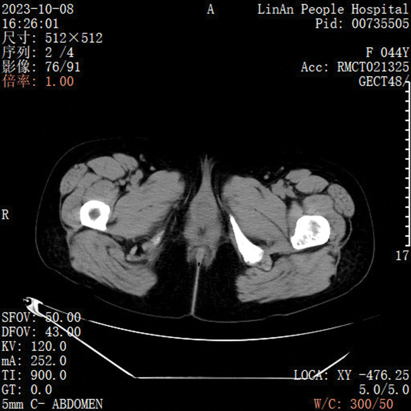
Interventions: Physical examination confirmed a solid urethral mass. CT scan and USG reports indicated that the mass originated from the mid-urethra with vascularity at the base. We performed a complete resection of the urethral mass. The patient was discharged after 3 days of observation.
Outcome: During a follow-up after 1 month, the patient reported improved urinary flow and no occurrence of hematuria. The patient recovered well after discharge.
Lesson: Urethral leiomyoma is a rare benign tumor that is often misdiagnosed in clinical practice. Diagnosis requires careful clinical examination. Surgical removal usually works well. It is important to remember that in some cases of acute urinary retention, it can be caused by a complete obstruction of a mass in the urethra. Urologists should be more cautious and experienced in handling such cases.
Copyright © 2024 the Author(s). Published by Wolters Kluwer Health, Inc.
Conflict of interest statement
The authors have no funding and conflicts of interest to disclose.
Figures


Similar articles
-
Leiomyoma: a case report of a rare benign tumor of the female urethra.Pan Afr Med J. 2015 Oct 8;22:111. doi: 10.11604/pamj.2015.22.111.7785. eCollection 2015. Pan Afr Med J. 2015. PMID: 26848358 Free PMC article.
-
Urethral fibroid: clinical insights and analysis.BMJ Case Rep. 2025 May 22;18(5):e264079. doi: 10.1136/bcr-2024-264079. BMJ Case Rep. 2025. PMID: 40409765
-
Leiomyoma of the urethra: a rare benign tumor.J Int Med Res. 2022 Sep;50(9):3000605221126662. doi: 10.1177/03000605221126662. J Int Med Res. 2022. PMID: 36168731 Free PMC article.
-
Leiomyoma of the female urethra with upper tract dilation and treatment with transurethral resection: a case report and literature review.Tech Urol. 2000 Sep;6(3):223-5. Tech Urol. 2000. PMID: 10963495 Review.
-
Leiomyoma of the female urethra. A case report.Urol Int. 2003;71(2):224-5. doi: 10.1159/000071854. Urol Int. 2003. PMID: 12890968 Review.
Cited by
-
A rare case of female urinary retention caused by urethral leiomyoma: A case report.Int J Surg Case Rep. 2025 Feb;127:110849. doi: 10.1016/j.ijscr.2025.110849. Epub 2025 Jan 7. Int J Surg Case Rep. 2025. PMID: 39793332 Free PMC article.
-
Urethral Leiomyoma in Pregnancy: A Rare Case of Symptomatic Pelvic Mass With Successful Surgical Management.Cureus. 2025 Jun 25;17(6):e86720. doi: 10.7759/cureus.86720. eCollection 2025 Jun. Cureus. 2025. PMID: 40718265 Free PMC article.
-
Incidental Discovery of a Urethral Leiomyoma: A Rare Case of an Asymptomatic Benign Tumor With Successful Surgical Management.Cureus. 2025 May 23;17(5):e84662. doi: 10.7759/cureus.84662. eCollection 2025 May. Cureus. 2025. PMID: 40546463 Free PMC article.
References
Publication types
MeSH terms
LinkOut - more resources
Full Text Sources
