. 2024 May 17;14(1):11325.
doi: 10.1038/s41598-024-59677-1.

Isobutyric acid enhances the anti-tumour effect of anti-PD-1 antibody

Affiliations

Isobutyric acid enhances the anti-tumour effect of anti-PD-1 antibody

Masakazu Murayama et al. Sci Rep. .

Abstract

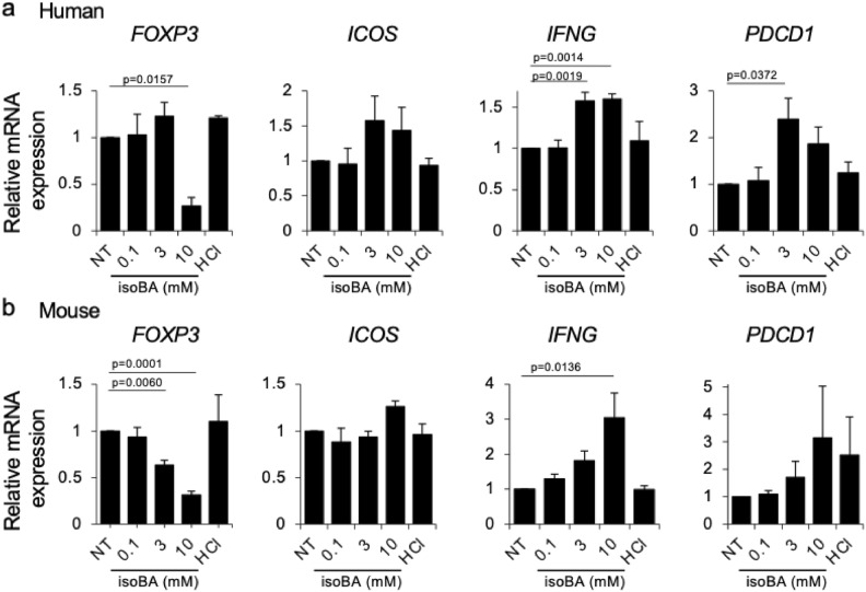
The low response rate of immune checkpoint inhibitors (ICIs) is a challenge. The efficacy of ICIs is influenced by the tumour microenvironment, which is controlled by the gut microbiota. In particular, intestinal bacteria and their metabolites, such as short chain fatty acids (SCFAs), are important regulators of cancer immunity; however, our knowledge on the effects of individual SCFAs remains limited. Here, we show that isobutyric acid has the strongest effect among SCFAs on both immune activity and tumour growth. In vitro, cancer cell numbers were suppressed by approximately 75% in humans and mice compared with those in controls. Oral administration of isobutyric acid to carcinoma-bearing mice enhanced the effect of anti-PD-1 immunotherapy, reducing tumour volume by approximately 80% and 60% compared with those in the control group and anti-PD-1 antibody alone group, respectively. Taken together, these findings may support the development of novel cancer therapies that can improve the response rate to ICIs.


Conflict of interest statement

The authors declare no competing interests.

Figures

Figure 1
Figure 1
Preferential inhibition of cancer cell survival by isobutyric acid over that of T cells. (a) Human T cells (5 × 105 cells) and T3M-1 Clone2 oral cancer cells (5 × 104 cells) were co-cultured for 72 h with isobutyric acid at the indicated concentrations. The number of cancer and T cells was analysed by flow cytometry using fluorescent counting beads. CD45-positive cells were defined as T cells and all others as cancer cells. The graphs in the left, centre, and right represent cancer/T cell co-culture, T cell mono-culture, and cancer cell mono-culture, respectively. (b) Mouse T cells and CT-26 cancer cells were co-cultured for 72 h with isobutyric acid at the indicated concentrations. The number of cancer and T cells was analysed using flow cytometry. To examine the effect of acidic conditions, hydrochloric acid was added to induce a low pH as that induced by 10 mM isobutyric acid. Data are representative of three independent experiments. Error bars represent S.E.M. NT no treatment, HCl hydrochloric acid, isoBA isobutyric acid.
Figure 2
Figure 2
Characterisation of T cell populations upon treatment with isobutyric acid. Human or mouse T cells (5 × 105 cells) were co-cultured with T3M-1 Clone2 oral cancer cells or CT-26 cells (5 × 104 cells) respectively in the presence of different concentrations of isobutyric acid for 72 h. Afterwards, the T cell populations were evaluated based on the expression of surface markers using flow cytometry. (a) Percentage of CD4+ T cells, CD8+ T cells, and Treg cells present in the cancer/T cell co-cultures with isobutyric acid. (bd) Expression of PD-1 (b) and HLA-DR (c) and ICOS (d) in CD4 + T cells, CD8 + T cells, and Treg cells. To examine the effect of acidic condition, hydrochloric acid was added to induce a low pH as that induced by 10 mM isobutyric acid. Data are mean of three independent experiments. Error bars represent S.E.M. NT no treatment, HCl 10 mM hydrochloric acid, isoBA isobutyric acid, ns not significant, *p < 0.05, **p < 0.01, ***p < 0.001, ****p < 0.0001 vs NT.
Figure 3
Figure 3
Direct effects of isobutyric acid on gene expression in T cells. Human and mouse T cells were cultured with various concentrations of isobutyric acid for 72 h before RNA extraction. Gene expression of IFNG, ICOS, PDCD1, and FOXP3 in human (a) and mouse (b) T cells was measured using quantitative PCR. Data are presented as relative values against non-treated cells and are mean of three independent experiments. Error bars represent S.E.M. NT no treatment, HCl hydrochloric acid, isoBA isobutyric acid.
Figure 4
Figure 4
Anti-cancer effect of isobutyric acid in mice treated with an anti-PD-1 antibody. (a) BALB/C mice with CT26 cancers (n = 40) were pre-treated with orally administered 100 mM isobutyric acid or pH-matched water and intraperitoneally injected with 150 μg/150 μL of anti-PD-1 antibody or IgG on days 4, 11, 18, and 25 after cancer cell inoculation. (b) Tumour volume was analysed on day 28 after inoculation of tumour cells. (c) CT26 tumour growth curves. (d) Pictures of removed tumours. Pictures without a tumour on it represent a tumour that has disappeared. (e) Objective response rate (ORR). (f,g) Haematoxylin and eosin (H&E) staining, and CD3 immunostaining of excised cancer tissues. Scale bar 100 μm. (h) IFNG, ICOS, PDCD1, CTLA-4, CD69, and CD95 levels in explanted cancer tissues. Data were obtained from two independent experiments. Statistical analysis was performed using ANOVA with Dunnett’s multiple comparison test. ns not significant. Error bars represent S.E.M.
Figure 5
Figure 5
Overview of the effects of isobutyric acid on cancer and T cells in the tumour microenvironment. Isobutyric acid exerts a direct anti-tumour effect on cancer cells. Isobutyric acid suppresses Tregs, activates effector T cells, and increases the number of T cells expressing PD-1. Isobutyric acid enhances the efficacy of anti-PD-1 immunotherapy through its direct action on cancer cells and activation of T cells.

References

    1. Bagchi S, et al. Immune checkpoint inhibitors for the treatment of cancer: Clinical impact and mechanisms of response and resistance. Annu. Rev. Pathol. 2021;16:223–249. doi: 10.1146/annurev-pathol-042020-042741. - DOI - PubMed
    1. Binnewies M, et al. Understanding the tumour immune microenvironment (TIME) for effective therapy. Nat. Med. 2018;24:541–550. doi: 10.1038/s41591-018-0014-x. - DOI - PMC - PubMed
    1. Zheng D, et al. Interaction between microbiota and immunity in health and disease. Cell Res. 2020;30:492–506. doi: 10.1038/s41422-020-0332-7. - DOI - PMC - PubMed
    1. Vetizou M, et al. Anticancer immunotherapy by CTLA-4 blockade relies on the gut microbiota. Science. 2015;350:1079–1084. doi: 10.1126/science.aad1329. - DOI - PMC - PubMed
    1. Sivan A, et al. Commensal Bifidobacterium promotes antitumour immunity and facilitates anti-PD-L1 efficacy. Science. 2015;350:1084–1089. doi: 10.1126/science.aac4255. - DOI - PMC - PubMed

MeSH terms