. 2024 May 17;22(1):265.
doi: 10.1186/s12951-024-02542-0.

Functional extracellular vesicles from SHEDs combined with gelatin methacryloyl promote the odontogenic differentiation of DPSCs for pulp regeneration

Affiliations

Functional extracellular vesicles from SHEDs combined with gelatin methacryloyl promote the odontogenic differentiation of DPSCs for pulp regeneration

Hui Lu et al. J Nanobiotechnology. .

Abstract

Background: Pulp regeneration is a novel approach for the treatment of immature permanent teeth with pulp necrosis. This technique includes the combination of stem cells, scaffolds, and growth factors. Recently, stem cell-derived extracellular vesicles (EVs) have emerged as a new methodology for pulp regeneration. Emerging evidence has proven that preconditioning is an effective scheme to modify EVs for better therapeutic potency. Meanwhile, proper scaffolding is of great significance to protect EVs from rapid clearance and destruction. This investigation aims to fabricate an injectable hydrogel loaded with EVs from pre-differentiated stem cells from human exfoliated deciduous teeth (SHEDs) and examine their effects on pulp regeneration.

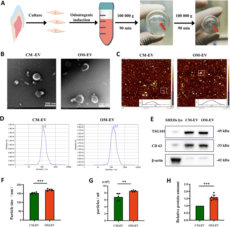
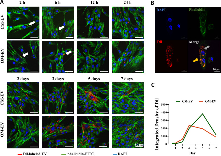
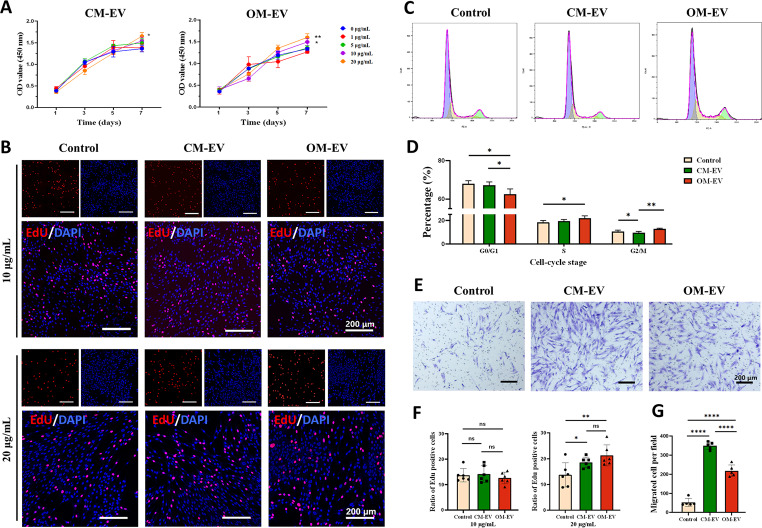
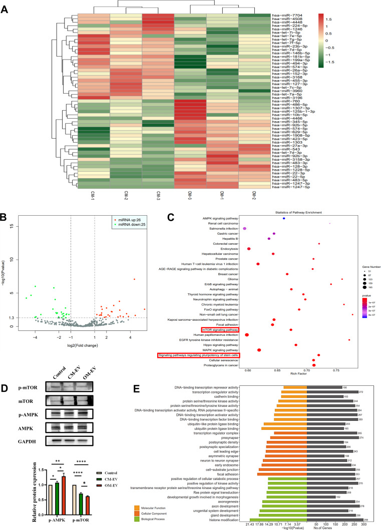
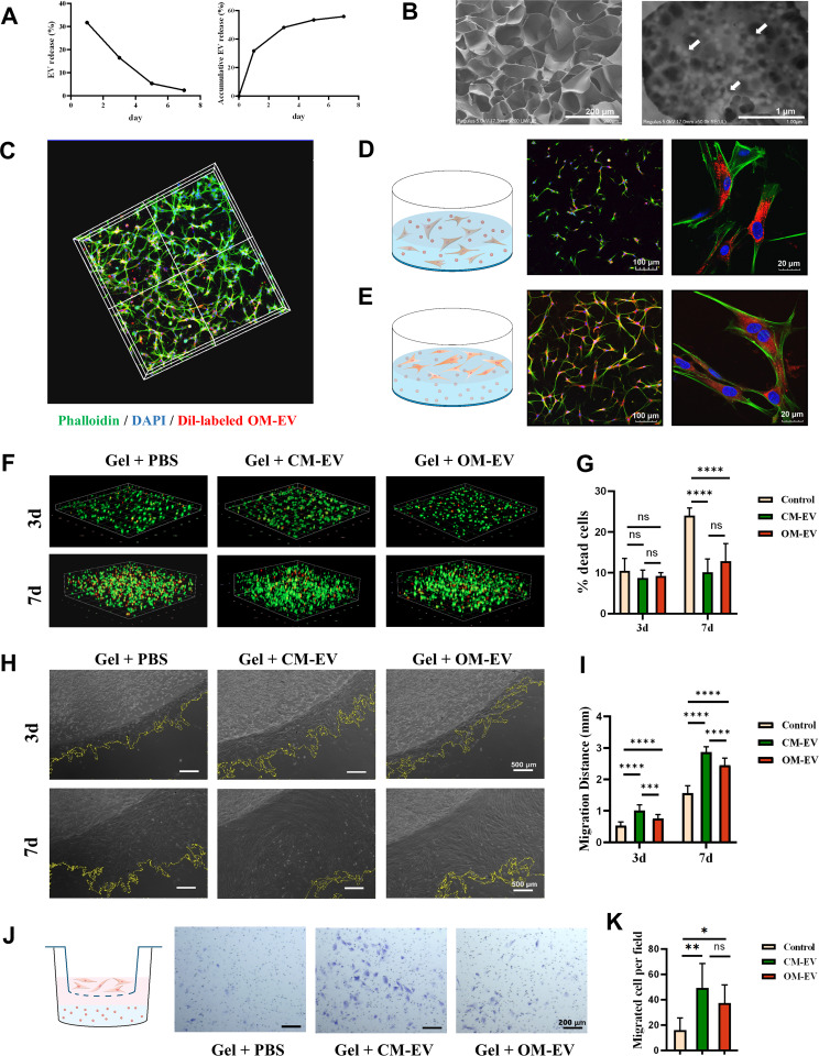
Results: We successfully employed the odontogenic induction medium (OM) of SHEDs to generate functional EV (OM-EV). The OM-EV at a concentration of 20 µg/mL was demonstrated to promote the proliferation and migration of dental pulp stem cells (DPSCs). The results revealed that OM-EV has a better potential to promote odontogenic differentiation of DPSCs than common EVs (CM-EV) in vitro through Alizarin red phalloidin, alkaline phosphatase staining, and assessment of the expression of odontogenic-related markers. High-throughput sequencing suggests that the superior effects of OM-EV may be attributed to activation of the AMPK/mTOR pathway. Simultaneously, we prepared a photocrosslinkable gelatin methacryloyl (GelMA) to construct an OM-EV-encapsulated hydrogel. The hydrogel exhibited sustained release of OM-EV and good biocompatibility for DPSCs. The released OM-EV from the hydrogel could be internalized by DPSCs, thereby enhancing their survival and migration. In tooth root slices that were subcutaneously transplanted in nude mice, the OM-EV-encapsulated hydrogel was found to facilitate dentinogenesis. After 8 weeks, there was more formation of mineralized tissue, as well as higher levels of dentin sialophosphoprotein (DSPP) and dentin matrix protein-1 (DMP-1).

Conclusions: The effects of EV can be substantially enhanced by preconditioning of SHEDs. The functional EVs from SHEDs combined with GelMA are capable of effectively promoting dentinogenesis through upregulating the odontogenic differentiation of DPSCs, which provides a promising therapeutic approach for pulp regeneration.

Keywords: Extracellular vesicles; Hydrogel; Odontogenic differentiation; Pulp regeneration; Stem cells from human exfoliated deciduous teeth.


Conflict of interest statement

The authors declare no competing interests.

Figures

Fig. 1
Fig. 1
Isolation and identification of OM-EV and CM-EV: A Schematic representation showing the preparation and isolation of OM-EV and CM-EV. B Morphology of OM-EV and CM-EV under TEM (note: Scale bar represents 200 nm). C Saucer-like morphology of OM-EV and CM-EV under AFM. D The presented particle distributions of OM-EV and CM-EV by NTA. E Detection of EV surface markers of TSG101 and CD63 by Western Blotting. F Comparison of particle size. G Comparison of particle concentration (EVs from 9 mL supernatant were resuspended in 2.5 mL ddH2O). H Comparison of protein amount. (note: Error bars represent the values of “mean ± s.d.”; ** P < 0.01, *** P < 0.001)
Fig. 2
Fig. 2
The uptake of OM-EV and CM-EV by DPSCs: A The internalization of Dil-labeled OM-EV and CM-EV (white arrow) by DPSCs observed in 7 days (note: Scale bar represents 50 μm). B A representative cross-section of the 3D reconstruction showing the localization of EVs inside (gray arrow) and outside (yellow arrow) the cell on day 3 (note: Scale bar represents 20 μm). C Integrated density of Dil in DPSCs treated with OM-EV and CM-EV in 7 days
Fig. 3
Fig. 3
The proliferation and migration of DPSCs promoted by the uptake of OM-EV. A Proliferation of DPSCs cocultured with various concentrations of CM-EV or OM-EV for 7 days assessed by CCK-8 assay (* P < 0.05, ** P < 0.01 vs. the control group). B Effects of 10 and 20 µg/mL CM-EV and OM-EV on the proliferation of DPSCs detected by EdU proliferation assay (note: Scale bar represents 200 μm). C Effect of 20 µg/mL CM-EV or OM-EV on the cell cycle of DPSCs. D Distribution of cell cycle stage in each group. E Representative images showing the capacity of DPSC migration under the treatment of 20 µg/mL CM-EV or OM-EV tested by the transwell assay (note: Scale bar represents 200 μm). F The ratio of EdU-positive cells under the treatment of 10 and 20 µg/mL CM-EV and OM-EV. G. Comparison of migrated cells per field (note: Error bars represent the values of “mean ± s.d.”; * P < 0.05, ** P < 0.01, *** P < 0.001, and **** P < 0.0001)
Fig. 4
Fig. 4
Assessment of odontogenic potential of DPSCs after culture with odontogenic induction medium containing OM-EV. A Gross appearance and microscopic images of Alizarin red staining for 7 days (note: Scale bar represents 200 μm). B Quantitation analysis of Alizarin red staining for 7 days. C Gross appearance and microscopic images of ALP staining for 7 days (note: Scale bar represents 200 μm). D Quantitative detection of ALP activities after 7 days. E Expression levels of odontogenic-related proteins for 7 days. F Quantification of the gray signal intensity based on the Western blotting at day 7. G Expression levels of odontogenic genes tested by qRT-PCR at day 7. H Gross appearance and microscopic images of Alizarin red staining for 14 days (note: Scale bar represents 200 μm). I Quantitation analysis of Alizarin red staining for 14 days. J Expression levels of odontogenic-related proteins for 14 days. K Quantification of the gray signal intensity based on the Western blotting at day 14. L Expression levels of odontogenic genes tested by qRT-PCR at day 14 (note: * P < 0.05, ** P < 0.01, *** P < 0.001, and **** P < 0.0001)
Fig. 5
Fig. 5
High-throughput sequencing for OM-EV and CM-EV. A Heat map of microRNA expression in OM-EV and CM-EV (n = 3). B Volcano plot showing significantly upregulated (red dots) and downregulated (green dots) microRNAs in OM-EV, compared with CM-EV. 51 microRNAs significantly changed, of which 26 microRNAs increased and 25 microRNAs decreased. C Kyoto Encyclopedia of Genes and Genomes (KEGG) pathway enrichment analysis. D Western blotting showing the expression of AMPK pathway-related proteins in DPSCs. E Gene Ontology (GO) analysis of the target mRNAs of the differentially expressed miRNAs (note: * P < 0.05, ** P < 0.01, and **** P < 0.0001)
Fig. 6
Fig. 6
Characterization of OM-EV-encapsulated hydrogel. A Release curves of OM-EV obtained from the GelMA hydrogel. B Microstructure of OM-EV-encapsulated hydrogel characterized by SEM (note: white arrow shows the encapsulated OM-EV. The corresponding scale bars represent 200 μm and 1 μm, respectively). C Distribution of OM-EV and DPSCs in the OM-EV-encapsulated hydrogel demonstrated by 3D reconstruction with CLSM (note: OM-EV were labeled with Dil (red), while DPSCs and nuclei were stained with phallotoxins (green) and DAPI (blue), respectively). D Schematic representation and CLSM images illustrating the DPSCs embedded in the OM-EV-encapsulated hydrogel endocytosed OM-EVs released from the hydrogel (note: the corresponding scale bars are presented). E Schematic representation and CLSM images illustrating the DPSCs on the surface of the OM-EV-encapsulated hydrogel endocytosed OM-EVs released from the hydrogel (note: the corresponding scale bars are presented). F Live/Dead staining of DPSCs within the OM-EV-encapsulated hydrogel (note: live and dead cells have been labeled with green and red colors, respectively). G The proportion of dead cells within the OM-EV-encapsulated hydrogel. H The migration ability of DPSCs within the OM-EV-encapsulated hydrogel (note: the scale bar length is 500 μm). I Migration distance of DPSCs within the OM-EV-encapsulated hydrogel. J Schematic illustration and representative images showing the effect of OM-EV-encapsulated hydrogel on the migration ability of DPSCs tested by the transwell assay (note: Scale bar represents 200 μm). K Migrated cells per field in each group (note: Error bars represent the values of “mean ± s.d.”; * P < 0.05, ** P < 0.01, *** P < 0.001, and **** P < 0.0001)
Fig. 7
Fig. 7
OM-EV-encapsulated hydrogel increased the formation of pulp-dentin complex in vivo. A Steps used for subcutaneous transplantation of tooth root slices in nude mice. B General view of the tooth root slices before transplantation and after 8 weeks of subcutaneous transplantation. C Representative images of H&E staining of sections from tooth root slice (note: P: dental pulp-like tissue, D: dentin, M: mineralized tissue (black arrow); Scale bar lengths are set as 200 and 100 μm). D Representative images of Masson staining of sections from tooth root slice (note: the scale bar lengths are 200 μm and 100 μm, respectively). E Representative images of immunohistochemical staining showing the upregulated expression of odontogenic markers (DSPP and DMP-1) (note: scale bar represents 100 μm). F Quantification analysis of mineralized tissue area fraction. G Quantitative analysis of DSPP-positive cells. H Quantitative analysis of DMP-1-positive cells (note: Error bars represent the values of “mean ± s.d.”; * P < 0.05, ** P < 0.01, *** P < 0.001, and **** P < 0.0001)
Scheme 1
Scheme 1
Graphical abstract of the present study and the potential application of OM-EV-encapsulated hydrogel for pulp regeneration

References

    1. Zheng C, Chen J, Liu S, Jin Y. Stem cell-based bone and dental regeneration: a view of microenvironmental modulation. Int J Oral Sci. 2019;11:23. doi: 10.1038/s41368-019-0060-3. - DOI - PMC - PubMed
    1. Xuan K, Li B, Guo H, Sun W, Kou X, He X, et al. Deciduous autologous tooth stem cells regenerate dental pulp after implantation into injured teeth. Sci Transl Med. 2018;10:eaaf3227. - PubMed
    1. Lai H, Li J, Kou X, Mao X, Zhao W, Ma L. Extracellular vesicles for dental pulp and periodontal regeneration. Pharmaceutics. 2023;15:282. - PMC - PubMed
    1. Ding Y, Li Y, Sun Z, Han X, Chen Y, Ge Y, Mao Z, Wang W. Cell-derived extracellular vesicles and membranes for tissue repair. J Nanobiotechnol. 2021;19:368. doi: 10.1186/s12951-021-01113-x. - DOI - PMC - PubMed
    1. Wu J, Chen L, Wang R, Song Z, Shen Z, Zhao Y, Huang S, Lin Z. Exosomes secreted by stem cells from human exfoliated deciduous teeth promote alveolar bone defect repair through the regulation of angiogenesis and osteogenesis. Acs Biomater Sci Eng. 2019;5:3561–71. doi: 10.1021/acsbiomaterials.9b00607. - DOI - PubMed

MeSH terms

Substances