Review
. 2024 May 18;23(1):107.
doi: 10.1186/s12943-024-02004-z.

Neutrophils in Cancer immunotherapy: friends or foes?

Affiliations
Review

Neutrophils in Cancer immunotherapy: friends or foes?

Xueqin Huang et al. Mol Cancer. .

Abstract

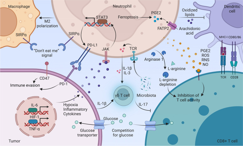
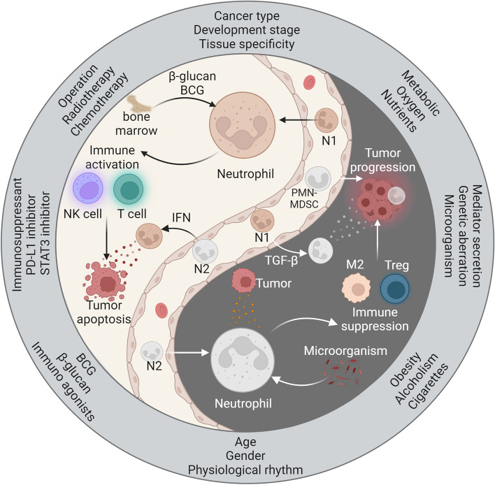
Neutrophils play a Janus-faced role in the complex landscape of cancer pathogenesis and immunotherapy. As immune defense cells, neutrophils release toxic substances, including reactive oxygen species and matrix metalloproteinase 9, within the tumor microenvironment. They also modulate the expression of tumor necrosis factor-related apoptosis-inducing ligand and Fas ligand, augmenting their capacity to induce tumor cell apoptosis. Their involvement in antitumor immune regulation synergistically activates a network of immune cells, bolstering anticancer effects. Paradoxically, neutrophils can succumb to the influence of tumors, triggering signaling cascades such as JAK/STAT, which deactivate the immune system network, thereby promoting immune evasion by malignant cells. Additionally, neutrophil granular constituents, such as neutrophil elastase and vascular endothelial growth factor, intricately fuel tumor cell proliferation, metastasis, and angiogenesis. Understanding the mechanisms that guide neutrophils to collaborate with other immune cells for comprehensive tumor eradication is crucial to enhancing the efficacy of cancer therapeutics. In this review, we illuminate the underlying mechanisms governing neutrophil-mediated support or inhibition of tumor progression, with a particular focus on elucidating the internal and external factors that influence neutrophil polarization. We provide an overview of recent advances in clinical research regarding the involvement of neutrophils in cancer therapy. Moreover, the future prospects and limitations of neutrophil research are discussed, aiming to provide fresh insights for the development of innovative cancer treatment strategies targeting neutrophils.

Keywords: Antitumor activity; Cancer; Immunotherapy; Neutrophils; Protumor activity.


Conflict of interest statement

The authors declare no competing interests.

Figures

Fig. 1
Fig. 1
The development, mobilization, and clearance of neutrophils in the tumor microenvironment. Hematopoietic progenitor stem cells (HSCs) in the bone marrow differentiate into common myeloid progenitor cells (CMPs), which give rise to granulocyte-monocyte progenitor cells (GMPs) and eventually mature segmented neutrophils. Tumor-derived mediators such as granulocyte colony-stimulating factor (G-CSF) and S100 calcium-binding protein A9 (S100A9) promote the differentiation of the neutrophil and monocyte lineages while leading to systemic dendritic cell deficiency in vivo. Chemokines trigger the mobilization of mature and immature neutrophils into the circulation. Immature neutrophils, known as polymorphonuclear myeloid-derived suppressor cells (PMN-MDSCs), are considered in this context. During transendothelial migration, the interaction between integrin α9β1 on neutrophils and vascular cell adhesion molecule 1 (VCAM-1) on endothelial cells stimulates the release of granulocyte-macrophage colony-stimulating factor (GM-CSF) from the latter, prolonging neutrophil lifespan. Neutrophils that extravasate into the tumor tissue adopt antitumor (type N1) or protumor (type N2) phenotypes, influenced by growth factor-β (TGF-β) and type 1 interferon (IFN), respectively. After fulfilling their functions, neutrophils undergo senescence due to intrinsic programs (CXCR2 or CXCR4) and extrinsic factors (microbiota) and are subsequently cleared by macrophages in the bone marrow, spleen, liver, and lungs. Neutrophils also undergo programmed death to form neutrophil extracellular traps (NETs)
Fig. 2
Fig. 2
Direct cytotoxic effects of neutrophils on cancer cells. Neutrophils, when exposed to β-glucan, rely on the memory of bone marrow precursors and exert antitumor effects. The chemokines CXCL1, CXCL2, and CXCL5, secreted by primary tumors, facilitate the recruitment of neutrophils to the tumor site. Under hypoxic conditions, activated neutrophils induce reactive oxygen species (ROS) and matrix metalloproteinase (MMP-9) degradation of the epithelial basement membrane, which ultimately restricts tumor development. Neutrophils directly secrete ROS, myeloperoxidase (MPO), and interferon γ (IFN-γ) to inhibit tumor progression. The interaction between the ligand hepatocyte growth factor (HGF) and receptor tyrosine protein kinase (MET) on neutrophils leads to the release of nitric oxide (NO) by MET+ neutrophils, which exerts antitumor effects. Neutrophils secrete hydrogen peroxide (H2O2), inducing apoptosis in tumor cells through Ca2+ influx via the transient receptor potential cation channel, subfamily M, member 2 (TRPM2). Neutrophil elastase (NE) hydrolyzes and releases the CD95 death structure domain (DD), selectively killing cancer cells. Additionally, NE has distant effects on CD8+ T cells. Neutrophils with enhanced expression of TNF-related apoptosis-inducing ligand (TRAIL) and Fas ligand (FasL) induce apoptosis in cancer cells through direct contact
Fig. 3
Fig. 3
Neutrophils regulate immune cells to drive antitumor immune responses. When exposed to IFN-γ and GM-CSF, immature neutrophils can differentiate into hybrid neutrophils with antigen-presenting cell (APC) characteristics. Thereafter, dendritic cells and APC-like neutrophils can pick up tumor antigens and migrate to lymph nodes (LNs). In LNs, these antigen-presenting cells cross-present tumor antigens to T cells using MHC molecules and costimulatory ligands (CD80/CD86 on dendritic cells and APC-like neutrophils and CD28 on T cells), thereby stimulating antitumor T cell responses. Activated T cells then exit the LN and specifically target and eliminate tumor cells. Tumor cells can undergo antigenic variations, leading to the formation of tumor variants with diverse antigenic profiles. In response, activated T cells can secrete chemokines to recruit neutrophils, which contribute to the elimination of antigenically heterogeneous tumors by releasing NO. Interferons play critical roles in inducing antitumor immune responses in neutrophils. IL-12, secreted by dendritic cells and macrophages, triggers the type I activation of T cells and αβ T cells, resulting in the production of IFN-γ. Subsequently, activated neutrophils can further stimulate macrophages to release IL-12, amplifying the antitumor immune response
Fig. 4
Fig. 4
Neutrophils promote tumor initiation, proliferation, metastasis, and angiogenesis. Neutrophils promote tumor progression by releasing proinflammatory particles (miR-23a, miR-155) and reactive ROS, which cause DNA damage and support carcinogenesis. They contribute to tumor proliferation through the release of NE and prostaglandin E2 (PGE2). Neutrophils also secrete interleukin-1 receptor antagonist (IL-1RA) to protect tumors from senescence and secrete IL-17, TGF-β, and NE, promoting epithelial-mesenchymal transition (EMT) and facilitating metastasis. Tumor-derived HMGB1 exosomes induce neutrophil autophagy, leading to the release of IL-1β and oncostatin M (OSM), which promote tumor migration. Amyloid β from cancer-associated fibroblasts (CAFs) stimulates the formation of NETs, releasing NE and MMP-9 to degrade the extracellular matrix (ECM) and revive dormant cancer cells. Lung mesenchymal cells (MCs) trigger lipid stores in neutrophils, providing nutrients to disseminated tumor cells. NETs also trap circulating tumor cells, protecting them from immunotoxic effects mediated by natural killer (NK) cells. Interactions between neutrophils (via β2 integrin) and tumor cells (through intercellular adhesion molecule-1) enable tumor evasion from blood shear. Additionally, OSM released by neutrophils induces tumor cells to secrete vascular endothelial growth factor (VEGF), promoting angiogenesis. Neutrophils themselves release Bv8, VEGF, and liver fibroblast growth factor 2 (FGF2), directly contributing to tumor angiogenesis. Neutrophil-derived MMP-9 and heparin degrade the ECM, releasing VEGF and FGF2, further supporting tumor angiogenesis
Fig. 5
Fig. 5
Neutrophils regulate immune cells to drive protumor immune responses. Neutrophil-produced IL-10 promotes M2 polarization in macrophages. Tumors evade immune attack through CD47, a “don’t eat me” signal that interacts with SIRPα on macrophages and neutrophils. Tumor-derived mediators (TNF-α, IL-6, GM-CSF, HMGB1, and CCL20) and hypoxia activate JAK-STAT3, upregulating PD-L1 in neutrophils. Microbiota-stimulated neutrophils secrete IL-1β and IL-3, along with tumor-derived IL-1β, activating γδ T cells. Activated γδ T cells produce IL-17, recruiting neutrophils to suppress CD8 T cells. Neutrophils expressing FATP2 enhance PGE2 biosynthesis by promoting arachidonic acid uptake. Neutrophils undergoing ferroptosis release lipid mediators, including PGE2. PGE2, ROS, RNS, and NO released by neutrophils limit T cell activity. Neutrophil-derived MPO drives lipid peroxidation, impairing dendritic cell antigenic cross-presentation. Neutrophil-released ARG1 reduces L-arginine availability, limiting T cell function. Tumors compete with T cells for glucose uptake, depleting T cells and evading the immune response
Fig. 6
Fig. 6
The Yin and Yang profiles of neutrophils in the progression of cancer. β-glucan and BCG stimulate neutrophils to develop antitumor innate immune memory in the bone marrow. Antitumor neutrophils (N1) induce tumor apoptosis by activating T cells and NK cells and releasing cytokines. However, tumor-derived factors and microbes can influence neutrophils to adopt a protumorigenic phenotype (N2). N2 neutrophils contribute to an immunosuppressive microenvironment by recruiting M2-type macrophages and Tregs. They also secrete mediators that directly promote tumor progression. IFN and TGF-β drive the formation of N1 and N2 neutrophil phenotypes, respectively. Moreover, neutrophil polarization varies based on tumor characteristics and patient status. Discontinuation of surgery, chemotherapy, and radiotherapy can lead to neutrophil-induced tumor recurrence. Adjuvant therapy using immunoagonists (e.g., β-glucan, BCG vaccine) and immunosuppressants (e.g., PD-L1, STAT3 inhibitors) enhances the antitumor effect of neutrophils. Poor patient habits, endogenous factors, and tumor metabolic reprogramming influence the tumor-promoting effect of neutrophils

References

    1. Liew PX, Kubes P. The Neutrophil's role during health and disease. Physiol Rev. 2019;99:1223–1248. doi: 10.1152/physrev.00012.2018. - DOI - PubMed
    1. Mayadas TN, Cullere X, Lowell CA. The multifaceted functions of neutrophils. Annu Rev Pathol. 2014;9:181–218. doi: 10.1146/annurev-pathol-020712-164023. - DOI - PMC - PubMed
    1. Yvan-Charvet L, Ng LG. Granulopoiesis and neutrophil homeostasis: a metabolic, daily balancing act. Trends Immunol. 2019;40:598–612. doi: 10.1016/j.it.2019.05.004. - DOI - PubMed
    1. Carnevale S, Di Ceglie I, Grieco G, Rigatelli A, Bonavita E, Jaillon S. Neutrophil diversity in inflammation and cancer. Front Immunol. 2023;14:1180810. doi: 10.3389/fimmu.2023.1180810. - DOI - PMC - PubMed
    1. Raftopoulou S, Valadez-Cosmes P, Mihalic ZN, Schicho R, Kargl J. Tumor-mediated neutrophil polarization and therapeutic implications. Int J Mol Sci. 2022;23 - PMC - PubMed