. 2024 Oct 21;16(5):mjae021.
doi: 10.1093/jmcb/mjae021.

CCT6A alleviates pulmonary fibrosis by inhibiting HIF-1α-mediated lactate production

Affiliations

CCT6A alleviates pulmonary fibrosis by inhibiting HIF-1α-mediated lactate production

Peishuo Yan et al. J Mol Cell Biol. .

Abstract

Idiopathic pulmonary fibrosis (IPF) is a lethal progressive fibrotic lung disease. The development of IPF involves different molecular and cellular processes, and recent studies indicate that lactate plays a significant role in promoting the progression of the disease. Nevertheless, the mechanism by which lactate metabolism is regulated and the downstream effects remain unclear. The molecular chaperone CCT6A performs multiple functions in a variety of biological processes. Our research has identified a potential association between CCT6A and serum lactate levels in IPF patients. Herein, we found that CCT6A was highly expressed in type 2 alveolar epithelial cells (AEC2s) of fibrotic lung tissues and correlated with disease severity. Lactate increases the accumulation of lipid droplets in epithelial cells. CCT6A inhibits lipid synthesis by blocking the production of lactate in AEC2s and alleviates bleomycin-induced pulmonary fibrosis in mice. In addition, our results revealed that CCT6A blocks HIF-1α-mediated lactate production by driving the VHL-dependent ubiquitination and degradation of HIF-1α and further inhibits lipid accumulation in fibrotic lungs. In conclusion, we propose that there is a pivotal regulatory role of CCT6A in lactate metabolism in pulmonary fibrosis, and strategies aimed at targeting these key molecules could represent potential therapeutic approaches for pulmonary fibrosis.

Keywords: CCT6A; HIF-1α; IPF; lactate signaling; metabolism.


Figures

Figure 1
Figure 1
CCT6A is highly expressed in lung tissues of IPF and BLM-induced lung fibrosis in mice. (A) Publicly available array data (GSE47460) showing CCT6A expression levels in lung tissues of IPF (n = 160) and control subjects (n = 108). (B) Correlation between CCT6A expression level and the diffusion capacity for carbon monoxide, determined by Pearson Correlation. (C) Representative images of immunohistochemical analysis of CCT6A level in human lung tissue samples (n = 4). Boxed regions are magnified from the panel. Scale bar, 50 μm. (D) Immunoblot analysis of Cct6a, α-SMA, and Col1a1 levels in lung tissues of BLM-induced lung fibrosis (n = 3) and control group (n = 3). (E) Representative images of immunohistochemistry analysis of Cct6a level in mouse lung tissue samples (n = 4). Boxed regions are magnified from the panel. Scale bar, 50 μm. (F) Representative images of co-immunostaining of Cct6a and SPC (an AT2 marker) in mouse lung tissue samples (n = 5). Scale bar, 50 μm. ***< 0.001, ****P <0.0001.
Figure 2
Figure 2
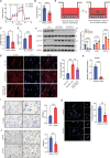
CCT6A suppresses lactate production and reverses the lipid accumulation in alveolar epithelial cells. (A and B) Real-time ECAR (mpH/min) analysis of cells transfected with CCT6A plasmid under basal conditions followed by addition of glucose (10 mM), oligomycin (1 μM), and 2-DG (50 mM) as indicated. (C and D) Extracellular lactate production in AEC2s transfected with CCT6A plasmid or CCT6A plasmid. (E) Schematic of a co-culture system of transfected epithelial cells and fibroblasts. (F) Western blotting and quantification of α-SMA and COL1A1 protein expression in fibroblasts co-cultured with siCCT6A-transfected epithelial cells. (G) Immunofluorescence staining and quantification of α-SMA in fibroblasts co-cultured with siCCT6A-transfected epithelial cells in the presence or absence of ARC155858. Scale bar, 50 μm. (H) qPCR analysis of HCAR1 mRNA expression levels in transfected epithelial cells. (I and J) Oil Red O staining and quantification of epithelial cells treated with 20 mM lactate or transfected epithelial cells following lactate induction. Boxed regions are magnified from the panel. Scale bar, 50 μm. (K) Immunofluorescence staining and quantification of lipid droplet in transfected epithelial cells following lactate induction. Scale bar, 50 μm. *< 0.05, **P <0.01, ***< 0.001, ****P <0.0001.
Figure 3
Figure 3
Activation of lactate signal promotes pulmonary fibrosis in mice. (A) Timeline of GPR81 agonist-treated mouse lung fibrosis model. (B) Quantification of hydroxyproline content in mouse right inferior lobe (n = 4 mice per group). (C) Quantification of total cells in BALF (n = 3). (D) Protein concentration in BALF (n = 3). (E) Representative images of H&E and Masson's Trichrome staining in mouse lung sections (n = 3). Scale bar, 50 μm. The Ashcroft score was determined to indicate the severity of fibrosis. (F) Western blotting and quantification of fibrosis marker expression in whole lung lysates (n = 3). (G and H) qPCR analysis of α-SMA, Col1a1, and Fn1 mRNA expression levels and immunofluorescence staining of lipid droplet in mouse lung tissue samples (n = 4). Scale bar, 50 μm. *< 0.05, **P <0.01, ***< 0.001, ****P <0.0001.
Figure 4
Figure 4
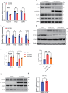
CCT6A mediates a reduction in lactate levels by inhibiting HIF-1α-mediated glycolysis. (A) qPCR analysis of mRNA expression levels of glycolysis-related genes GLUT1, HK2, and LDHA in epithelial cells. (BE) Western blotting and quantification of glycolysis-related protein expression in A549 cells. (F) Extracellular lactate production in siCCT6A-transfected epithelial cells in the presence or absence of PX-478 (15 μM). (G and H) Western blotting of β-catenin (> 0.05) and C-MYC (> 0.05) protein expression and qPCR analysis of HIF-1α mRNA expression levels (> 0.05) in epithelial cells. *< 0.05, **P <0.01, ***< 0.001, ****P <0.0001. ns, not significant.
Figure 5
Figure 5
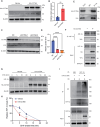
CCT6A interacts with VHL and stimulates the ubiquitination of HIF-1α. (A and B) Western blotting and quantification of VHL in epithelial cells transfected with vehicle and HA-CCT6A. (C and D) Western blotting and quantification of VHL in A549 cells transfected with siNC and siCCT6A. (E) Co-IP of CCT6A and VHL in epithelial cells. (F) Co-IP of HIF-1α and VHL in transfected epithelial cells. (G and H) A549 cells transfected with HA-CCT6A or vehicle were treated with CHX and collected at the indicated time points for western blotting and quantification of HIF-1α protein expression. (I) HEK293T cells were transfected with the indicated plasmids and assayed for the ubiquitination level of HIF-1α by IP and western blotting. **P <0.01, ****P <0.0001.
Figure 6
Figure 6
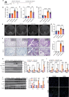
Cct6a restores BLM-induced lung fibrosis in mice. (A) Timeline of AAV-treated mouse lung fibrosis model. (B) Hydroxyproline in the right inferior lobe (Vehicle Sal n = 7, Cct6a Sal n = 8, Vehicle BLM n = 8, Cct6a BLM n = 7). (C) Total cell counts in BALF (n = 7 mice per group). (D) Protein concentration in BALF (Vehicle Sal n = 7, Cct6a Sal n = 7, Vehicle BLM n = 8, Cct6a BLM n = 9). (E) Representative images of micro CT scans of mouse lung density (n = 3). (F) Representative images of H&E and Masson's Trichrome staining in mouse lung sections (n = 3). Boxed regions in the right superior panel are panoramic images of the lung tissue sections. Scale bar, 50 μm. The Ashcroft score was determined to indicate the severity of fibrosis. (G) Lactate level in mouse serum (Vehicle Sal n = 8, Cct6a Sal n = 10, Vehicle BLM n = 9, Cct6a BLM n = 10). (H) Western blotting and quantification of fibrosis marker expression in whole lung lysates (n = 3). (I) qPCR analysis of Col1a1, Col3a1, Fn1, and Ctgf mRNA expression levels in the lung homogenate (n = 6). (J) Immunoblot analysis of Vhl, Hif-1α, and Ldha protein levels in whole lung lysates (n = 3). (K) Immunofluorescence staining of lipid droplet in mouse lung sections (n = 3). Scale bar, 50 μm. *< 0.05, **P <0.01, ***< 0.001, ****P <0.0001.
Figure 7
Figure 7
Mechanism of CCT6A preventing the progression of pulmonary fibrosis. In the fibrotic lung, metabolic abnormalities in alveolar epithelial cells lead to an increase in HIF-1α-mediated lactate production, which induces the activation of interstitial fibroblasts, thereby accelerating the progression of pulmonary fibrosis. High expression of CCT6A stabilizes the cellular VHL protein level and thus promotes VHL-mediated ubiquitination and degradation of HIF-1α, thereby inhibiting the increased lactate level in alveolar epithelial cells and alleviating pulmonary fibrosis.

References

    1. Ahmed K., Tunaru S., Tang C. et al. (2010). An autocrine lactate loop mediates insulin-dependent inhibition of lipolysis through GPR81. Cell Metab. 11, 311–319. - PubMed
    1. Brackley K.I., Grantham J. (2009). Activities of the chaperonin containing TCP-1 (CCT): implications for cell cycle progression and cytoskeletal organisation. Cell Stress Chaperones 14, 23–31. - PMC - PubMed
    1. Brooks G.A. (2009). Cell-cell and intracellular lactate shuttles. J. Physiol. 587, 5591–5600. - PMC - PubMed
    1. Brooks G.A. (2020). Lactate as a fulcrum of metabolism. Redox. Biol. 35, 101454. - PMC - PubMed
    1. Bueno M., Calyeca J., Rojas M. et al. (2020). Mitochondria dysfunction and metabolic reprogramming as drivers of idiopathic pulmonary fibrosis. Redox. Biol. 33, 101509. - PMC - PubMed

Publication types

MeSH terms

Substances