. 2024 May 18;147(1):87.
doi: 10.1007/s00401-024-02729-7.

Identification of high-performing antibodies for the reliable detection of Tau proteoforms by Western blotting and immunohistochemistry

Affiliations

Identification of high-performing antibodies for the reliable detection of Tau proteoforms by Western blotting and immunohistochemistry

Michael J Ellis et al. Acta Neuropathol. .

Abstract

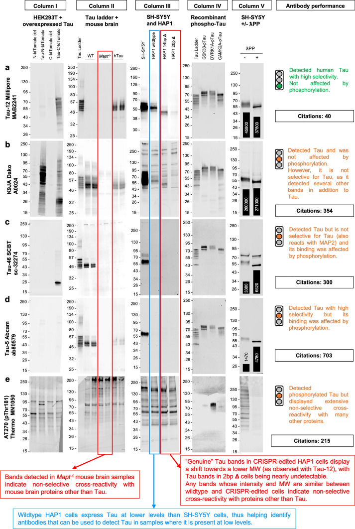
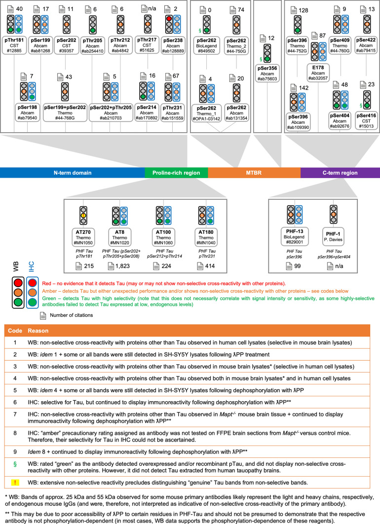
Antibodies are essential research tools whose performance directly impacts research conclusions and reproducibility. Owing to its central role in Alzheimer's disease and other dementias, hundreds of distinct antibody clones have been developed against the microtubule-associated protein Tau and its multiple proteoforms. Despite this breadth of offer, limited understanding of their performance and poor antibody selectivity have hindered research progress. Here, we validate a large panel of Tau antibodies by Western blot (79 reagents) and immunohistochemistry (35 reagents). We address the reagents' ability to detect the target proteoform, selectivity, the impact of protein phosphorylation on antibody binding and performance in human brain samples. While most antibodies detected Tau at high levels, many failed to detect it at lower, endogenous levels. By WB, non-selective binding to other proteins affected over half of the antibodies tested, with several cross-reacting with the related MAP2 protein, whereas the "oligomeric Tau" T22 antibody reacted with monomeric Tau by WB, thus calling into question its specificity to Tau oligomers. Despite the presumption that "total" Tau antibodies are agnostic to post-translational modifications, we found that phosphorylation partially inhibits binding for many such antibodies, including the popular Tau-5 clone. We further combine high-sensitivity reagents, mass-spectrometry proteomics and cDNA sequencing to demonstrate that presumptive Tau "knockout" human cells continue to express residual protein arising through exon skipping, providing evidence of previously unappreciated gene plasticity. Finally, probing of human brain samples with a large panel of antibodies revealed the presence of C-term-truncated versions of all main Tau brain isoforms in both control and tauopathy donors. Ultimately, we identify a validated panel of Tau antibodies that can be employed in Western blotting and/or immunohistochemistry to reliably detect even low levels of Tau expression with high selectivity. This work represents an extensive resource that will enable the re-interpretation of published data, improve reproducibility in Tau research, and overall accelerate scientific progress.

Keywords: Antibody validation; Immunohistochemistry; Phosphorylation; Splice isoforms; Tau; Western blot.


Conflict of interest statement

This work was partly funded by a collaborative agreement between the University of Oxford and UCB BioPharma. UCB staff were not involved in the design of the experiments, analysis of the data or interpretation of the results. The authors declare no other competing interests.

Figures

Fig. 1
Fig. 1
Overview of the human MAPT gene, Tau protein, antibody epitopes and antibody validation experimental strategies. a Diagram of the human MAPT gene structure with currently described exons depicted as rectangles and introns depicted as connecting lines. Exon numbering shown above. The canonical transcription start site (ATG) located in exon 1 is indicated (black arrow). Non-coding exonic regions are shown in light grey. Constitutively included exons (1, 4, 5, 7, 9, 11, 12 and 13) are shown in white. Alternatively spliced exons included in common brain Tau isoforms, Big Tau isoforms or not included in any human Tau isoforms described to date are shown in yellow (exon 2,3 and 10), light grey (exons 4A and 6) and black (exon 8), respectively. b Diagram of the human Tau protein (2N4R isoform, longest canonical Tau isoform), showing the four main protein domains: N-terminal acidic region (blue), proline-rich mid region (green), microtubule-binding repeat region (orange) and C-terminal region (purple). Amino acid residues marking the domain boundaries are shown above. Protein regions encoded by different exons are indicated. Amino acid residues marking exon–exon boundaries are shown below. c Schematic depiction of the epitopes targeted by the Tau antibodies included in this study: “total” Tau antibodies (above, black), phospho-Tau antibodies (below, blue), other PTM-dependent antibodies (magenta) and isoform-specific antibodies (green)
Fig. 2
Fig. 2
Overview of the experimental approaches employed in this study to validate Tau antibodies using WB (a) and IHC-IF (b)
Fig. 3
Fig. 3
Representative antibody validation results for WB. a–e WB data illustrating the performance of different Tau antibodies: Tau-12 Millipore #MAB2241 (a), K9JA Dako #A0024 (b), Tau-46 SCBT #sc-32274 (c), Tau-5 Abcam #ab80579 (d), AT270 (pThr181) ThermoFisher Scientific #MN1050 (e). For each antibody, overall performance was categorised based on a traffic light system, as defined in Figs. 5–8. Number of citations for each antibody clone as of 9 Oct 2023 is indicated. WBs shown in columns I to V are as follows: WB of lysates from HEK293T cells overexpressing 0N3R human Tau and corresponding control cells (column I); WB of recombinant human Tau ladder (5 ng/isoform/lane), plus adult mouse brain lysates from wildtype, Mapt−/− and hTau mice (column II); WB of lysates from SH-SY5Y neuroblastoma cells, plus HAP1 cells: parental (wildtype) and two cell lines carrying either a 14 bp deletion (14 bp Δ) or a 2-bp deletion (2 bp Δ) in MAPT exon 4 (column III); WB of recombinant human Tau ladder (50 ng/isoform/lane) plus recombinant 2N4R Tau that has been phosphorylated by one of three known Tau kinases: GSK3ꞵ, DYRK1A or CAMKIIA (column IV); WB of lysates from SH-SY5Y neuroblastoma cells that have been either untreated (-) or treated ( +) with λPP. Where applicable, quantifications of the Tau signal intensity for each lane are shown superimposed as a bar chart, with the respective values [a.u.] printed on or above each bar of the chart (column V)
Fig. 4
Fig. 4
Representative antibody validation results for IHC-IF. a–g’: IHC-IF data illustrating the performance of different Tau antibodies: Tau-12 Millipore #MAB2241 (ab’’), Tau-46 SCBT #sc-32274 (cd’’), Tau-5 Abcam #ab80579 (ef’’), pSer396 Abcam #ab109390 (gh'’), pSer199 Abcam #ab81268 (ij'’), 77E9 BioLegend #814,402 (kk’). For each antibody, overall performance was categorised based on a traffic light system, as defined in the text and in Figs. 5–8. a-a’, c–c’, e-e’, g-g’, i-i', k-k': Fluorescence micrographs of serial FFPE brain sections from 9-month old rTg4510 mice, either untreated (control; a, c, e, g, i and k) or treated (λPP-treated; a’, c’, e’, g’, i' and k’) with λPP, immunolabelled with Tau antibodies. b-b”, d-d’’, f-f”, h–h'’, j-j'’: Fluorescence micrographs of FFPE brain sections from 5-month-old wildtype (b, d, f, h and j), Mapt−/− (b’, d’, f’ and j’) and hTau (b”, d”, f” and j’’) mice immunolabelled with Tau antibodies. Brain regions imaged are cortex (a-a’, c–c’, e-e’, g-g’, i-i' and k-k') and CA1 region of the hippocampus (b-b”, d-d”, f-f”, h–h'’ and j-j'’). Tau labelling is shown in grayscale. Scale bars = 100 µm
Fig. 5
Fig. 5
Summary of results for “total” Tau antibodies. Diagram of the Tau protein showing the locations of the epitopes of “total” Tau antibodies. In this and the following two figures: (i) residue numbering is based on 2N4R Tau (441 amino acids; Uniprot ID 10636–8); (ii) traffic lights summarise the performance of each antibody in WB (black outline) and IHC (blue outline); (iii) antibody performance was classified as either: Green = detects Tau with high selectivity; Amber = detects Tau but either unexpected performance was observed and/or antibody displayed non-selective cross-reactivity with other proteins (see codes); or Red = no evidence that the antibody detects Tau (may or may not show non-selective cross-reactivity with other proteins); (iv) the total number of studies citing the use of each antibody clone (aggregated for all formulations and vendors) is displayed
Fig. 6
Fig. 6
Summary of results for phosphorylation-dependent, other PTM-dependent and isoform-specific Tau antibodies. Diagram of the Tau protein showing the locations of the epitopes of phospho-Tau antibodies
Fig. 7
Fig. 7
Summary of results for other PTM-dependent and isoform-specific Tau antibodies. Diagram of the Tau protein showing the locations of the epitopes of other-PTM dependent antibodies, as well as isoform-specific Tau antibodies
Fig. 8
Fig. 8
Antibodies binding to the N-term, mid- and MTBR-domains reveal presence of C-term-cleaved Tau variants in human brain extracts from both control and tauopathy donors. a Diagram of the main domains of the Tau protein showing the locations of the epitopes of the various antibodies. Antibodies raised against specific splice isoforms are shown in magenta. b Tau was detected by WB in human brain protein extracts from control and diagnosed tauopathy donors with antibodies binding close to the N-terminus (SP70), in the mid-domain (Tau-1) or close the C-terminus (D1M9X) of the protein. MW range of full-length Tau isoforms, Tau oligomers and higher order Tau aggregates are indicated. Blue and red boxes outline the MW range where N-terminal-truncated and C-terminal-truncated Tau variants were observed, respectively. c Tau was detected in dephosphorylated human brain extracts by WB with a panel of antibodies with epitopes spanning the entire length of the protein. The antibody used to probe each WB membrane is indicated above each respective image. Magenta asterisks indicate putative Tau fragments that lack both the N- and C-termini. dg Human brain protein extracts were either dephosphorylated via treatment with λPP ( +) or mock-treated (-) and analysed by fluorescent WB with combinations of mouse (green) + rabbit (red) Tau antibody pairs to assess the overlap between the bands detected with each antibody: 0N Tau isoform-specific antibody (Abcam reagent) + Tau-12 “total” Tau N-term antibody (d); 2N Tau Tau isoform-specific antibody (Abcam reagent) + Tau-12 “total” Tau N-term antibody (e); D1M9X “total” Tau C-term antibody + Tau-12 “total” Tau N-term antibody (f); 4R Tau isoform-specific antibody (CST reagent) + RD3 (3R Tau) isoform-specific antibody (g). Individual channels are shown in grayscale (red channel, left; green channel, right)

Similar articles

Cited by

References

    1. Agholme L, Lindström T, Kågedal K, Marcusson J, Hallbeck M. An in vitro model for neuroscience: differentiation of SH-SY5Y cells into cells with morphological and biochemical characteristics of mature neurons. J Alzheimers Dis. 2010;20:1069–1082. doi: 10.3233/JAD-2010-091363. - DOI - PubMed
    1. Algenäs C, Agaton C, Fagerberg L, Asplund A, Björling L, Björling E, Kampf C, Lundberg E, Nilsson P, Persson A, Wester K, Pontén F, Wernérus H, Uhlén M, Ottosson Takanen J, Hober S. Antibody performance in western blot applications is context-dependent. Biotechnol J. 2014;9:435–445. doi: 10.1002/biot.201300341. - DOI - PubMed
    1. Alquezar C, Arya S, Kao AW. Tau post-translational modifications: Dynamic transformers of tau function, degradation, and aggregation. Front Neurol. 2020;11:595532. doi: 10.3389/fneur.2020.595532. - DOI - PMC - PubMed
    1. ALZFORUM antibodies database: Tau antibodies. https://www.alzforum.org/antibodies/search?category%5B616%5D=Tau&page=0. Accessed 30 Mar 2023
    1. Andersson S, Sundberg M, Pristovsek N, Ibrahim A, Jonsson P, Katona B, Clausson C-M, Zieba A, Ramström M, Söderberg O, Williams C, Asplund A. Insufficient antibody validation challenges oestrogen receptor beta research. Nat Commun. 2017;8:15840. doi: 10.1038/ncomms15840. - DOI - PMC - PubMed

Publication types

Substances