

Inflammatory myofibroblastic tumor of the liver after adrenal neuroblastoma surgery: a case report
- PMID: 38761221
- PMCID: PMC11102410
- DOI: 10.1007/s12672-024-01039-4
Inflammatory myofibroblastic tumor of the liver after adrenal neuroblastoma surgery: a case report
Abstract
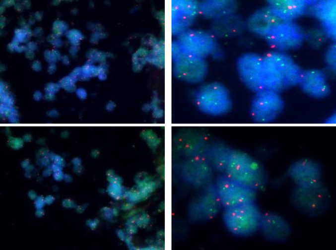
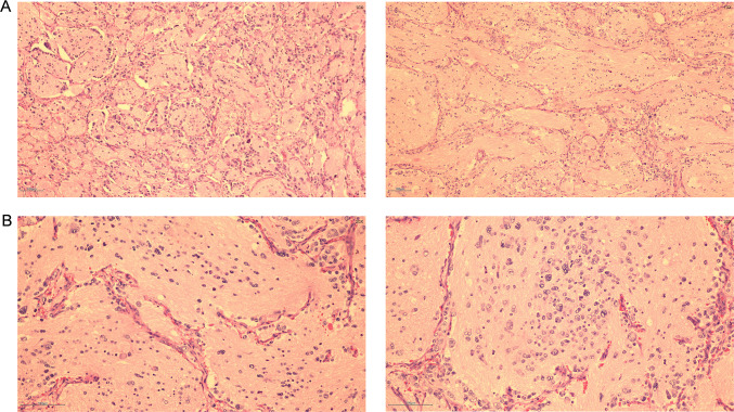
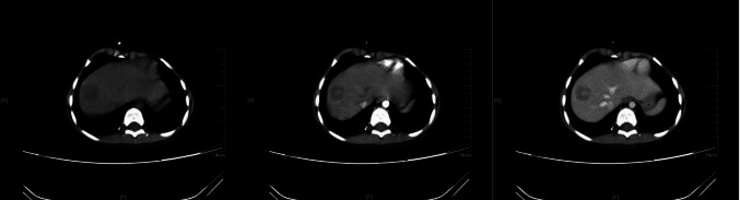
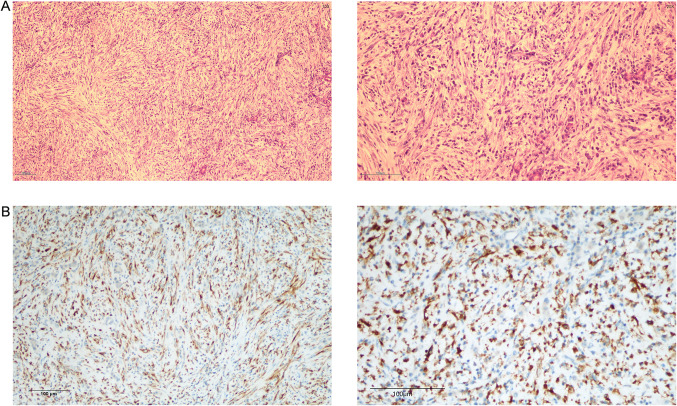
A boy aged 55 months was diagnosed with stage IV Neuroblastoma (NB) of the right adrenal gland 2 years ago. Preoperative chemotherapy was given and he was then treated with retroperitoneal tumor resection and lymph node dissection. After surgery, the children were transferred to the Hemato-Oncology Department for chemotherapy according to the high-risk group NB, with outpatient follow-up every 6 months. In the second postoperative year, abdominal computed tomography (CT) scan revealed a rounded hypodense area in the upper part of the right posterior lobe of the liver, with marked inhomogeneous enhancement in the venous phase after enhancement, which was surgically resected, and postoperative pathology confirmed inflammatory myofibroblastic tumor (IMT) of liver. The patient was not given any special treatment after surgery. In this study, whole transcriptome sequencing was performed on the postoperative specimen of adrenal NB and the specimen of IMT of liver. This unusual case emphasizes the need for close monitoring of second tumor development in NB survivors even in the absence of known predisposing factors.
Keywords: ICG; Inflammatory myofibroblastic tumor; Neuroblastoma; Secondary tumor; Whole transcriptome sequencing.
© 2024. The Author(s).
Conflict of interest statement
The authors declare that the research was conducted in the absence of any commercial or financial relationships that could be construed as a potential competing interests.
Figures

Similar articles
-
Unusual Techniques for Preserving Surgical and Oncologic Safety in Hepatectomy of Advanced Adrenal Malignancy with Vena Cava and Liver Invasion.Ann Surg Oncol. 2018 Oct;25(11):3324-3325. doi: 10.1245/s10434-018-6657-5. Epub 2018 Jul 17. Ann Surg Oncol. 2018. PMID: 30019302
-
[Comprehensive treatment of neuroblastoma in children associated with opsoclonus-myoclonus-ataxia syndrome].Zhonghua Er Ke Za Zhi. 2014 Jul;52(7):540-3. Zhonghua Er Ke Za Zhi. 2014. PMID: 25224062 Chinese.
-
[Adrenal cavernous hemangioma: A case report and literature review].Beijing Da Xue Xue Bao Yi Xue Ban. 2021 Aug 18;53(4):808-810. doi: 10.19723/j.issn.1671-167X.2021.04.032. Beijing Da Xue Xue Bao Yi Xue Ban. 2021. PMID: 34393250 Free PMC article. Review. Chinese.
-
Removal of pediatric stage IV neuroblastoma by robot-assisted laparoscopy: A case report and literature review.World J Clin Cases. 2019 Jun 26;7(12):1499-1507. doi: 10.12998/wjcc.v7.i12.1499. World J Clin Cases. 2019. PMID: 31363479 Free PMC article.
-
Adult renal neuroblastoma: A case report and literature review.Medicine (Baltimore). 2018 Apr;97(14):e0345. doi: 10.1097/MD.0000000000010345. Medicine (Baltimore). 2018. PMID: 29620664 Free PMC article. Review.
Cited by
-
Clinical diagnosis and treatment of abdominal inflammatory myofibroblastic tumors.Discov Oncol. 2025 Apr 17;16(1):554. doi: 10.1007/s12672-025-02343-3. Discov Oncol. 2025. PMID: 40244559 Free PMC article.
-
Therapeutic strategy and prognostic analysis of inflammatory myofibroblastic tumor in the head and neck: a retrospective study.PeerJ. 2025 Apr 18;13:e19315. doi: 10.7717/peerj.19315. eCollection 2025. PeerJ. 2025. PMID: 40260197 Free PMC article.
-
Indocyanine Green Fluorescence Navigation in Pediatric Hepatobiliary Surgery: Systematic Review.Children (Basel). 2025 Jul 18;12(7):950. doi: 10.3390/children12070950. Children (Basel). 2025. PMID: 40723143 Free PMC article. Review.
References
-
- Baird DC, Meyers GJ, Hu JS. Testicular cancer: diagnosis and treatment [J] Am Fam Phys. 2018;97(4):261–268. - PubMed
LinkOut - more resources
Full Text Sources