. 2024 Jun 6;187(12):2919-2934.e20.
doi: 10.1016/j.cell.2024.04.033. Epub 2024 May 17.

Vaccine induction of heterologous HIV-1-neutralizing antibody B cell lineages in humans

Affiliations

Vaccine induction of heterologous HIV-1-neutralizing antibody B cell lineages in humans

Wilton B Williams et al. Cell. .

Abstract

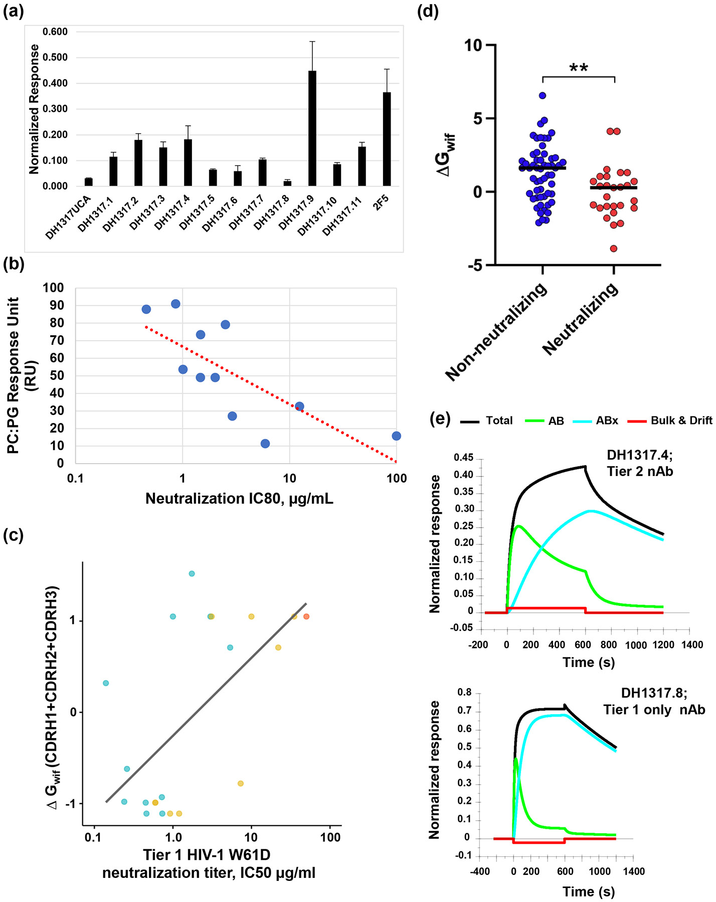
A critical roadblock to HIV vaccine development is the inability to induce B cell lineages of broadly neutralizing antibodies (bnAbs) in humans. In people living with HIV-1, bnAbs take years to develop. The HVTN 133 clinical trial studied a peptide/liposome immunogen targeting B cell lineages of HIV-1 envelope (Env) membrane-proximal external region (MPER) bnAbs (NCT03934541). Here, we report MPER peptide-liposome induction of polyclonal HIV-1 B cell lineages of mature bnAbs and their precursors, the most potent of which neutralized 15% of global tier 2 HIV-1 strains and 35% of clade B strains with lineage initiation after the second immunization. Neutralization was enhanced by vaccine selection of improbable mutations that increased antibody binding to gp41 and lipids. This study demonstrates proof of concept for rapid vaccine induction of human B cell lineages with heterologous neutralizing activity and selection of antibody improbable mutations and outlines a path for successful HIV-1 vaccine development.


Conflict of interest statement

Declaration of interests B.F.H. and S.M.A. have US patents 9402917, 9402893, 9717789, and 10588960 and US patent application 63/540482. B.F.H., S.M.A., and B.K. have US patent 10076567. B.F.H. and K.O.S. have patent applications PCT/US2023/077677 and PCT/US2023/077686. C.B.F. has patents on PEGylated liposomes.

Figures

Figure 1.
Figure 1.. HVTN 133 Clinical Trial Elicited a Polyclonal MPER+ Antibody Response.
(a) Immunization (IMM) schedule for HVTN 133. The timeline for IMMs in months (M) is indicated. B cell repertoire analyses performed on PBMCs 2 weeks post-3rd IMM. Trial was halted after one anaphylactic reaction. (b) Vaccine recipient groups based on peptide dose. (c) Schema for B cell repertoire analysis performed on PBMCs from 5 HVTN 133 vaccinees who received three IMMs. Boxed area: single cell sorting of MPER+ B cells. The top 4 VH genes used by MPER+ antibodies were VH2-5, VH3-49, VH5-51 and VH7-4-1. (d) Frequency of immunoglobulin (Ig) genes used by MPER+ antibodies. Data shown reflect individual Ig gene usages; unpaired heavy and light chain genes. (e) Analysis of MPER+ antibodies isolated post-3rd immunization. (f) Mutation frequency of the predominant VH genes used by MPER+ antibodies. Error bars represent the median and interquartile range (GraphPad Prism, v9.0). (g) CDR3 length distributions of predominant VH genes used by MPER+ antibodies. See also Figures S1, and Tables S1-S2.
Figure 2.
Figure 2.. Neutralization Breadth Profile of Vaccine-induced Antibodies.
(a) Schematic outlining maturation pathway for MPER bnAbs. (b) Flowchart summarizing the neutralization screens for MPER+ recombinant antibodies isolated from HVTN133 vaccine recipients post-3rd immunization. (c) Neutralization profile of 49/80 MPER+ Abs that neutralized tier 1 or 2 clade B HIV-1 strains in TZM-bl/FcγRI or TZM-bl assays. (d) Neutralization curves against tier 1 and 2 clade B HIV-1 strains for DH1317.4 and DH1317.9 antibodies. Data shown are from single or mean titers of duplicate experiments. (e) Summary of neutralization breadth for DH1317.4 NAb. See also Figures S2-S3, and Tables S3-S4.
Figure 3.
Figure 3.. MPER+ NAbs Mapped to Proximal BnAb Epitopes.
(a) (Left) Neutralization profile and heavy chain gene usage by the MPER+ NAbs. (Right) Mapping of MPER+ NAbs to proximal MPER bnAb epitope. Binding was measured in ELISA and reported as Log AUC as shown in the heatmap. Binding sensitivity or epitope mapping to a specific residue tested were based on ≥50% reduction in binding to single alanine-scanned mutant peptides. (b) Neutralization titers of DH1317.4, DH1317.9 and MPER proximal bnAbs m66.6 and 2F5 tested against a panel of clade B HIV-1 strains from the AMP trial and non-clade B HIV-1 strains in the TZM-bl/FcγRI assay. See Figure S2 for data from TZM-bl assay. (c) NSEM of 11 MPER-directed Abs. The Env trimer, excluding the MPER domain (gray), the MPER region with partial Fab density (green), and the MPER Fabs are colored: VH5-51 = pink; VH2-5 = sky blue; VH3-49 = gold, and VH7-4-1 = brown. See also Figures S2 and S6, and Table S5.
Figure 4.
Figure 4.. Lipid Reactivity Profiles of DH1317 Lineage Antibodies.
(a) Binding at 100μg/mL to POPC:DOPG (25:75) liposomes. Values and error bars represent the average and standard deviation of three measurements. (b) Plot of phosphatidylglycerol lipid (PC:PG) binding response in RU versus W61D pseudovirus neutralization IC80 values of each of the DH1317 lineage Abs. The plotted PC:PG binding responses are mean values of three independent experiments. Kendall’s Tau correlation coefficient =0.6363, P=0.0054 for both IC80 and IC50 values. (c) Computed lipid insertion propensity scores ΔGwif of HCDR1, 2 and 3 (summed) versus IC50 (μg/mL) W61D neutralization in the TZM-bl assay. Computed lipid insertion propensity scores ΔGwif (kcal/mol) for HCDR1, 2 and 3 (summed) is positively correlated with log W61D neutralization potency for DH1317 clonal members (R = 0.61, P = 0.0033, Pearson correlation). Isolated members of the DH1317 clone are shown in blue, inferred intermediate Abs in yellow, and inferred UCA in orange. DH1317I6 and I7 appear as a single dot due to identical ΔGwif scores (−0.99) and almost identical neutralization values (0.598 and 0.595, respectively). Comprehensive ΔGwif scores of all antibodies within the DH1317 clonal lineage are detailed in Mendeley Data, and corresponding neutralization values (Table S4). (d) Computed lipid insertion propensity scores ΔGwif (kcal/mol) for HCDR1, 2 and 3 (summed) are significantly higher (less favorable for lipid insertion) in W61D nnAbs (blue) than NAbs (red). ** p<0.01; Mann-Whitney test. (e) Biolayer interferometry (BLI) binding of DH1317.4 heterologous tier 2 NAb (top) or DH1317.8 (bottom) tier 1 only NAb Fabs to MPER liposomes fitted to the 2-step conformational change model. See also Figure S4 and Table S4.
Figure 5.
Figure 5.. Structural Analysis of the Polyclonal VH7-4-1 Antibody Response to the MPER Peptide-Liposome in Vaccinee 133-23.
(a) Structures of NAbs DH1317.8, DH1322.1, and DH1346 in complex with MPER peptides. (b) Helical wheel representations of MPER with NAb contacting faces shown in red. (c). Overlay of antibody complexes based on alignment of common VH7-4-1 regions, shown in same orientations and colors as panel a. (d) Salt bridge interactions between MPER residue D664 and NAbs DH1317.8, DH1322.1, and DH1346. (e) Paratope interfaces on DH1317.8, DH1322.1, and DH1346 mapped orange on heavy chains and dark blue on light chains, shown in same orientation as in panel a (top). (f) Sequence alignment of the heavy chain regions of DH1317.4, DH1317.8, DH1322.1, and DH1346 against VH7-4-1 germline gene, with interface residue BSAs plotted as bars above. (g) VH7-4-1-encoded HCDR1 and HCDR2 loops in DH1317.8, DH1322.1, and DH1346 adopt similar conformations and interact with the same set of MPER residues. See also Figure S7 and S8.
Figure 6.
Figure 6.. Structural Comparison of Antibody Recognition of Proximal Gp41 MPER.
(a) DH1317.8, DH1322.1, DH1346, DH570 (5DD0), m66 (4NRX), and 2F5 (1TJI)-bound conformations of proximal gp41 MPER (yellow), oriented based on Cα alignments to reference DH1322.1-bound MPER. R.m.s.d. values shown for residue ranges. The epitope of 2F5 is shown based on alignment to residues 664-666 of m66-bound MPER. (b) Antibody variable region structures bound to MPER, oriented based on bound MPER peptides as shown in panel a. (c) Overlay of MPER-bound antibody variable region structures shown in same orientations and colors as in panel b (top), and rotated ~90° (bottom). (d) Plots of interface residue BSA along proximal MPER for DH1317.8, DH1322.1, and DH1346 (top) and DH570, m66, and 2F5 (bottom). (e) Pairwise correlation analysis of BSA of shared MPER interface residues between VH7-4-1-using NAbs (upper panels) and with reference antibodies DH570, m66, and 2F5 (lower panels). See also Figure S7 and S8.

Similar articles

Cited by

References

    1. Haynes BF, Gilbert PB, McElrath MJ, Zolla-Pazner S, Tomaras GD, Alam SM, Evans DT, Montefiori DC, Karnasuta C, Sutthent R et al. 2012. Immune-correlates analysis of an hiv-1 vaccine efficacy trial. N Engl J Med. 366(14):1275–1286. - PMC - PubMed
    1. Rerks-Ngarm S, Pitisuttithum P, Nitayaphan S, Kaewkungwal J, Chiu J, Paris R, Premsri N, Namwat C, de Souza M, Adams E et al. 2009. Vaccination with alvac and aidsvax to prevent hiv-1 infection in thailand. N Engl J Med. 361(23):2209–2220. - PubMed
    1. Haynes BF, Wiehe K, Borrrow P, Saunders KO, Korber B, Wagh K, McMichael AJ, Kelsoe G, Hahn BH, Alt F et al. 2022. Strategies for hiv-1 vaccines that induce broadly neutralizing antibodies. Nat Rev Immunol. - PMC - PubMed
    1. Desrosiers RC. 2023. The failure of aids vaccine efficacy trials: Where to go from here. J Virol. 97(3):e0021123. - PMC - PubMed
    1. Gray GE, Bekker LG, Laher F, Malahleha M, Allen M, Moodie Z, Grunenberg N, Huang Y, Grove D, Prigmore B et al. 2021. Vaccine efficacy of alvac-hiv and bivalent subtype c gp120-mf59 in adults. N Engl J Med. 384(12):1089–1100. - PMC - PubMed

LinkOut - more resources