Review
. 2024 Jun;45(6):537-551.
doi: 10.1016/j.tips.2024.04.007. Epub 2024 May 17.

Emerging targets in lipid metabolism for cancer therapy

Affiliations
Review

Emerging targets in lipid metabolism for cancer therapy

Alexander R Terry et al. Trends Pharmacol Sci. 2024 Jun.

Abstract

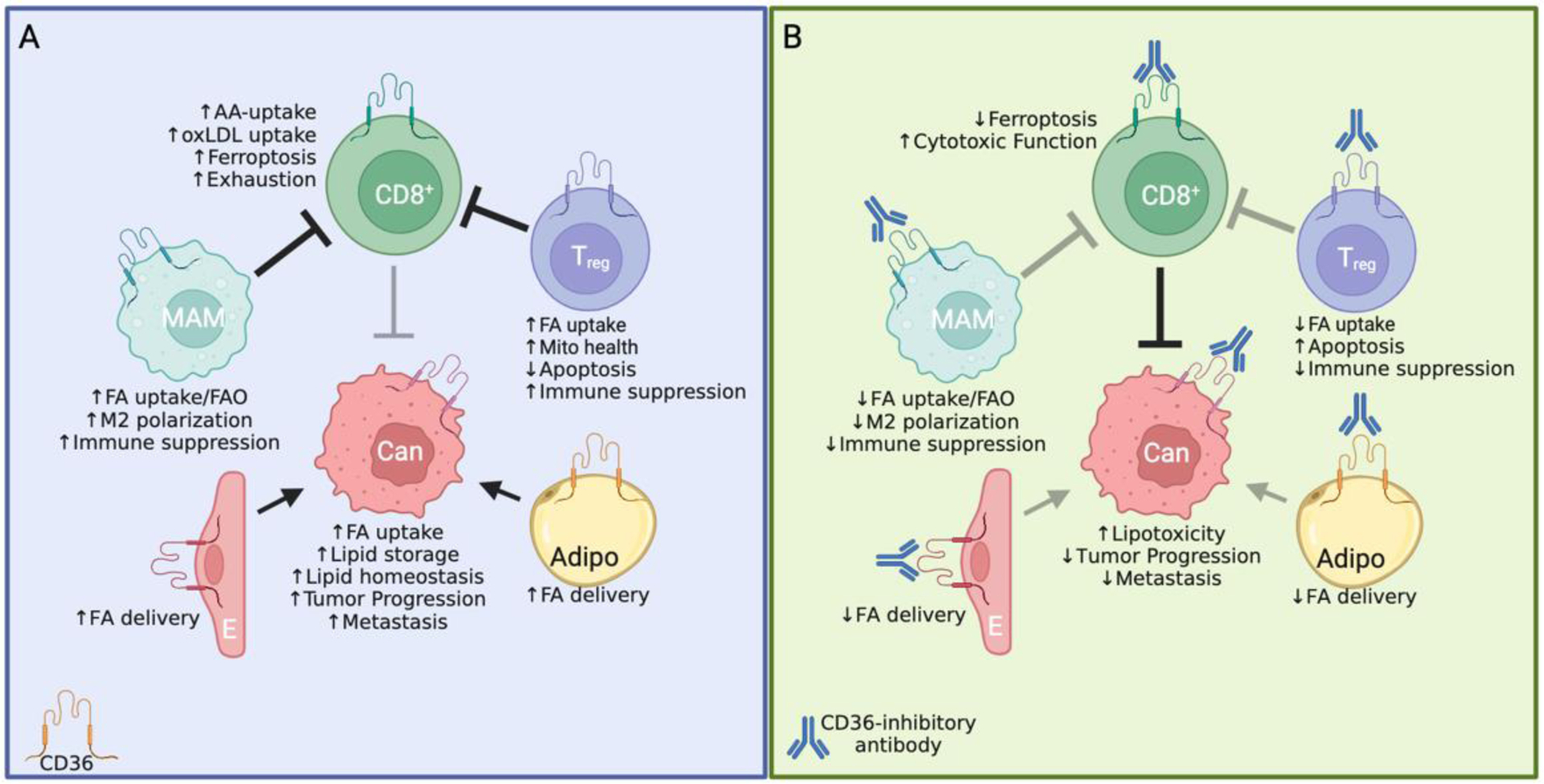
Cancer cells perturb lipid metabolic pathways for a variety of pro-tumorigenic functions, and deregulated cellular metabolism is a hallmark of cancer cells. Although alterations in lipid metabolism in cancer cells have been appreciated for over 20 years, there are no FDA-approved cancer treatments that target lipid-related pathways. Recent advances pertaining to cancer cell fatty acid synthesis (FAS), desaturation, and uptake, microenvironmental and dietary lipids, and lipid metabolism of tumor-infiltrating immune cells have illuminated promising clinical applications for targeting lipid metabolism. This review highlights emerging pathways and targets for tumor lipid metabolism that may soon impact clinical treatment.

Keywords: cancer metabolism; fatty acids; lipid metabolism.


Conflict of interest statement

Declaration of interests The authors have no competing interests to declare.

Figures

Figure 1:
Figure 1:. Fundamentals of lipid metabolism.
Cells synthesize fatty acids de novo (FAS) or acquire them through extracellular uptake. FAS occurs in the cytosol and is catalyzed by ACC and FASN enzymes, which produce palmitate, a 16-carbon SFA. MUFAs are produced by desaturation of SFAs by SCD1 or FADS2. The PUFAs, ALA and LA, are essential fatty acids and serve as building blocks for PUFA biosynthesis. Fatty acid uptake occurs through free fatty acid diffusion or CD36-mediated fatty acid uptake. When intracellular levels of palmitate are high, palmitoylated CD36 promotes preferential MUFA uptake. The composite intracellular fatty acid pool is comprised of SFAs, MUFAs, and PUFAs derived from FAS and extracellular uptake. Fatty acids are activated by ACSL enzymes and incorporated into glycerolipids and phospholipids to support membrane biogenesis and lipid storage. Fatty acid β-oxidation (FAO) occurs in the mitochondria and supports energy production by driving the TCA-cycle. Acetyl-CoA also supports the mevalonate pathway to produce cholesterol. Abbreviations: ALA: alpha-linoleic acid, LA: linoleic acid, PUFA: polyunsaturated fatty acid, MUFA: monounsaturated fatty acid, SFA: saturated fatty acid, CTP1: carnitine palmitoyl transferase 1, FAO: fatty acid oxidation, TCA Cycle: tricarboxylic acid cycle, ETC: electron transport chain, ACSS2: acetyl-CoA synthetase enzyme 2, ACLY: ATP citrate lyase, ACAT: acetyl-CoA acetyltransferase, HMGCS: hydroxyl-3-methylglutaryl-CoA synthetase, HMGCR: hydroxyl-3-methylglutaryl-CoA reductase, ACC: acetyl-CoA carboxylase, FASN: fatty acid synthase, SCD1: stearoyl-CoA desaturase 1, FADS2: fatty acid desaturase 2, ELOV: elongation of very-long-chain fatty acids protein, G3P: glycerol-3-phosphate, GPAT: glycerol-3-phosphate acyltransferase, LPA: lysophosphatidic acid, PA: phosphatidic acid, DAG: diacylglycerol, TAG: triacylglycerol, DGAT: diglyceride acyltransferase, LD: lipid droplet, PC: phosphatidylcholine, PE: phosphatidylethanolamine, PS: phosphatidylserine, PI: phosphatidylinositol.
Figure 2:
Figure 2:. Chemical structure of commons long chain fatty acids.
Fatty acid synthesis produces palmitic acid, which can be elongated by ELOV enzymes to produce stearic acid. The SFAs, palmitic acid and stearic acid, are substrates for oxygen-dependent SCD1, producing the MUFAs, palmitoleic acid and oleic acid, respectively. Palmitic acid is also a substrate for FADS2, which produces the MUFA, sapienic acid. Polyunsaturated fatty acids are derived from essential fatty acids linoleic acid (an n-6 PUFA) or alpha-linoleic acid (n-3 PUFA). Note, this figure shows examples but is not exhaustive of all PUFAs. For omega nomenclature, n-x, x indicates where the first double bond is from the methyl end of the fatty acid. Abbreviations: SCD1: stearoyl-CoA desaturase 1, FADS2: fatty acid desaturase 2, ELOV: elongation of very-long-chain fatty acids protein, SFA: saturated fatty acid, PUFA: polyunsaturated fatty acid, MUFA: monounsaturated fatty acid.
Figure 3:
Figure 3:. Saturated fatty acid-induced toxicity for cancer treatment.
A) ER stress kinases IRE1α and PERK are activated by misfolded proteins and increased membrane lipid saturation independent of their misfolded-protein domains (represented by truncated cartoon of each protein). Through a variety of mechanism, including SREBP-activation and IRE1α-dependent XBP1s-activation, adaptive ER stress signaling induces genes involved in de novo lipogenesis to promote membrane synthesis and ER homeostasis. For example, MYC-driven tumors rely on pro-survival ER stress signaling through XBP1s, and inhibition of XBP1s-activation inhibits tumor growth. B) During the same process, SCD1 inhibition drives accumulation of SFAs, which are incorporated in membrane lipids further activating ER stress and ultimately inducing IRE1α-dependent apoptosis. In MYC-driven tumors, inhibition of SCD1 phenocopies XBP1s-inhibition. C) In HCC, targeted activation of LXRα promotes induction of FAS genes including ACC, FASN, and SCD1. Raf inhibition disrupts its direct binding to SCD1, thereby destabilizing SCD1. When used together, saturated fatty acids accumulate to toxic levels, induce ER stress, and inhibit HCC growth in vivo. D) A calorie restricted diet decreases lipids in the serum and tumor microenvironment as well as decreases SCD1 protein level in tumor cells. This leads to toxic SFA accumulation which inhibits PDAC growth in vivo. E) A ketogenic diet increases serum and tumor microenvironmental lipids, so despite decreasing SCD1 protein, PDAC cells maintain a balance of the SFA/MUFA ratio through exogenous sources. F) A ketogenic diet designed with a higher SFA/MUFA ratio causes increased SFA levels intracellularly and inhibits PDAC growth in vivo. G) In ovarian cancer, targeting SCD1 in the presence of an SFA-rich diet promotes ER stress and inhibits peritoneal metastases. Abbreviations: SCD1: stearoyl-CoA desaturase 1, FASN: fatty acid synthase, ACC: acetyl-CoA carboxylase, XBP1s: spliced X-box protein 1, SREBP: sterol regulatory element binding protein, LXRα: liver X receptor α, FAS: fatty acid synthesis, HCC: hepatocellular carcinoma, PDAC: pancreatic ductal adenocarcinoma, SFA: saturated fatty acid, MUFA: monounsaturated fatty acid, PERK: protein kinase RNA-like ER kinase, IRE1α: inositol-requiring enzyme type 1.
Figure 4:
Figure 4:. Lipids determine sensitivity to ferroptosis.
Ferroptosis is initiated by the reaction between a PC containing two PUFAs (PC-PUFA2) and mitochondrial ROS to form a hydroperoxide species (PC-PUFA2-OOH). PUFA-peroxidation is propagated through PUFA-containing phospholipids, leading to plasma membrane disruption and cell death. PC-PUFA2 synthesis is promoted by increased PUFA uptake and activation by ACSL4, and this process is opposed by MUFAs following their activation by ACSL3. Lymphatics are rich in MUFAs and can promote metastasis by protecting cells from ferroptosis. Conversely, diets rich in PUFAs can inhibit tumor growth by inducing ferroptosis. FASN and SCD1 protect cells from ferroptosis by producing SFAs and MUFAs which outcompete PUFAs in membrane phospholipids. LDs protect cancer cells from ferroptosis by providing MUFAs and sequestering PUFAs to prevent their membrane incorporation. PUFA biosynthesis catalyzed by FADS1 and ELOV5 generates AA and DHA, which are PUFAs most sensitive to peroxidation. Both the GPX4/GSH and CoQ10/FSP1 pathway are endogenous inhibitors of ferroptosis. Erastin and RSL3 are examples of ferroptosis inducing agents which both act on the GPX4/GSH pathway. Blue lines indicated ferroptosis suppression; red lines indicate ferroptosis promotion. Abbreviations: PC: phosphatidylcholine, PUFA: polyunsaturated fatty acid, ROS: reactive oxygen species, ACSL: acyl-CoA synthetase long-chain family member, MUFA: monounsaturated fatty acid, FASN: fatty acid synthase, SCD1: stearoyl-CoA desaturase 1, SFA: saturated fatty acid, LD: lipid droplets, FADS1: fatty acid desaturase 1, ELOV5: elongation of very-long-chain fatty acids protein 5, AA: arachidonic acid, DHA: docosahexaenoic acid, LA: linoleic acid, ALA: alpha-linoleic acid, GPX4: glutathione peroxidase 4, GSH: glutathione, CoQ10: coenzyme Q10, FSP1: ferroptosis suppressor protein 1.
Figure 5:
Figure 5:. Targeting CD36 for cancer therapy.
A) CD36 promotes tumor progression cell-autonomously through numerous mechanisms. Importantly, these depend on fatty acid uptake and storage to promote lipid homeostasis and aggressive cancer phenotypes. In CD8+ T cells, which inhibit tumorigenesis by killing cancer cells, CD36 promotes uptake of oxidized LDL (oxLDL) and arachidonic acid (AA) leading to ferroptosis and T cell exhaustion. In Treg cells, which inhibit CD8+ T cells to promote tumorigenesis, CD36 promotes fatty acid uptake for maintenance of mitochondrial health and survival. In metastasis associated macrophages (MAM), CD36 increases fatty acid uptake and fatty acid oxidation (FAO) to drive M2 polarization leading to immunosuppression and increased liver metastases. CD36 expression in endothelial cells (E) or adipocytes (Adipo) promotes fatty acid transfer to tumor cells to increase tumor growth. B) Since fatty acid uptake activity of CD36 requires extracellular expression, antibodies can bind to CD36 to inhibit its function. Loss of CD36 inhibits tumor progression by reducing FA uptake and promoting lipotoxicity in cancer cells. CD36-inhibition enables CD8+ T cells by directly preventing their exhaustion and ferroptotic cell death. It also promotes apoptosis in Treg cells and inhibits M2 polarization in MAMs to decrease immunosuppression, further invigorating CD8+ T cells. Finally, loss of CD36 in endothelial cells and adipocytes decreases fatty acid transfer to cancer cells and inhibits tumorigenesis. Note: some studies utilized only genetic deletion models without inhibitory antibodies; however, CD36-targeting antibodies phenocopy CD36-deficiency in studies that use them.

References

    1. Menendez JA and Lupu R (2007) Fatty acid synthase and the lipogenic phenotype in cancer pathogenesis. Nat Rev Cancer 7, 763–777 - PubMed
    1. Ookhtens M, et al. (1984) Liver and adipose tissue contributions to newly formed fatty acids in an ascites tumor. Am J Physiol 247, R146–153 - PubMed
    1. Kamphorst JJ, et al. (2013) Hypoxic and Ras-transformed cells support growth by scavenging unsaturated fatty acids from lysophospholipids. Proc Natl Acad Sci U S A 110, 8882–8887 - PMC - PubMed
    1. Batchuluun B, et al. (2022) Lipogenesis inhibitors: therapeutic opportunities and challenges. Nat Rev Drug Discov 21, 283–305 - PMC - PubMed
    1. Scott JS, et al. (2022) Monounsaturated Fatty Acids: Key Regulators of Cell Viability and Intracellular Signaling in Cancer. Mol Cancer Res 20, 1354–1364 - PubMed

Publication types

MeSH terms