. 2024 May 18;15(1):4241.
doi: 10.1038/s41467-024-48606-5.

Stimulation of tumoricidal immunity via bacteriotherapy inhibits glioblastoma relapse

Affiliations

Stimulation of tumoricidal immunity via bacteriotherapy inhibits glioblastoma relapse

Yulin Zhang et al. Nat Commun. .

Abstract

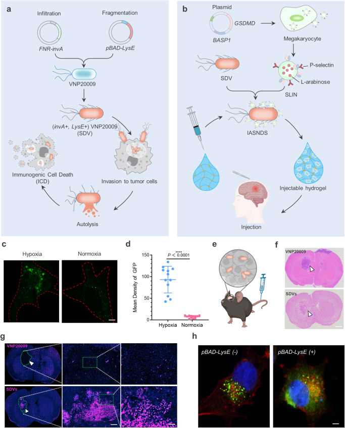
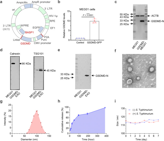
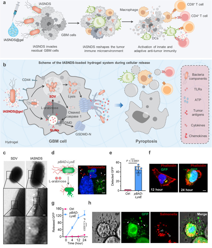
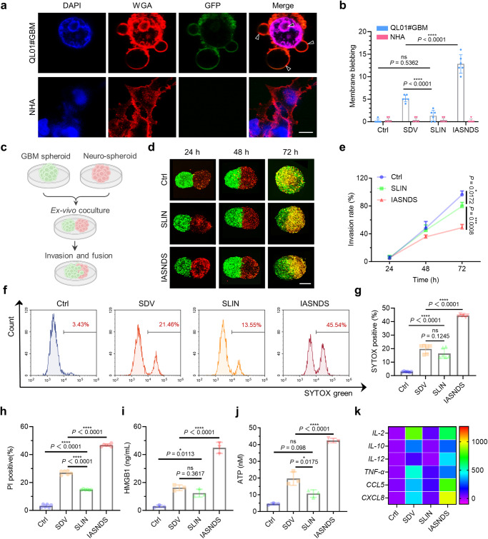
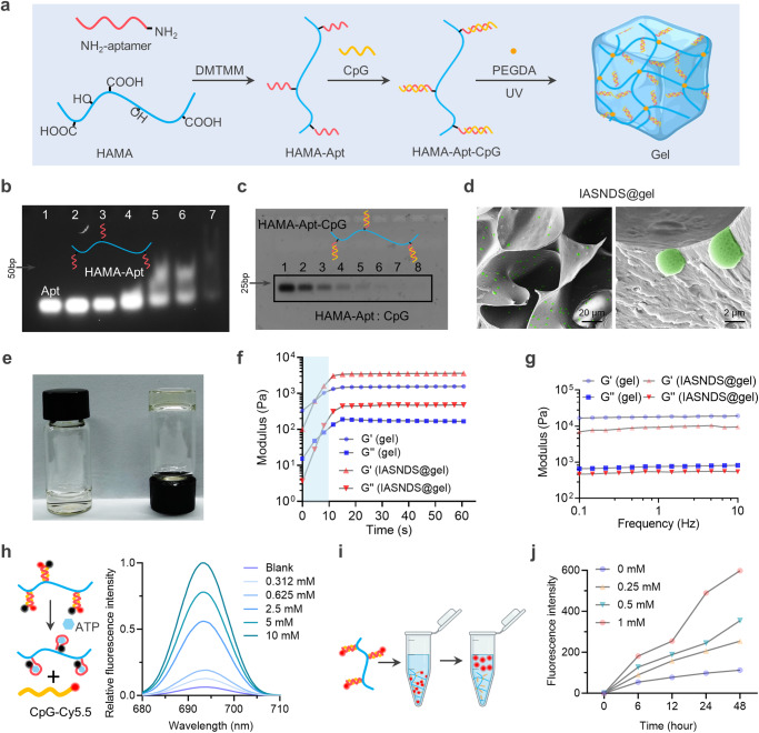
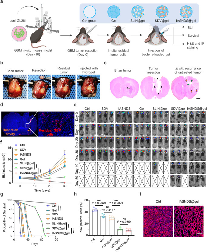
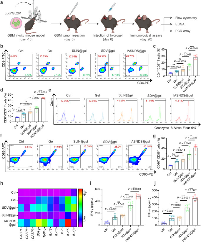
Glioblastoma multiforme (GBM) is a highly aggressive brain tumor characterized by invasive behavior and a compromised immune response, presenting treatment challenges. Surgical debulking of GBM fails to address its highly infiltrative nature, leaving neoplastic satellites in an environment characterized by impaired immune surveillance, ultimately paving the way for tumor recurrence. Tracking and eradicating residual GBM cells by boosting antitumor immunity is critical for preventing postoperative relapse, but effective immunotherapeutic strategies remain elusive. Here, we report a cavity-injectable bacterium-hydrogel superstructure that targets GBM satellites around the cavity, triggers GBM pyroptosis, and initiates innate and adaptive immune responses, which prevent postoperative GBM relapse in male mice. The immunostimulatory Salmonella delivery vehicles (SDVs) engineered from attenuated Salmonella typhimurium (VNP20009) seek and attack GBM cells. Salmonella lysis-inducing nanocapsules (SLINs), designed to trigger autolysis, are tethered to the SDVs, eliciting antitumor immune response through the intracellular release of bacterial components. Furthermore, SDVs and SLINs administration via intracavitary injection of the ATP-responsive hydrogel can recruit phagocytes and promote antigen presentation, initiating an adaptive immune response. Therefore, our work offers a local bacteriotherapy for stimulating anti-GBM immunity, with potential applicability for patients facing malignancies at a high risk of recurrence.


Conflict of interest statement

The authors declare no competing interests.

Figures

Fig. 1
Fig. 1. Mechanism, preparation, and characterization of the hydrogel-based autolysing bacterial delivery system.
a Schematic depicting the strategic transformation of Salmonella into the SDV and the induction of ICD in GBM cells via Salmonella VNP20009. b Illustration detailing the preparation of the IASNDS and synergistic localized treatment with the hydrogel for surgical cavity application. c Visualization of fluorescence (GFP+) in the SDV (green) under both normoxic and hypoxic conditions for 24 hours. Red dashed lines represent GL261 cell boundaries. (n = 3 independent experiments). d Statistical analysis showing the intensity of GFP fluorescence in normoxic and hypoxic cultures. (n = 12 images from three independent experiments). (exact P value: P =  2.42423E-09); ****P < 0.0001. e, f In vivo distribution assessment of 1 × 10^7 CFU SDVs (GFP-) injected into intracranial tumor-bearing mice via the tail vein. Brain GBM tissue sectioning was performed, with the SDVs depicted by a white arrow. g Additional fluorescence staining of brain tissue sections to observe the SDV distribution (GFP-, shown in red) in intracranial GBM tissues (white arrows). (n = 3 independent experiments). h Visualization of the green fluorescence distribution of the SDVs (GFP+) under pBAD-LysE (-) and pBAD-LysE (+) conditions induced by 500 μM L-arabinose for 24 hours in the cell culture medium (n = 3 independent experiments). The intracellular GFP distribution in the pBAD-LysE (+) condition indicates initiation of SDV autolysis in GL261 cells. Data are shown as the means ± SEMs. The statistical comparisons in d were performed with two-tailed, unpaired Student’s t tests, with asterisks indicating significant differences. c, f, g, and h show representative images of the corresponding independent biological samples. (n = 3 independent biological samples). The scale bar in c is 1 μm, the scale bar in f is 1.5 mm, the scale bars in g are 50 μm (middle) and 10 μm (right), and the scale bar in h is 1 μm. Source data are provided as a Source Data file.
Fig. 2
Fig. 2. Design and characterization of Salmonella lysis-inducing nanocapsules.
a Design of the gene construct illustrating GSDMD overexpression and attachment to the exosome membrane surface. b Verification of GSDMD overexpression to evaluate transfection efficiency in the MEG01 cell line through PCR (n = 4 independent experiments). (exact P value: P = 2.75356E-06); ****P < 0.0001. c Western blot analysis showing GSDMD-N protein expression in MEG01 (GSDMD+) cells (n = 3 independent experiments). d The collected exosomes were confirmed to express exosome-specific proteins by Western blotting (n = 3 independent experiments). e Western blot results confirming GSDMD-N protein expression in exosome-derived (GSDMD-EXO) samples (n = 3 independent experiments). f Transmission electron microscopy reveals the SLIN morphology and particle size distribution (g) using dynamic light scattering (DLS). (= 3 independent experiments). h Kinetic profile of L-arabinose release from SLINs in PBS, pH 7.4 (n = 3 independent experiments). i Particle size changes of SLINs loaded in a 10 nm dialysis bag and coincubated with 1 × 10^6 CFU SDVs for 7 days at 4 °C. (n = 3 independent experiments). Data are presented as the mean ± S.D. The statistical comparisons in b were performed with two-tailed, unpaired Student’s t tests, with asterisks indicating significant differences. cf show representative images of the corresponding independent biological samples. The scale bar in f is 50 nm. Source data are provided as a Source Data file.
Fig. 3
Fig. 3. Mechanism of immunogenic cell death induced by the autolysing SDV system.
a Illustration depicting the IASNDS system invading GBM cells, triggering cell death, activating antigen-presenting cells, and eliciting an immune response. b IASNDS cellular entry, intracellular autocleavage initiation, and tumor cell pyroptosis induction mechanism. c Transmission electron microscopy characterizing the structures of the SDVs and IASNDS, highlighting SLINs coupled to the IASNDS surface (white arrow). (n = 3 independent experiments). d GFP release from SDVs into the cytoplasm (green, black arrow) and the presence of lysed Salmonella membranes inside cells (red, white arrow) after coculture of 1 × 10^6 CFU of the IASNDS with QL01#GBM cells for 48 hours (n = 3 independent experiments). e Detection of the GFP fluorescence intensity inside tumor cells after coincubation of 1 × 10^6 CFU of the IASNDS with QL01#GBM cells for 48 hours (n = 9 images from three independent experiments). (exact P value: P = 2.491E−12); ****P < 0.0001. f, g Intratumor cell fluorescence intensity over 24 hours in QL01#GBM cells, illustrating IASND intracellular release behavior (n = 9 images from three independent experiments). (exact P value: P = 7.8029E-11); ****P < 0.0001. h Altered cell membrane permeability and cytoplasmic efflux post IASNDS treatment of QL01#GBM cells (green, white arrow), along with dispersed bacterial membranes after SDV cleavage (red, black arrow). (n = 3 independent experiments). Data are presented as the mean ± S.D. The statistical comparisons in e was performed with two-tailed, unpaired Student’s t tests, with asterisks indicating significant differences. c, d, f, and h are representative images of the corresponding independent biological samples. The scale bars in c are 150 nm (middle) and 50 nm (bottom), the scale bar in d is 0.5 μm, and the scale bars in f and h are 1 μm. Source data are provided as a Source Data file.
Fig. 4
Fig. 4. Validation of IASNDS-induced GBM pyroptosis and ICD-related changes in vitro.
a Visualization of cellular pyroptosis changes induced by the IASNDS under confocal microscopy, showing membrane pores and vacuole formation in QL01#GBM cells (n = 3 independent experiments). b Quantification of vacuoles in cells from different treatment groups to assess pyroptosis efficiency (n = 6 images from three independent experiments). (exact P values: SLIN vs. SDV P = 6.28201E−05, IASNDS vs. SDV P = 4.22634E−06); ****P < 0.0001. ce 3D neurosphere and QL01#GBM tumor sphere invasion experimental design, confocal images of the extent of GBM sphere invasion over time (n = 3 independent experiments). fh Flow cytometry analysis of stained cells (SYTOX, PI). (n = 6 independent experiments). (exact P values of g: Ctrl vs. SLIN P = 5.45783E−08, SDV vs. IASNDS P = 1.141E−12; exact P values of h: Ctrl vs. SLIN P = 1.24E−13, SDV vs. SLIN P = 7.5E−14,  SDV vs. IASNDS P = 1.9E−14); ****P < 0.0001. i, j HMGB1 and ATP release from QL01#GBM cells after 24 hours of PBS, SDV, SLIN, and IASNDS treatment, analyzed by flow cytometry (n = 3 independent experiments). (exact P value of i: SDV vs. IASNDS P = 5.8384E-06; exact P value of j: SDV vs. IASNDS P = 1.38256E-05); ****P < 0.0001. k PCR detection of cytokine and chemokine expression trends after 24 hours of PBS, SDV, SLIN, and IASNDS treatment of QL01#GBM cells. (n = 3 independent experiments). Data are presented as the mean ± S.D. The statistical comparisons in b, e, gj were performed using one-way ANOVA with Tukey’s post hoc test, with asterisks indicating significant differences (ns = no significance, *P < 0.05, **P < 0.01, ***P < 0.001, ****P < 0.0001). a, d show representative images of the corresponding independent biological samples. The scale bar in a is 5 μm, and the scale bar in d is 1 mm. Source data are provided as a Source Data file.
Fig. 5
Fig. 5. ATP-responsive hydrogel design and characterization.
a Schematic of the ATP-responsive hydrogel used to encapsulate the IASNDS. b PAGE analysis of HAMA-Apt synthetic conjugates formed from reaction mixtures of ATP nucleic acid aptamer (lane 1) and Apt with HAMA at molar ratios of 100:1 (lane 2), 50:1 (lane 3), 25:1 (lane 4), 12:1 (lane 5), 6:1 (lane 6), and 1:1 (lane 7). (n = 3 independent experiments). c PAGE analysis of the supernatant after binding of HAMA-Apt to CpG ODN with molar ratios of HAMA-Apt to CpG ODN of 1:0 (lane 8), 1:1 (lane 7), 1:2 (lane 6), 1:4 (lane 5), 1:8 (lane 4), 1:16 (lane 3), 1:32 (lane 2) and 1:64 (lane 1). (n = 3 independent experiments). d Representative scanning electron microscope image of the hydrogel (n = 3 independent experiments). e Representative photographs of hydrogels before and after gel formation. f Variation in the hydrogel modulus with time, measured at an angular frequency of 1 rad s-1. (n = 3 independent experiments). g Analysis of hydrogel variation with frequency, measured at a fixed strain of 0.5%. (n = 3 independent experiments). h Schematic of ATP-induced dehybridization of double-stranded BHQ3-modified Apt and Cy5.5-modified CpG structures, with fluorescence spectra showing the fluorescence recovery of Cy5.5 under ATP induction. (n = 3 independent experiments). i Schematic showing the release of Cy5-labeled CpG from the hydrogel. j Cumulative amount of CpG ODN released from the hydrogel under different concentrations of ATP. (n = 3 independent experiments). Data are presented as the mean ± S.D. The images in bd are representative images of the corresponding independent biological samples. The scale bars in d are 20 μm (left) and 2 μm (right). Source data are provided as a Source Data file.
Fig. 6
Fig. 6. Suppression of postoperative recurrence and prolonged survival via intracavity injection of IASNDS@gel.
a Illustration of the in vivo experimental design. b Upon reaching day 10 after initial inoculation, tumor-bearing GL261 mice underwent surgical tumor resection, followed by hydrogel injection. Subsequently, brain tissues from the excised mice were subjected to euthanasia. H&E (c) and immunofluorescence (d) staining was carried out to visualize residual GBM cells (labeled red with a Vimentin primary antibody). (n = 3 independent experiments). e In vivo bioluminescence images were captured (n = 3), and f the resulting signal intensity was quantified (n = 10). (exact P values: SLIN@gel vs. SDV@gel P = 1.05777E-09, SDV@gel vs. IASNDS@gel P = 6.4922E−11); ****P < 0.0001. g Mouse survival was assessed through Kaplan‒Meier survival curves for each treatment group (n = 10). Data analysis was performed employing the log-rank (Mantel‒Cox) test. (exact P values: Ctrl vs. Gel P = 2.55345E-08, SLIN@gel vs. SDV@gel P = 1.60341E−08, SDV@gel vs. IASNDS@gel P =  3.26493E−05); ****P < 0.0001. h The percentage of positive cells was tallied following immunohistochemical staining for Ki-67 in tumor tissues originating from various mouse groups (n = 5 images from three independent experiments). (exact P values: Ctrl vs. Gel P = 8.876E−05, SLIN@gel vs. SDV@gel P =  7.74109E−09); ****P < 0.0001. i Visualization of alterations in mouse brain tissues was achieved by applying WGA staining (red), enabling observation of tumor cell dynamics across the different treatment groups in mice (n = 3 independent experiments). Data are presented as the mean ± S.D. The statistical comparisons in f and h were performed using two-way ANOVA and one-way ANOVA by Tukey’s post hoc test, with asterisks indicating significant differences (***P < 0.001). The images in c, d, and i are representative images of the corresponding independent biological samples. The scale bar in c is 1.5 mm, and the scale bar in d and i is 50 μm. Source data are provided as a Source Data file.
Fig. 7
Fig. 7. Immunomodulatory efficacy of the Salmonella-loaded hydrogel system for mobilizing immunity against tumors via intracavity injection.
a Schematic of the experimental design for assessing immunomodulatory efficacy. b Flow cytometry results showing CD3+ cells in brain tissues after GBM removal in each treatment group. c Quantitative analysis of CD3+CD4+ cytotoxic T cells (n = 6 independent experiments). (exact P value: SDV@gel vs. IASNDS@gel P =  1.57104E-08); ****P < 0.0001. d Quantitative analysis of CD3+CD8+ cytotoxic T cells (n = 6 independent experiments). (exact P value: SDV@gel vs. IASNDS@gel P = 1.32267E−10); ****P < 0.0001. e Flow cytometry results indicating CD3+CD8+Granzyme B+ T cells in GBM tissues from different treatment groups. f, g Flow cytometry analysis and statistical analysis of CD80+CD86+ cells in GBM tissues after different treatments (n = 6 independent experiments). h Heatmap displaying the expression profiles of pyroptosis-related proteins, cytokines, and chemokines in brain tumor tissues. (n = 3 independent experiments). i, j Statistical analysis of IFN-γ expression and TNF-α expression in GBM tissues under different treatment conditions (n = 6 independent experiments). (exact P values of i: SLIN@gel vs. SDV@gel P =  1.22239E−07, SDV@gel vs. IASNDS@gel P =  8.7998E−07; exact P values of j: SLIN@gel vs. SDV@gel P =  2.02309E−07; SDV@gel vs. IASNDS@gel P = 3.4876E−07); ****P < 0.0001. Data are presented as the mean ± S.D. The statistical comparisons in c, d, g, i and j were performed using one-way ANOVA with Tukey’s post hoc test, with asterisks indicating significant differences (ns = no significance, *P < 0.05, **P < 0.01, ***P < 0.001, ****P < 0.0001). Source data are provided as a Source Data file.

Similar articles

Cited by

References

    1. Schaff LR, Mellinghoff IK. Glioblastoma and other primary brain malignancies in adults: a review. JAMA. 2023;329:574–587. doi: 10.1001/jama.2023.0023. - DOI - PMC - PubMed
    1. van den Bent, M. J. et al. Primary brain tumours in adults. Lancet402, 1564–1579 (2023). - PubMed
    1. Mair MJ, et al. Understanding the activity of antibody-drug conjugates in primary and secondary brain tumours. Nat. Rev. Clin. Oncol. 2023;20:372–389. doi: 10.1038/s41571-023-00756-z. - DOI - PubMed
    1. Knisely JPS, Fine HA. Reirradiation for recurrent glioblastoma: what we know and what we do not. J. Clin. Oncol.: Off. J. Am. Soc. Clin. Oncol. 2023;41:1183–1188. doi: 10.1200/JCO.22.01785. - DOI - PubMed
    1. Tan AC, et al. Management of glioblastoma: State of the art and future directions. CA: Cancer J. Clin. 2020;70:299–312. - PubMed