. 2024 May 18;14(1):204.
doi: 10.1038/s41398-024-02915-x.

The circulating proteome and brain health: Mendelian randomisation and cross-sectional analyses

Affiliations

The circulating proteome and brain health: Mendelian randomisation and cross-sectional analyses

Rosie M Walker et al. Transl Psychiatry. .

Abstract

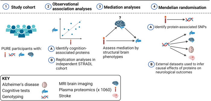
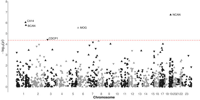
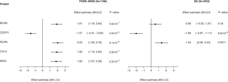
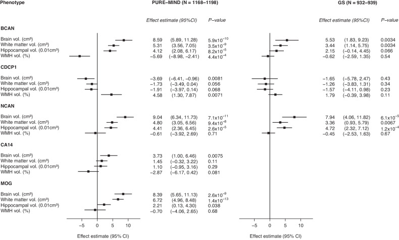
Decline in cognitive function is the most feared aspect of ageing. Poorer midlife cognitive function is associated with increased dementia and stroke risk. The mechanisms underlying variation in cognitive function are uncertain. Here, we assessed associations between 1160 proteins' plasma levels and two measures of cognitive function, the digit symbol substitution test (DSST) and the Montreal Cognitive Assessment in 1198 PURE-MIND participants. We identified five DSST performance-associated proteins (NCAN, BCAN, CA14, MOG, CDCP1), with NCAN and CDCP1 showing replicated association in an independent cohort, GS (N = 1053). MRI-assessed structural brain phenotypes partially mediated (8-19%) associations between NCAN, BCAN, and MOG, and DSST performance. Mendelian randomisation analyses suggested higher CA14 levels might cause larger hippocampal volume and increased stroke risk, whilst higher CDCP1 levels might increase intracranial aneurysm risk. Our findings highlight candidates for further study and the potential for drug repurposing to reduce the risk of stroke and cognitive decline.


Conflict of interest statement

MC is supported by a Canadian Institute of Health Research doctoral award and has received consulting fees from Bayer AG. MP is supported by the EJ Moran Campbell Internal Career Research Award from McMaster University. DAG is a part-time employee of Optima partners, a health data consultancy based at the Bayes centre, The University of Edinburgh. SH is an employee of Bayer AG. AMM has previously received speaker’s fees from Illumina and Janssen and research grant funding from The Sackler Trust. SY is supported by the Heart and Stroke Foundation/Marion W Burke Chair in Cardiovascular Disease. GP is supported by the CISCO Professorship in Integrated Health Systems. The other authors declare no competing interests.

Figures

Fig. 1
Fig. 1. Overview of the study design.
This study involved European (N = 3514), Latin (N = 4309), and Persian (N = 1332) PURE participants for whom genetic and plasma proteomic data were available. Observational analyses to detect plasma biomarkers of cognitive function were performed in the subset of these participants who were enrolled in the PURE-MIND sub-study (N = 1198), for whom plasma protein (N = 1060 proteins) and MRI measurements were available. Mediation analyses were performed to assess whether any observed associations between protein levels and cognitive function were mediated by structural brain phenotypes ascertained by MRI. Finally, two-sample Mendelian randomisation analyses were performed to assess potentially causal effects of genetically-predicted cognition-associated protein levels on genetically-predicted neurological outcomes. For these analyses, genetic instrumental variables for protein levels were identified in the European, Latin, and Persian PURE participants, and associations with neurological outcomes were assessed using external (non-PURE) datasets. Created with BioRender.com.
Fig. 2
Fig. 2. Manhattan plot indicating associations between the levels of plasma proteins and performance on the DSST in participants from the PURE-MIND cohort (N = 1198).
Each protein is represented by a triangle with upwards-facing triangles indicating a positive association with DSST performance and downwards-facing triangles indicating a negative association with DSST performance. The position of each protein on the x-axis is determined by the genomic location of its corresponding gene and the position on the y-axis is determined by the –log10 p-value. The dashed horizontal line indicates the Bonferroni-corrected significance threshold (p = 4.31 × 10⁻⁵) required to maintain a 5% type I error rate.
Fig. 3
Fig. 3. Forest plot indicating the association between protein levels and DSST performance for significantly associated proteins.
For each protein, the difference in DSST score associated with a standard deviation higher level of protein is shown, together with the 95% confidence interval. Abbreviations: BCAN brevican, CA14 carbonic anhydrase 14, CDCP1 CUB-domain containing protein 1, CI confidence interval, GS Generation Scotland imaging subsample, MOG myelin oligodendrocyte glycoprotein, NCAN neurocan, PURE Prospective Urban and Rural Epidemiology study.
Fig. 4
Fig. 4. Forest plots indicating the association between the levels of DSST-associated proteins and DSST-associated structural brain phenotype.
For each protein, the effect estimate (change in brain volume (cm3 or %) per standard deviation increase in protein expression) is shown, together with the 95% confidence interval. Abbreviations: BCAN brevican; CA14 carbonic anhydrase 14, CDCP1 CUB-domain containing protein 1, CI confidence interval, GS Generation Scotland imaging subsample, MOG myelin oligodendrocyte glycoprotein, NCAN neurocan, PURE Prospective Urban and Rural Epidemiology study, WMH white matter hyperintensity.

References

    1. Martin GM. Defeating Dementia. Nature. 2004;431:247–8. doi: 10.1038/431247b. - DOI
    1. Rostamian S, Mahinrad S, Stijnen T, Sabayan B, de Craen AJ. Cognitive impairment and risk of stroke: a systematic review and meta-analysis of prospective cohort studies. Stroke. 2014;45:1342–8. doi: 10.1161/STROKEAHA.114.004658. - DOI - PubMed
    1. Valenzuela MJ, Sachdev P. Brain reserve and dementia: a systematic review. Psychol Med. 2006;36:441–54. doi: 10.1017/S0033291705006264. - DOI - PubMed
    1. Pettigrew C, Soldan A. Defining Cognitive Reserve and Implications for Cognitive Aging. Curr Neurol Neurosci Rep. 2019;19:1. doi: 10.1007/s11910-019-0917-z. - DOI - PMC - PubMed
    1. Harris SE, Cox SR, Bell S, Marioni RE, Prins BP, Pattie A, et al. Neurology-related protein biomarkers are associated with cognitive ability and brain volume in older age. Nat Commun. 2020;11:800. doi: 10.1038/s41467-019-14161-7. - DOI - PMC - PubMed