

Spontaneous regression of an interhemispheric arachnoid cyst: illustrative case
- PMID: 38762563
- PMCID: PMC11269485
- DOI: 10.1007/s00381-024-06464-y
Spontaneous regression of an interhemispheric arachnoid cyst: illustrative case
Abstract
Background: Intracranial arachnoid cysts are benign collections of cerebrospinal fluid that are often asymptomatic and discovered incidentally. An interhemispheric location of these lesions is rare, with only a few such cases reported in the literature. Though spontaneous regression of arachnoid cysts has been described in other locations, to date this phenomenon has not been reported in interhemispheric fissure cysts.
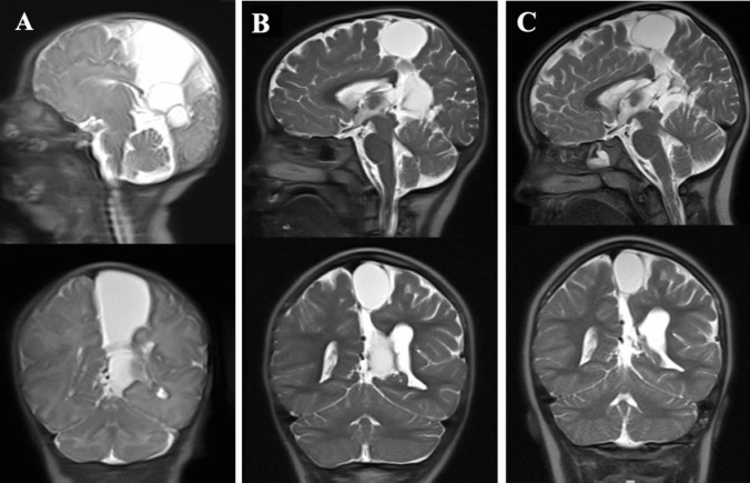
Observations: In this report, we describe a patient with a large, multiloculated interhemispheric arachnoid cyst diagnosed on prenatal ultrasound. She did not exhibit neurologic deficits or signs of increased intracranial pressure and was observed with serial imaging. After several years of observation, imaging revealed spontaneous and progressive decrease in the cyst size.
Lessons: We illustrate a case of regression of an interhemispheric arachnoid cyst in a pediatric patient. To our knowledge, this is the first reported case of spontaneous shrinkage of an arachnoid cyst in this location. Although the current presentation is rare, this reporting adds to the current understanding of natural history of arachnoid cysts and provides an example of radiographical improvement without intervention of a cyst located within the interhemispheric fissure.
Keywords: Arachnoid cyst; Interhemispheric cyst; Pediatric; Spontaneous regression.
© 2024. The Author(s).
Conflict of interest statement
The authors have no competing interests to declare that are relevant to the content of this article.
Figures
Similar articles
-
Arachnoid cysts: using prenatal imaging and need for pediatric neurosurgical intervention to better understand their natural history and prognosis.J Matern Fetal Neonatal Med. 2022 Dec;35(24):4728-4733. doi: 10.1080/14767058.2020.1863361. Epub 2021 Jan 4. J Matern Fetal Neonatal Med. 2022. PMID: 33397177
-
Spontaneous disappearance of two asymptomatic arachnoid cysts in two different locations.Minim Invasive Neurosurg. 2003 Apr;46(2):110-2. doi: 10.1055/s-2003-39337. Minim Invasive Neurosurg. 2003. PMID: 12761683
-
Spontaneous Resolution of Asymptomatic Pediatric Suprasellar Arachnoid Cysts: Report of 2 Cases and Review of the Literature.Pediatr Neurosurg. 2020;55(1):62-66. doi: 10.1159/000504262. Epub 2019 Nov 26. Pediatr Neurosurg. 2020. PMID: 31770757 Review.
-
Interhemispheric arachnoid cyst in the elderly: case report and review of the literature.Surg Neurol. 2003 Jan;59(1):68-74. doi: 10.1016/s0090-3019(02)00876-5. Surg Neurol. 2003. PMID: 12633971 Review.
-
Spontaneous resolution of arachnoid cysts: review and features of an unusual case.Acta Neurochir (Wien). 2007 Jan;149(1):75-8; discussion 78. doi: 10.1007/s00701-006-1073-1. Epub 2006 Dec 21. Acta Neurochir (Wien). 2007. PMID: 17180304 Review.
References
-
- Kotil K, Balci N, Bilge T (2007) Intracranial symptomatic giant arachnoid cyst of the interhemispheric fissure presenting with frontal lobe syndrome. Turk Neurosurg 17(2):147–151 - PubMed
Publication types
MeSH terms
LinkOut - more resources
Full Text Sources
