. 2024 May 18;7(1):596.
doi: 10.1038/s42003-024-06310-6.

TBC9, an essential TBC-domain protein, regulates early vesicular transport and IMC formation in Toxoplasma gondii

Affiliations

TBC9, an essential TBC-domain protein, regulates early vesicular transport and IMC formation in Toxoplasma gondii

Ming Sun et al. Commun Biol. .

Abstract

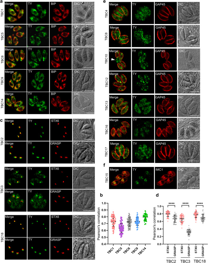
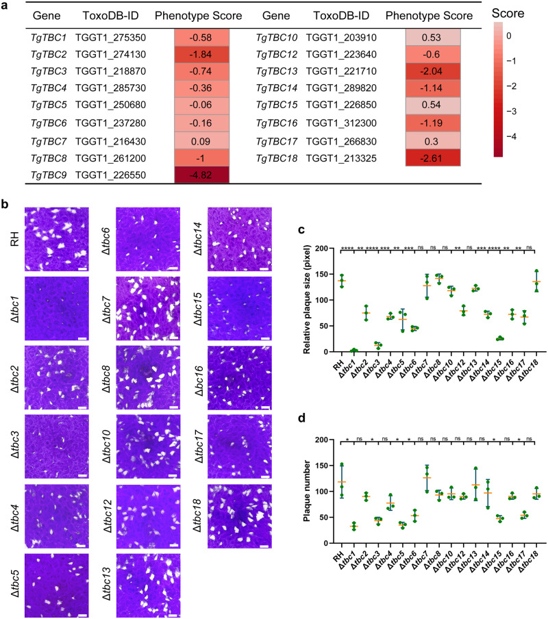
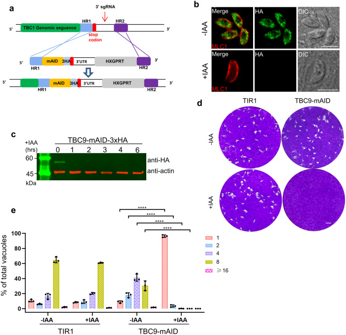
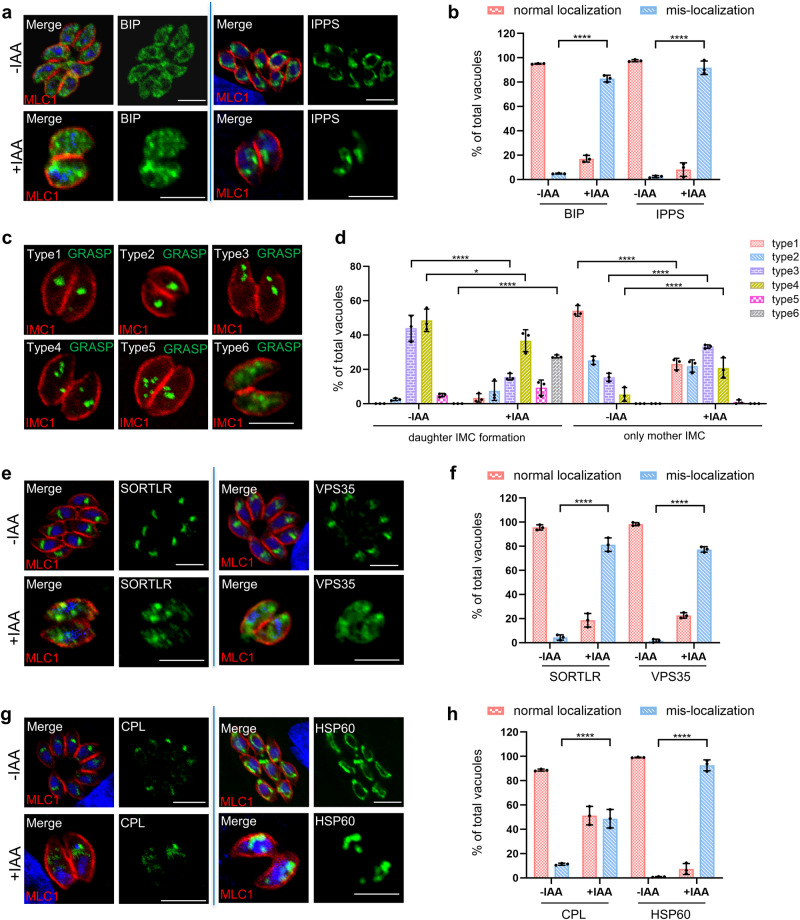
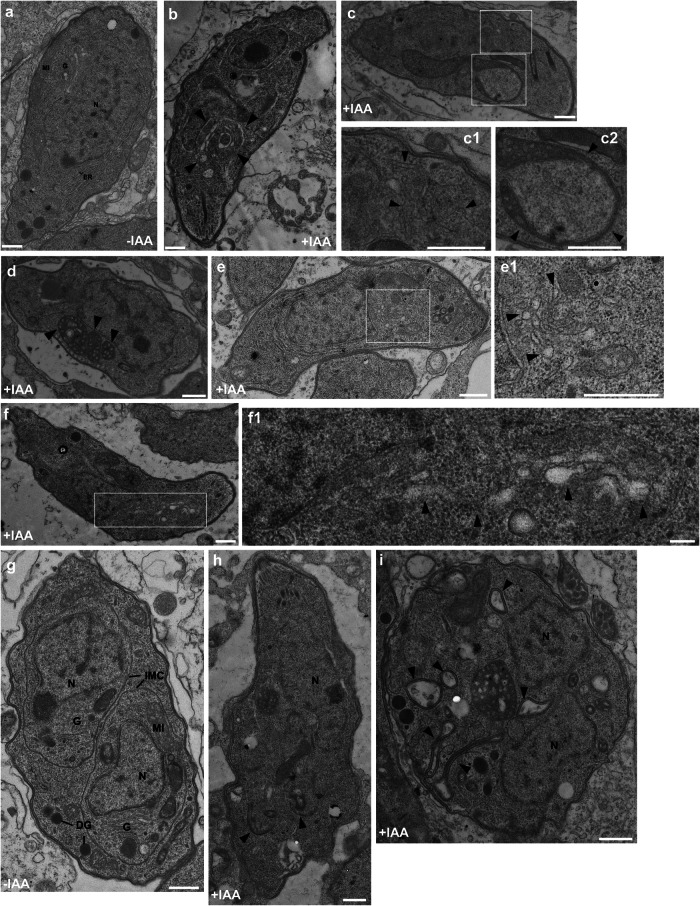
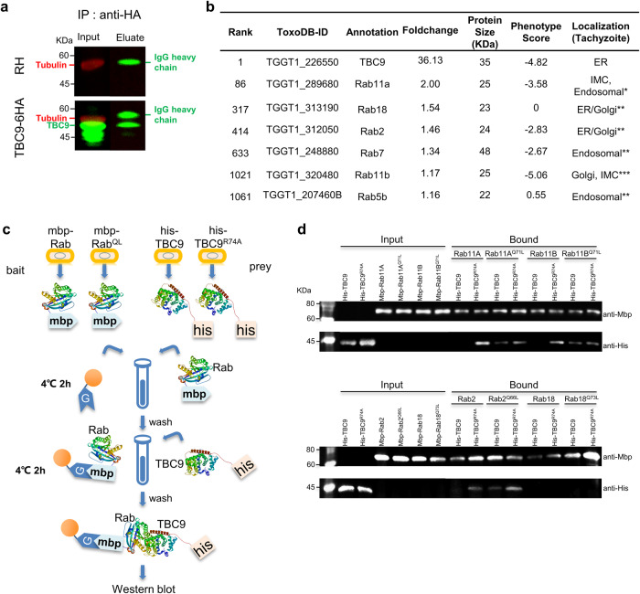
Apicomplexan parasites harbor a complex endomembrane system as well as unique secretory organelles. These complex cellular structures require an elaborate vesicle trafficking system, which includes Rab GTPases and their regulators, to assure the biogenesis and secretory of the organelles. Here we exploit the model apicomplexan organism Toxoplasma gondii that encodes a family of Rab GTPase Activating Proteins, TBC (Tre-2/Bub2/Cdc16) domain-containing proteins. Functional profiling of these proteins in tachyzoites reveals that TBC9 is the only essential regulator, which is localized to the endoplasmic reticulum (ER) in T. gondii strains. Detailed analyses demonstrate that TBC9 is required for normal distribution of proteins targeting to the ER, and the Golgi apparatus in the parasite, as well as for the normal formation of daughter inner membrane complexes (IMCs). Pull-down assays show a strong protein interaction between TBC9 and specific Rab GTPases (Rab11A, Rab11B, and Rab2), supporting the role of TBC9 in daughter IMC formation and early vesicular transport. Thus, this study identifies the only essential TBC domain-containing protein TBC9 that regulates early vesicular transport and IMC formation in T. gondii and potentially in closely related protists.


Conflict of interest statement

The authors declare no competing interests.

Figures

Fig. 1
Fig. 1. TBC domain-containing proteins in Toxoplasma gondii.
The schematic diagram illustrates the domain structure and other features of the 17 TBC proteins as predicted by the InterPro (https://www.ebi.ac.uk/interpro/). TBC - Tre2-Bub2-Cdc16 domain; CC - coiled coil domain; PK - protein kinase domain; Transmembrane domain - region of a membrane-bound protein predicted to be embedded in the membrane; TLDc - a domain that includes two conserved domains: TBC and LysM; Sec7 - a conserved domain of GEF (guanine nucleotide exchange factors). A scale of 300 amino acids length was shown for the protein length displayed in the schematic.
Fig. 2
Fig. 2. Localization of TBC protein candidates in the parasite.
Immunofluorescence assays (IFAs) of endogenously epitope-tagged TBC1-10, 12–18 parasites were performed for parasites grown for 24 h. a Confocal co-localization of the epitope Ty fused at TBC1, 5, 6, 9, and 14 (green) with the ER marker BIP (red). b Statistics of Pearson correlation coefficient (PCC). The PCC values over the merged fluorescent region of TBC1, 5, 6, 9, and 14 with BIP were analyzed using the colocalization and ROI intensity analysis module in NIS software. c Confocal co-localization of the epitope Ty fused at TBC2, 3, and 18 (green) with the TGN marker STX6 (red) and the cis-Golgi marker GRASP (red). d Statistics of Pearson correlation coefficient (PCC). The PCC values over the merged fluorescent foci of TBC2, 3, and 18 with STX6 and GRASP. e TBC4, 8, 12, 13, 16, and 17 are localized to the vesicles of the cytosol and TBC10 are localized to the cytosol and the PV (arrowhead). The IFA analyses were achieved using anti-Ty antibodies (green), and the outline of the parasites was visualized using anti-GAP45 antibodies (red). f Confocal co-localization of the epitope Ty fused at TBC15 (green) with the IMC marker IMC1 (red). Data are presented as mean ± SD (n = 50 parasites), with the Mann–Whitney test used for the PCC of TBC2 and 3, and the unpaired t-test used for the PCC of TBC18 (****p < 0.0001). Scale bar = 5 µm.
Fig. 3
Fig. 3. Plaque formation of the knockout lines.
a The table lists the phenotype scores of 17 TBC genes. The phenotype scores were retrieved from a CRISPR-library screening. b The parental line RHΔku80Δhxgprt (RH) and knockout lines of TBC1-8, 10, and 12–18 (Δtbc1-8, 10, and 12–18) were grown on HFF monolayers in 6-well plates, followed by fixation and staining with crystal violet after 7 days of culturing of parasites. The representative images for each line were shown. Scale bar = 2 mm. c, d The plaque size and numbers for the parasite lines examined in (b) were measured and plotted. Data (N = 3 independent experiments; n = 3 replicates) were shown with means ± SD and analyzed by the unpaired t test. (ns, not significant; *0.01 ≤ p < 0.05; **0.001 ≤ p < 0.01; ***0.0001 ≤ p < 0.001; ****p < 0.0001).
Fig. 4
Fig. 4. TBC9 is essential for parasite replication.
a Schematic diagram for generation of the TBC9-mAID fusion by tagging the gene at the C-terminus with mAID-3xHA using a CRISPR/Cas9 approach in the parental line RHΔku80Δhxgprt/TIR1. The homologous regions (HR) were retrieved from the upstream of the stop codon and the downstream of the 3’ sgRNA targeting site, while the HXGPRT represents the resistant expression cassette. b The TBC9-mAID-3xHA parasite line was grown in IAA or ethanol for 24 h, followed by fixation for IFA analyses, using antibodies against MLC1 (red) and the epitope tag HA (green). Scale bar = 5 µm. c Western blot analysis for the TBC9-mAID-3xHA line grown for induction of auxin for different times (hrs) as indicated. Actin served as a load control. d Plaque formation of the TIR1 and TBC9-mAID lines. Parasites were grown on HFF monolayers in ± IAA, followed by fixation and staining with crystal violet after 7 days of culturing. Scale bar = 2 mm. e Parasite replication of the TIR1 and TBC9-mAID lines. Parasites were grown in ±IAA for 24 h, followed by IFA analysis of parasites by antibodies against GAP45. Vacuoles with different parasite numbers (1, 2, 4, 8, and ≥16) were counted (≥200 vacuoles for each replicate). Data (N = 3 independent experiments; n = 3 replicates) were shown with means ± SD, and were analyzed by two-way ANOVA with Tukey’s multiple comparison test. (****p < 0.0001, and no significant differences were observed for other situations).
Fig. 5
Fig. 5. Depletion of TBC9 affects the early vesicle transport pathway and its related proteins in T. gondii.
Immunofluorescence analysis of normal or mis-localization of protein sorting pathway proteins and organelle marker proteins upon depletion of TBC9 in T. gondii. TBC9-mAID parasites were grown on HFF monolayers for 16 h, followed by ± IAA treatment for 13 h before processing for IFA analyses using the corresponding antibodies. MLC1 or IMC1 (red) for the inner membrane complex, and Hoechst (blue) dye was used to stain the nucleus. a BIP and IPPS staining for the ER. b Vacuoles with normal localization or mis-localization of ER were counted (≥200 vacuoles for each replicate). Two-way ANOVA with Tukey’s multiple comparison test was performed. c GRASP was tagged with Ty using a CRISPR approach in the TBC9-mAID line. Six distinct types of Golgi were found for the GRASP staining. d Vacuoles with different GRASP types were specifically counted during either daughter IMC formation or only mother IMC (≥200 vacuoles for each replicate). Two-way ANOVA with Sidak’s multiple comparison test was performed. e Staining of SORTLR and VPS35 tagged with Ty in TBC9-mAID line. f Vacuoles with normal localization or mis-localization of SORTLR or VPS35 were counted (≥200 vacuoles for each replicate). Two-way ANOVA with Tukey’s multiple comparison test was performed. g Staining of CPL tagged with Ty in TBC9-mAID line, and HSP60 staining for the mitochondrion. h Vacuoles with normal localization or mis-localization of CPL or HSP60 were counted (≥200 vacuoles for each replicate). Two-way ANOVA with Tukey’s multiple comparison test was performed. Data (N = 3 independent experiments; n = 3 replicates) were shown with means ± SD. (*0.01 ≤ p < 0.05; ****p < 0.0001). Scale bar = 5 µm.
Fig. 6
Fig. 6. Depletion of the TBC9 affects daughter bud formation and organelles duplication.
a, b TBC9-mAID parasites were grown on HFF monolayers for 16 h, followed by ± IAA treatment for 13 h before processing for IFA analyses. Hoechst (blue) dye was used to stain the nucleus. Using antibody combinations against IMC1 (red) and GAP45 (green), IFA results were obtained for morphologically normal parasites without daughter bud formation (a, white arrowheads) and morphologically fragmented parasites with daughter bud formation (b, green arrowheads), which were classified into four types (type A-D) by daughter formation and the number of nucleus (yellow arrowheads). c Vacuoles with 4 types in (A-D) were counted (≥200 vacuoles for each replicate). Two-way ANOVA with Sidak’s multiple comparison test was performed. d TBC9-mAID parasites were grown for 6 h, followed by ± IAA treatment for 16 h before processing for IFA analysis. Antibodies against IMC1 (green) and GAP45 (red) were used for staining the inner membrane complex. e Vacuoles with 4 types (the same as in a, b) in (d) were counted (≥200 vacuoles for each replicate). Two-way ANOVA with Sidak’s multiple comparison test was performed. f Staining of Ty-tagged GRASP in the TBC9-mAID line, and the six distinct types of Golgi are consistent with those shown in Fig. 5c. g Vacuoles with different GRASP types in (f) were counted (≥200 vacuoles for each replicate). Two-way ANOVA with Sidak’s multiple comparison test was performed. h Staining of CEN1 for the centrosome and ACP for the apicoplast. They were classified into three types based on the replication status: not replicated (type 1), elongated with budding (type 2), and fully divided into two (type 3). i Vacuoles with different centrosome or apicoplast types were specifically counted (≥200 vacuoles for each replicate). Two-way ANOVA with Tukey’s multiple comparison test was performed. Data (N = 3 independent experiments; n = 3 replicates) were shown with means ± SD. (ns, not significant; *0.01 ≤ p < 0.05; ***0.0001 ≤ p < 0.001; ****p < 0.0001). Scale bar = 5 µm.
Fig. 7
Fig. 7. Transmission electron micrographs of TBC9-mAID parasites treated with or without IAA.
Transmission electron micrographs of parasites grown for 16 h, followed by treatment with (bf) or without (a) IAA for 13 h. a Longitudinal section through a control sample showing morphologically normal endoplasmic reticulum, mitochondria, and Golgi. The longitudinal sections of the sample treated with IAA show abnormally folded ER (b arrowheads) and disrupted ER network (c, c1 arrowheads). Mitochondria show varying degrees of folding (c, c2 arrowheads; d arrowheads). Golgi exhibits dispersed vesicular structures (e, e1 arrowheads), some of which extend and diffuse into larger areas of the cytoplasm (f, f1 arrowheads). Transmission electron micrographs of parasites grown for 6 h, followed by treatment with (h, i) or without (g) IAA for 16 h. The longitudinal sections of control samples reveal morphologically normal daughter buds with intact IMC, Golgi, and nuclei in the mother parasite (g). The longitudinal sections of samples treated with IAA show a slightly elongated parasite slice with the presence of only two conical structures representing the initiation of daughter formation, but no evidence of other daughter organizations formation (h, arrowheads). Spherical parasites with two nuclei and invaginated mother parasite cell membranes (i, arrowheads). N, nucleus; MI, mitochondria; G, Golgi; ER, endoplasmic reticulum; DG, dense granule; IMC, inner membrane complexes. Scale bar = 500 nm.
Fig. 8
Fig. 8. The dual-finger catalytic sites are essential for the activity of TBC9 in the parasite.
ac IFA detection of TBC9-mAID-3xHA line complemented with TBC9wt-2TY (TBC9wt). Parasites were grown on HFF monolayers for 16 h, followed by ± IAA treatment for 13 h before processing for IFA analyses using antibodies against the proteins indicated in the images: MLC1, GAP45, and IMC1 for the inner membrane complex; Tubulin for the subpellicular microtubules; HSP60 for the mitochondrion, and IPPS for the ER. Scale bar = 5 µm. d Diagram illustrating the TBC dual-finger catalytic sites of TBC9. The arginine and glutamine in the sites were mutated into alanine, respectively. Yellow boxes depict arginine (R finger) and glutamine (Q finger), red boxes depict residues that were mutated to alanine (A). e IFA detection of the TBC9-mAID-3xHA line complemented with TBC9R74A-2TY or TBC9Q101A-2TY. Parasites were grown on HFF monolayers for 16 h, followed by ± IAA treatment for 13 h before processing for IFA analyses using antibodies against the TY (green) and MLC1 (red). Hoechst (blue) dye was used to stain the nucleus. Scale bar = 5 µm. f Parasite replication of the TBC9-mAID, TBC9wt, TBC9R74A and TBC9Q101A lines. Vacuoles with different parasite numbers (1, 2, 4, 8, and ≥16) were counted (≥200 vacuoles for each replicate). Two-way ANOVA with Tukey’s multiple comparison test was performed. g Plaque formation of the TBC9-mAID, TBC9wt, TBC9R74A and TBC9Q101A lines. Scale bar = 2 mm. h The plaque sizes for the parasite lines examined in (g) were measured and plotted. Two-way ANOVA with Sidak’s multiple comparison test was performed. Data (N = 3 independent experiments; n = 3 replicates) were shown with means ± SD. (ns, not significant; ****p < 0.0001).
Fig. 9
Fig. 9. Identification of protein interaction between TBC9 and Rab GTPases.
a The parasite line TBC9-6HA was harvested for the Co-immunoprecipitation (Co-IP), and the parental line RHΔku80Δhxgprt (RH) was used as control. Western blot detection of proteins purified by immunoprecipitation of TBC9 using antibodies against the epitope HA. Western blot was performed using the input samples (Input) and eluted material (Eluate). Tubulin served as a load control. b Mass spectrometry analysis of the proteins immunoprecipitated in (a). Table shows TBC9 and RabGTPase detected by the mass-spectrometry, and the hits were ranked by foldchanges obtained by comparing the mass-spectrometry area from the TBC9-6HA to that from the parental line. The phenotype scores were obtained from a CRISPR library screening. The localization data for the hits can be found in previous studies (references indicated by *; **; ***). The complete dataset can be found in Supplementary Data 2. c Schematic of pull-down assay. The proteins mbp-Rab, mbp-RabQL (GTP-locked mutants), TBC9, and TBC9R74A (GAP-inactive mutants) were expressed in E. coli and purified respectively. The mbp-Rab or mbp-RabQL proteins were used as bait and incubated with MBP agarose resin in tubes. After washing, the resin bound with mbp-Rab or mbp-RabQL was individually incubated with TBC9 or TBC9R74A. Subsequently, the samples were thoroughly washed and subjected to Western blot analysis. d Samples of MBP- Rabs (MBP- Rab11A, MBP- Rab11AQ71L, MBP- Rab11B, MBP- Rab11BQ71L, MBP- Rab2, MBP-Rab2Q66L, MBP-Rab18, or MBP-Rab18Q73L) were incubated with MBP agarose beads, respectively, followed by mixtures with his-TBC9 or his-TBC9R74A in the pull-down assays. Proteins bound to the beads were analyzed by Western blot using antibodies against the His tag and MBP tag. These proteins were expressed in E. coli and purified using affinity standard protocols.

Similar articles

Cited by

References

    1. Tenter AM, Heckeroth AR, Weiss LM. Toxoplasma gondii: from animals to humans. Int. J. Parasitol. 2000;30:1217–1258. doi: 10.1016/S0020-7519(00)00124-7. - DOI - PMC - PubMed
    1. Dubey JP, Lindsay DS, Speer CA. Structures of Toxoplasma gondii tachyzoites, bradyzoites, and sporozoites and biology and development of tissue cysts. Clin. Microbiol. Rev. 1998;11:267–299. doi: 10.1128/CMR.11.2.267. - DOI - PMC - PubMed
    1. Jeffers V, Tampaki Z, Kim K, Sullivan WJ. A latent ability to persist: differentiation in Toxoplasma gondii. Cell. Mol. Life Sci. 2018;75:2355–2373. doi: 10.1007/s00018-018-2808-x. - DOI - PMC - PubMed
    1. Sin-Yew W, Remington JS. Toxoplasmosis in pregnancy. Clin. Infect. Dis. 1994;18:853–861. doi: 10.1093/clinids/18.6.853. - DOI - PubMed
    1. Nishi M, Hu K, Murray JM, Roos DS. Organellar dynamics during the cell cycle of Toxoplasma gondii. J. Cell Sci. 2008;121:1559–1568. doi: 10.1242/jcs.021089. - DOI - PMC - PubMed

Publication types

MeSH terms