

Iron parameters analysis in dogs with myxomatous mitral valve disease
- PMID: 38762716
- PMCID: PMC11102178
- DOI: 10.1186/s12917-024-04071-2
Iron parameters analysis in dogs with myxomatous mitral valve disease
Abstract
Background: Myxomatous mitral valve disease (MMVD) is the most common acquired cardiovascular disease in small breed dogs. In contrast to human patients with heart failure (HF), iron deficiency (ID) prevalence in dogs with MMVD is weakly known. The study aimed to assess the usability of ID markers in serum and reticulocyte parameters from whole blood of dogs with MMVD to evaluate early ID symptoms.
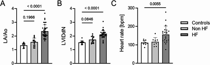
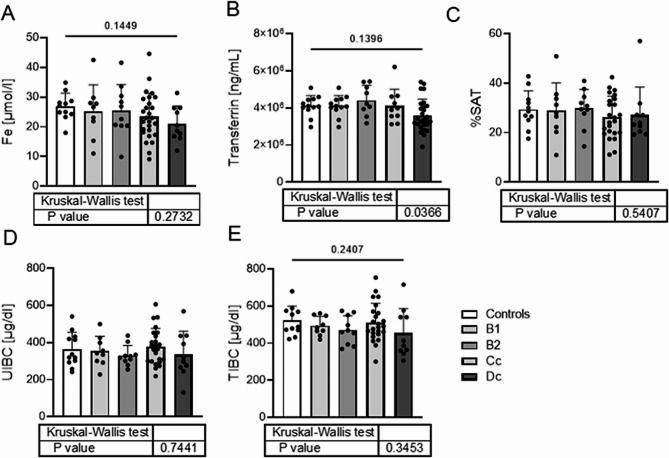
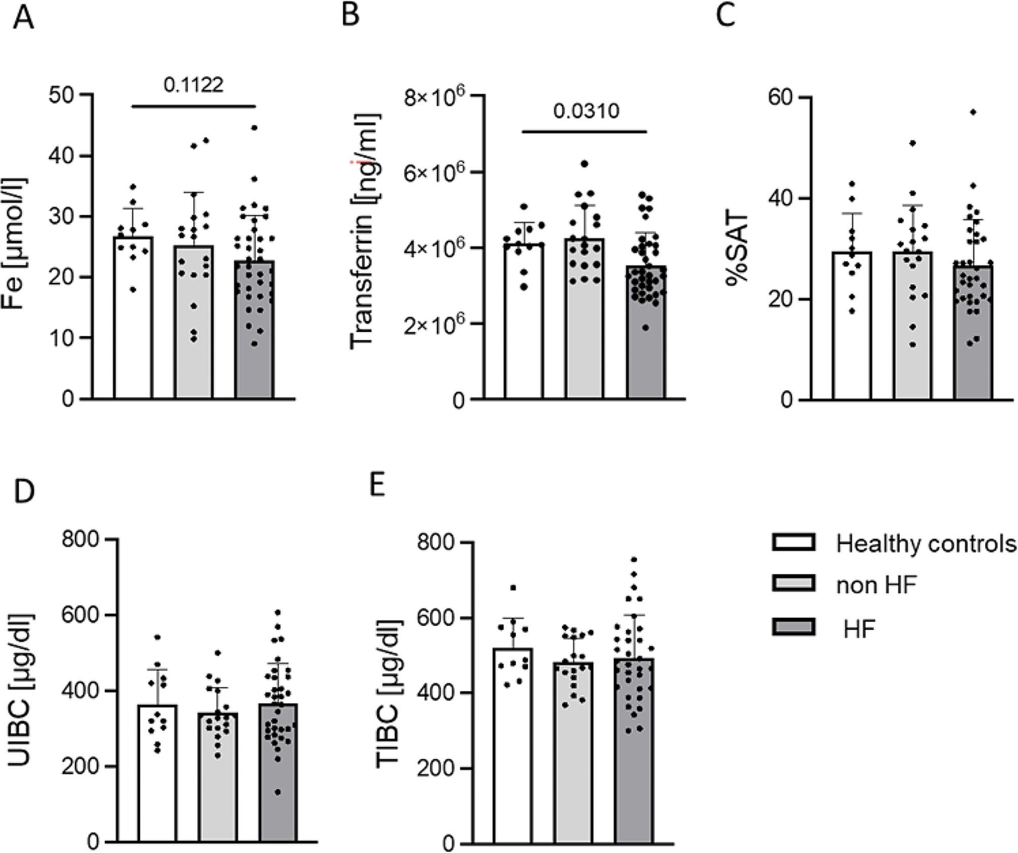
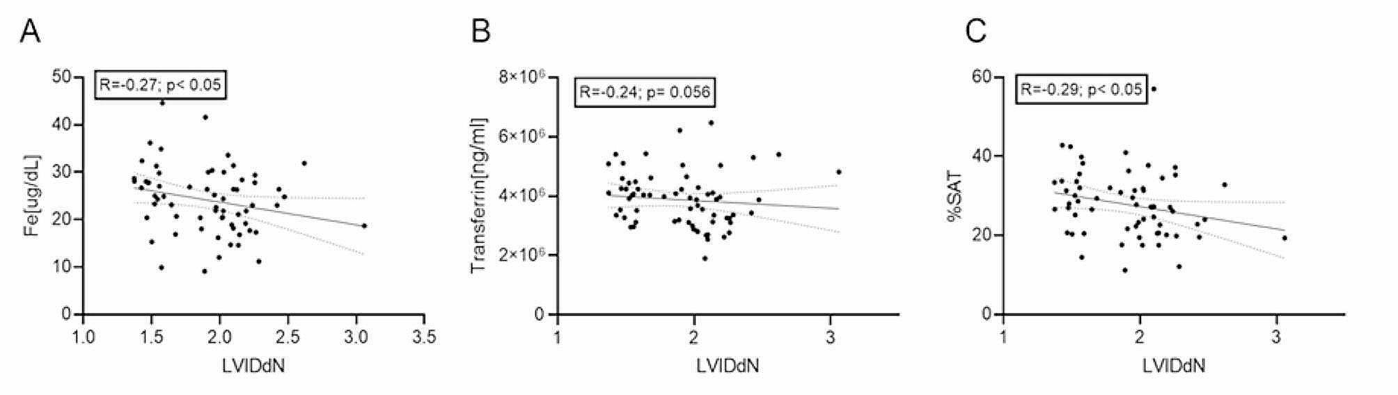
Results: Sixty-eight dogs (43 male and 25 female) were included in the study. MMVD dogs were assigned according to the 2019 ACVIM guidelines for groups B1 (n = 9), B2 (n = 10), C (n = 27) and D (n = 10). Groups were also combined into B1 and B2 as non-symptomatic HF and C with D as symptomatic HF. Healthy controls were 12 dogs. Serum iron concentration below the reference range in dogs with MMVD was 12.5%. Other ID indices, such as %SAT, UIBC, and TIBC were similar in the MMVD groups and healthy controls (p > 0.05 for all parameters). Statistical comparison between control group and 4 groups of different stages of MMVD showed that significant differences occur only in serum transferrin. The assessment of ferritin and soluble transferrin receptors using Western Blotting did not show differences between control (n = 7) and MMVD (n = 33) dogs. Study has shown positive correlation between ID parameters and echocardiographic indices such as LA/Ao and LVIDdN, and some biochemical parameters. A significant increase in reticulocytes percentage, assessed manually, was observed in the HF group of animals (p = 0.027) compared to the control group.
Conclusions: Studies have shown that ID parameters in serum are not significantly different in dogs with MMVD compared to healthy dogs. However, there is a clear correlation between atrial size and normalised left ventricular size to body size and some biochemical parameters, including ID parameters and therefore the severity of MMVD.
Keywords: Dogs; Heart failure; Iron status; Myxomatous mitral valve disease.
© 2024. The Author(s).
Conflict of interest statement
The authors declare no competing interests.
Figures
Similar articles
-
Diagnostic value of atrial natriuretic peptide (ANP), B-type natriuretic peptide (BNP) and their correlation with lipoproteins in dogs with myxomatous mitral valve disease.BMC Vet Res. 2022 Dec 23;18(1):448. doi: 10.1186/s12917-022-03548-2. BMC Vet Res. 2022. PMID: 36564735 Free PMC article.
-
Iron status in dogs with myxomatous mitral valve disease.Pol J Vet Sci. 2018 Sep;21(3):507-515. doi: 10.24425/122625. Pol J Vet Sci. 2018. PMID: 30468339
-
Symmetric dimethylarginine in dogs with myxomatous mitral valve disease at various stages of disease severity.PLoS One. 2020 Sep 1;15(9):e0238440. doi: 10.1371/journal.pone.0238440. eCollection 2020. PLoS One. 2020. PMID: 32870923 Free PMC article.
-
Genetics of canine myxomatous mitral valve disease.Anim Genet. 2021 Aug;52(4):409-421. doi: 10.1111/age.13082. Epub 2021 May 24. Anim Genet. 2021. PMID: 34028063 Review.
-
Myxomatous mitral valve disease in dogs: does size matter?J Vet Cardiol. 2012 Mar;14(1):19-29. doi: 10.1016/j.jvc.2012.01.006. Epub 2012 Feb 20. J Vet Cardiol. 2012. PMID: 22356836 Free PMC article. Review.
References
-
- Abbott J. Acquired valvular disease. In: Tilley LP, Smith FWK Jr, Oyama MA, Sleeper MM, editors. Manual of canine and feline cardiology. 4. St. Louis, USA: Saunders Elsevier; 2008. pp. 110–38.
MeSH terms
LinkOut - more resources
Full Text Sources
Medical
Research Materials
Miscellaneous
