. 2024 Sep;20(9):2041-2054.
doi: 10.1080/15548627.2024.2356488. Epub 2024 May 26.

VCP/p97 UFMylation stabilizes BECN1 and facilitates the initiation of autophagy

Affiliations

VCP/p97 UFMylation stabilizes BECN1 and facilitates the initiation of autophagy

Zhifeng Wang et al. Autophagy. 2024 Sep.

Abstract

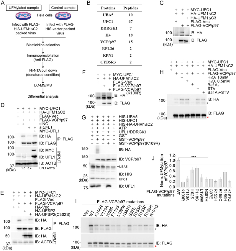
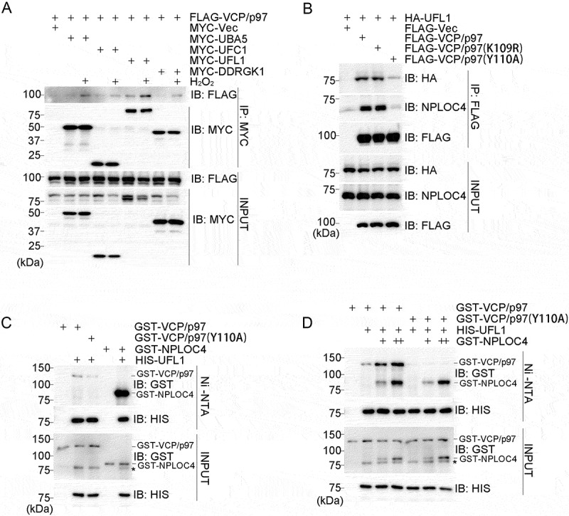
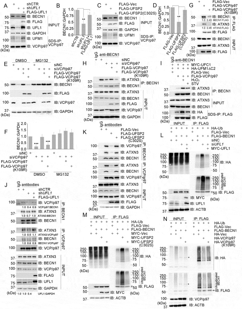
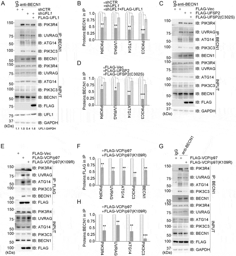
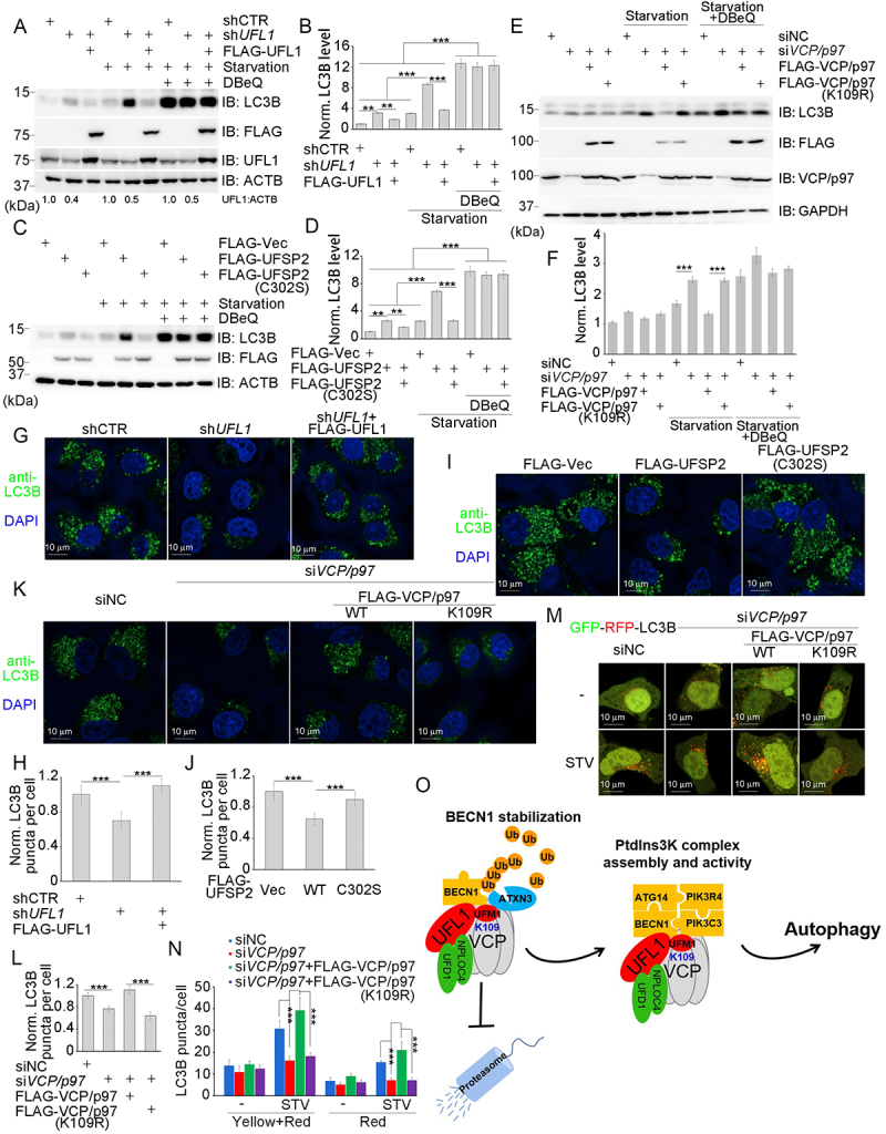
Macroautophagy/autophagy is essential for the degradation and recycling of cytoplasmic materials. The initiation of this process is determined by phosphatidylinositol-3-kinase (PtdIns3K) complex, which is regulated by factor BECN1 (beclin 1). UFMylation is a novel ubiquitin-like modification that has been demonstrated to modulate several cellular activities. However, the role of UFMylation in regulating autophagy has not been fully elucidated. Here, we found that VCP/p97 is UFMylated on K109 by the E3 UFL1 (UFM1 specific ligase 1) and this modification promotes BECN1 stabilization and assembly of the PtdIns3K complex, suggesting a role for VCP/p97 UFMylation in autophagy initiation. Mechanistically, VCP/p97 UFMylation stabilizes BECN1 through ATXN3 (ataxin 3)-mediated deubiquitination. As a key component of the PtdIns3K complex, stabilized BECN1 facilitates assembly of this complex. Re-expression of VCP/p97, but not the UFMylation-defective mutant, rescued the VCP/p97 depletion-induced increase in MAP1LC3B/LC3B protein expression. We also showed that several pathogenic VCP/p97 mutations identified in a variety of neurological disorders and cancers were associated with reduced UFMylation, thus implicating VCP/p97 UFMylation as a potential therapeutic target for these diseases. Abbreviation: ATG14:autophagy related 14; Baf A1:bafilomycin A1;CMT2Y: Charcot-Marie-Toothdisease, axonal, 2Y; CYB5R3: cytochromeb5 reductase 3; DDRGK1: DDRGK domain containing 1; DMEM:Dulbecco'smodified Eagle's medium;ER:endoplasmic reticulum; FBS:fetalbovine serum;FTDALS6:frontotemporaldementia and/or amyotrophic lateral sclerosis 6; IBMPFD1:inclusion bodymyopathy with early-onset Paget disease with or withoutfrontotemporal dementia 1; LC-MS/MS:liquid chromatography tandem mass spectrometry; MAP1LC3B/LC3B:microtubule associated protein 1 light chain 3 beta; MS: massspectrometry; NPLOC4: NPL4 homolog, ubiquitin recognition factor;PIK3C3: phosphatidylinositol 3-kinase catalytic subunit type 3;PIK3R4: phosphoinositide-3-kinase regulatory subunit 4; PtdIns3K:phosphatidylinositol 3-kinase; RPL26: ribosomal protein L26; RPN1:ribophorin I; SQSTM1/p62: sequestosome 1; UBA5: ubiquitin likemodifier activating enzyme 5; UFC1: ubiquitin-fold modifierconjugating enzyme 1; UFD1: ubiquitin recognition factor in ERassociated degradation 1; UFL1: UFM1 specific ligase 1; UFM1:ubiquitin fold modifier 1; UFSP2: UFM1 specific peptidase 2; UVRAG:UV radiation resistance associated; VCP/p97: valosin containingprotein; WT: wild-type.

Keywords: BECN1/beclin 1; PtdIns3K complex; UFL1; UFMylation; VCP/p97.


Conflict of interest statement

No potential conflict of interest was reported by the author(s).

Figures

Figure 1.
Figure 1.
VCP/p97 is UFMylated on lysine 109. (A) Schematic diagram of the approach used to identify UFMylated proteins. (B) List of UFMylated substrates identified by MS. Peptides: the total counts of the peptides of one protein per test per sample. (C-F and H-J) HEK293T cells were transfected and treated as indicated before collection. After FLAG-VCP/p97 immunoprecipitation under denaturing conditions, proteins were detected by Western blotting with the indicated antibodies. (C) VCP/p97 is UFMylated. HEK293T cells were co-transfected with FLAG-VCP/p97, HA-UFM1ΔC2 or HA-UFM1ΔC3 and MYC-UFC1 before collection. HA-UFM1ΔC3: negative control. Asterisk: non-specific bands. (D) VCP/p97 UFMylation is regulated by UFL1. HEK293T cells transfected with UFL1-targeted siRNA (siUFL1) or negative control siRNA (siNC), or siUFL1 plus MYC-UFL1 cDNA were co-transfected with FLAG-VCP/p97, HA-UFM1ΔC2 and MYC-UFC1 before collection. (E) VCP/p97 UFMylation is regulated by UFSP2. HEK293T cells transfected with HA-UFSP2, HA-UFSP2C302S or HA-vector were co-transfected with FLAG-VCP/p97, HA-UFM1ΔC2 and MYC-UFC1 before collection. (F) VCP/p97 UFMylation is abolished by the K109R mutation. HEK293T cells were co-transfected with FLAG-VCP/p97 WT or K109R, HA-UFM1ΔC2 and MYC-UFC1 before collection. Asterisk: non-specific bands. (G) UFMylation of VCP/p97 in vitro. The reactions were analyzed by immunoblot with the indicated antibodies. (H) VCP/p97 UFMylation in response to oxidative stress was detected as described in (C) after treatment as indicated (10 mM H2O2 for 15 min, 0.5 mM H2O2 for 4 h, 2 μM Baf A1 for 4 h, nutrient-starving medium for 3 h, Baf A1+nutrient starving medium). Asterisk: non-specific bands. Arrow: degraded FLAG-VCP/p97. (I and J) Pathogenic mutations impair VCP/p97 UFMylation. HEK293T cells were co-transfected with FLAG-VCP/p97 WT or mutations, HA-UFM1ΔC2 and MYC-UFC1 before collection. Asterisk: non-specific bands. Arrow: degraded FLAG-VCP/p97. (J) Bar graph of the ratio of HA:FLAG, calculated based on the grayscale values. Data represent the mean±SD of three independent experiments; ***p < 0.001. IB: immunoblot. IP: immunoprecipitation. Vec: vector. ACTB: loading control. STV: starvation.
Figure 2.
Figure 2.
VCP/p97 is presented to UFL1 by NPLOC4. (A) FLAG-VCP/p97 was co-transfected into HEK293T cells with MYC-UBA5, MYC-UFC1, MYC-UFL1, or MYC-DDRGK1. Cells 48 h later were treated with 10 mM H2O2 for 15 min and harvested and immunoprecipitated with anti-FLAG antibody, respectively. The immunocomplex of FLAG-VCP/p97 was immunoblotted with MYC and FLAG antibodies to determine the interactions between MYC-tagged proteins and FLAG-VCP/p97. (B) the VCP/p97Y110A mutation impairs its interaction with UFL1 and NPLOC4. HEK293T cells co-transfected with FLAG-VCP/p97 WT, K109R or Y110A and HA-UFL1 were harvested and immunoprecipitated with anti-FLAG antibody. The interactions between VCP/p97 and UFL1, or VCP/p97 and NPLOC4 were analyzed with HA, NPLOC4 and FLAG antibodies, respectively, after immunoprecipitation of FLAG-VCP/p97. (C) the interactions between UFL1 and VCP/p97 or NPLOC4 in vitro. Asterisk: HIS-UFL1. (D) NPLOC4 enhanced the interaction between UFL1 and VCP/p97. GST-VCP/p97, GST-VCP/p97Y110A, GST-NPLOC4 and HIS-UFL1 proteins purified from E. coli were incubated in vitro as indicated and were then pulled down with Ni-NTA agarose resin. The interactions were analyzed by western blotting. IB: immunoblot. IP: immunoprecipitation. Vec: vector. Asterisk: HIS-UFL1. ++: protein level greater than +.
Figure 3.
Figure 3.
VCP/p97 UFMylation stabilizes BECN1 through ATXN3-mediated VCP/p97 deubiquitination. (A and B) UFL1 knockdown decreased VCP/p97 UFMylation and BECN1 protein levels. HeLa cells stably expressing shCTR (control), shUFL1 (UFL1 depleted with shRNA) and FLAG-UFL1 (UFL1 re-expressed in shUFL1 cells) were subjected to endogenous VCP/p97 immunoprecipitation under denaturing conditions (SDS-IP). (B) Bar graph of the ratio of BECN1:GAPDH. (C and D) UFSP2 overexpression decreased VCP/p97 UFMylation and BECN1 protein levels. HeLa cells stably expressing FLAG-vector, FLAG-UFSP2 and FLAG-UFSP2C302S were subjected to endogenous VCP/p97 immunoprecipitation under denaturing conditions (SDS-IP). (D) Bar graph of the ratio of BECN1:GAPDH. (A-D) the immunoprecipitates (for VCP/p97 UFMylation) and INPUT (for BECN1 protein levels) were analyzed by immunoblot with the indicated antibodies. (E and F) Deficiency of VCP/p97 or its UFMylation decreased BECN1 protein levels. HeLa cells stably expressing FLAG-VCP/p97, FLAG-VCP/p97K109R, and control cells were transfected with siNC or siVCP/p97 as indicated and the cells were treated or not with MG132 for 9 h before collection. The harvested cells were lysed by 2 × sample buffer and the lysates were then analyzed by immunoblot with the indicated antibodies. (F) Bar graph of the ratio of BECN1:GAPDH. All the protein ratios in (A-F) were calculated based on the grayscale values. Data represent the mean±SD of three independent experiments; **p < 0.01; ***p < 0.001. (G) VCP/p97 UFMylation enhanced its interaction with BECN1 and ATXN3. HeLa cells stably expressing FLAG-vector, FLAG-VCP/p97 and FLAG-VCP/p97K109R were harvested and subjected to immunoprecipitation with anti-FLAG antibody and the immunoprecipitates were then analyzed by immunoblot with the indicated antibodies. (H) VCP/p97 and its UFMylation enhanced the interaction between BECN1 and ATXN3. HeLa cells stably expressing FLAG-VCP/p97, FLAG-VCP/p97K109R, and control cells were transfected with siNC or siVCP/p97 as indicated. The cells were then subjected to endogenous BECN1 immunoprecipitation and the immunoprecipitates were analyzed by immunoblot with the indicated antibodies. (I) H2O2 and nutrient starvation enhanced the UFMylation of VCP/p97, thereby promoting the interaction between BECN1 and ATXN3. HEK293T cells co-transfected with FLAG-VCP/p97, HA-UFM1ΔC2 and MYC-UFC1 were treated with H2O2 (10 mM for 15 min) and nutrient starvation (3 h) before collection. The harvested cells were aliquoted into two parts; one for immunoprecipitation to isolate endogenous BECN1 and the other to detect the UFMylation of VCP/p97 under denaturing conditions (SDS-IP). The immunoprecipitates were then analyzed by immunoblot with the indicated antibodies. (J and K) UFMylation depletion reduced the interactions among VCP/p97, BECN1 and ATXN3. HeLa cells stably expressing shCTR, shUFL1 and FLAG-UFL1 (UFL1 re-expressed in shUFL1 cells) (J) or FLAG-vector, FLAG-UFSP2 and FLAG-UFSP2C302S (K) were subjected to immunoprecipitation with the indicated antibodies. The immunoprecipitates were then analyzed by immunoblot with the indicated antibodies. (J) UFL1 knockdown reduced the interactions among VCP/p97, BECN1 and ATXN3. (K) UFSP2 overexpression reduced the interactions among VCP/p97, BECN1 and ATXN3. (L-N) VCP/p97 UFMylation inhibits BECN1 ubiquitination. HEK293T cells transfected as indicated were subjected to denaturing immunoprecipitation with anti-FLAG antibody and the FLAG-BECN1 immunoprecipitates were analyzed by immunoblot with the indicated antibodies. (L) UFL1 knockdown increased BECN1 ubiquitination. HEK293T cells co-transfected with FLAG-BECN1, HA-Ub and MYC-UFL1 or not were transfected with the indicated siRnas before collection. (M) UFSP2 overexpression increased BECN1 ubiquitination. HEK293T cells were co-transfected with FLAG-BECN1, HA-Ub and MYC-UFSP2 or MYC-UFSP2C302S before collection. (N) VCP/p97 UFMylation reduced BECN1 ubiquitination. HEK293T cells were co-transfected with FLAG-BECN1, HA-Ub and HA-VCP/p97 or HA-VCP/p97K109R before collection. Asterisk: HA-VCP/p97. All immunoprecipitated FLAG-BECN1 in (L-N) were detected first with anti-HA antibody (ubiquitination) and then with anti-FLAG antibody (BECN1) on the same PVDF membrane by western blotting. Arrows: FLAG-BECN1 signals. IB: immunoblot. IP: immunoprecipitation. Vec: vector. NC: negative control. CTR: control. SDS-IP: denaturing IP. GAPDH: loading control.
Figure 4.
Figure 4.
VCP/p97 UFMylation aids PtdIns3K complex assembly. (A-D) UFMylation enhances the interactions between BECN1 and other components of the PtdIns3K complex. The indicated stable HeLa cells were subjected to immunoprecipitation with anti-BECN1 antibody and the immunoprecipitates were then analyzed by immunoblot with the indicated antibodies. (A and B) UFL1 knockdown decreased the interactions between BECN1 and other components of the PtdIns3K complex. (B) Bar graph of the ratios of PIK3R4:BECN1, UVRAG:BECN1, ATG14:BECN1 and PIK3C3:BECN1 in the immunoprecipitation complex. (C and D) UFSP2 overexpression decreased the interactions between BECN1 and other components of the PtdIns3K complex. (D) Bar graph of the ratios of PIK3R4:BECN1, UVRAG:BECN1, ATG14:BECN1 and PIK3C3:BECN1 in the immunoprecipitation complex. (E and F) VCP/p97 UFMylation enhanced its interactions with components of the PtdIns3K complex. HeLa cells stably expressing FLAG-vector, FLAG-VCP/p97 and FLAG-VCP/p97K109R were subjected to immunoprecipitation with anti-FLAG antibody. The immunoprecipitates were then analyzed by immunoblot with the indicated antibodies. (F) Bar graph of the ratios of PIK3R4:FLAG, UVRAG:FLAG, ATG14:FLAG, PIK3C3:FLAG and BECN1:FLAG in the immunoprecipitation complex. (G and H) VCP/p97 UFMylation enhanced the interactions between BECN1 and other components of the PtdIns3K complex. HeLa cells stably expressing FLAG-VCP/p97, FLAG-VCP/p97K109R and control cells were subjected to immunoprecipitation with anti-BECN1 antibody. The immunoprecipitates were then analyzed by immunoblot with the indicated antibodies. (H) Bar graph of the ratios of PIK3R4:BECN1, UVRAG:BECN1, ATG14:BECN1 and PIK3C3:BECN1 in immunoprecipitation complex. All the protein ratios were calculated based on the grayscale values. Data represent the mean±SD of three independent experiments; *p < 0.05; **p < 0.01; ***p < 0.001. IB: immunoblot. IP: immunoprecipitation. Vec: vector. GAPDH: loading control.
Figure 5.
Figure 5.
VCP/p97 UFMylation promotes autophagy initiation. (A-F) VCP/p97 UFMylation regulates total LC3B levels. The indicated stable HeLa cells were treated or not with starvation (nutrient starvation medium for 2 h) or DBeQ (12 h). Protein levels were analyzed by western blotting with the indicated antibodies. All the protein ratios were calculated based on the grayscale values. (A and B) UFL1-mediated regulation of total LC3B levels after nutrient starvation are dependent on VCP/p97. (B) Bar graph of the data shown in (A) representing LC3B:ACTB ratios, normalized to the control without treatment. (C and D) UFSP2-mediated regulation of total LC3B levels after nutrient starvation are dependent on VCP/p97. (D) Bar graph of the data shown in (C) representing LC3B:ACTB ratios, normalized to the control without treatment. (E and F) VCP/p97 and its UFMylation regulate total LC3B levels. (F) Bar graph of the data shown in (E) representing LC3B:ACTB ratios, normalized to the control without treatment. (G) Representative images of immunofluorescence analysis of LC3B puncta formation influenced by UFL1. Cell lines stably expressing shCTR, shUFL1 or FLAG-UFL1 were treated with nutrient starvation medium for 2 h before fixation. LC3B puncta were analyzed by anti-LC3B immunofluorescence staining. (H) Bar graph of the data shown in (G) representing LC3B puncta, normalized to the control. (I) Representative images of immunofluorescence analysis of LC3B puncta formation influenced by UFSP2. Cell lines stably transfected with FLAG-vector, FLAG-UFSP2 or FLAG-UFSP2C302S were treated with nutrient starvation medium for 2 h before fixation. LC3B puncta were analyzed by anti-LC3B immunofluorescence staining. (J) Bar graph of the data shown in (I) representing LC3B puncta, normalized to the vector control. (K) Representative images of immunofluorescence analysis of LC3B puncta formation influenced by VCP/p97 and its UFMylation. Cell lines stably transfected with FLAG-vector, FLAG-VCP/p97 WT or K109R were transfected by VCP/p97 or negative control siRnas and, after 48 h, treated with nutrient starvation medium for 2 h before fixation. LC3B puncta were analyzed by anti-LC3B and immunofluorescence staining. (L) Bar graph of the data shown in (K) representing LC3B puncta, normalized to control. Green: LC3B; Blue: DAPI, nucleus. (M) Representative images of GFP-RFP-LC3B puncta influenced by VCP/p97 and its UFMylation. Cell lines stably transfected with FLAG-vector, FLAG-VCP/p97 WT or K109R were transfected with VCP/p97 or negative control siRnas and then with GFP-RFP-LC3B. After 48 h, cells were treated with nutrient starvation medium for 4 h before fixation. (N) Bar graph of the data shown in (M) representing GFP-RFP-LC3B puncta. Yellow+Red: total LC3B puncta; Red: the LC3B fused with lysosome. STV: starvation. IB: immunoblot. CTR: control. Vec: vector. NC: negative control. ACTB and GAPDH: loading control. Data represent the mean±SD of three independent experiments; **p < 0.01, ***p < 0.001. (O) Schematic diagram of the proposed mechanism by which UFMylation controls the function of VCP/p97 in autophagy. UFL1 promotes VCP/p97 UFMylation at K109 with the aid of NPLOC4, which stabilizes the interaction between ATXN3 and BECN1. ATXN3 de-ubiquitinates BECN1, thereby protecting BECN1 against degradation. Stabilized BECN1 promotes PtdIns3K complex assembly with UFMylated VCP/p97 functioning as a scaffolding protein to initiate autophagy initiation.

Similar articles

Cited by

References

    1. Wang Z, Gong Y, Peng B, et al. MRE11 UFMylation promotes ATM activation. Nucleic Acids Res. 2019 May 7;47(8):4124–4135. doi: 10.1093/nar/gkz110 - DOI - PMC - PubMed
    1. Wang X, Xu X, Wang Z.. The post-translational role of ufmylation in physiology and disease. Cells. 2023 Oct 29;12(21):2543. doi: 10.3390/cells12212543 - DOI - PMC - PubMed
    1. Millrine D, Cummings T, Matthews SP, et al. Human UFSP1 is an active protease that regulates UFM1 maturation and UFMylation. Cell Rep. 2022 Aug 2;40(5):111168. doi: 10.1016/j.celrep.2022.111168 - DOI - PMC - PubMed
    1. Liang Q, Jin YQ, Xu SW, et al. Human UFSP1 translated from an upstream near-cognate initiation codon functions as an active UFM1-specific protease. J Biol Chem. 2022. Jun;298(6):102016. doi: 10.1016/j.jbc.2022.102016 - DOI - PMC - PubMed
    1. Tatsumi K, Sou YS, Tada N, et al. A novel type of E3 ligase for the Ufm1 conjugation system. J Biol Chem. 2010 Feb 19;285(8):5417–5427. doi: 10.1074/jbc.M109.036814 - DOI - PMC - PubMed

Publication types

LinkOut - more resources