. 2024 Oct;20(10):2133-2145.
doi: 10.1080/15548627.2024.2356505. Epub 2024 Jun 30.

Bunyavirus SFTSV NSs utilizes autophagy to escape the antiviral innate immune response

Affiliations

Bunyavirus SFTSV NSs utilizes autophagy to escape the antiviral innate immune response

Ze-Min Li et al. Autophagy. 2024 Oct.

Abstract

Severe fever with thrombocytopenia syndrome virus (SFTSV) nonstructural protein (NSs) is an important viral virulence factor that sequesters multiple antiviral proteins into inclusion bodies to escape the antiviral innate immune response. However, the mechanism of the NSs restricting host innate immunity remains largely elusive. Here, we found that the NSs induced complete macroautophagy/autophagy by interacting with the CCD domain of BECN1, thereby promoting the formation of a BECN1-dependent autophagy initiation complex. Importantly, our data showed that the NSs sequestered antiviral proteins such as TBK1 into autophagic vesicles, and therefore promoted the degradation of TBK1 and other antiviral proteins. In addition, the 8A mutant of NSs reduced the induction of BECN1-dependent autophagy flux and degradation of antiviral immune proteins. In conclusion, our results indicated that SFTSV NSs sequesters antiviral proteins into autophagic vesicles for degradation and to escape antiviral immune responses.

Keywords: Autophagy; SFTSV; TBK1; immune escape; inclusion bodies.


Conflict of interest statement

No potential conflict of interest was reported by the author(s).

Figures

Figure 1.
Figure 1.
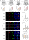
SFTSV NSs induces autophagy. (A-B) HEK293T and HeLa cells were transfected with pCAGGS or HA-NSs at doses of 0.25 μg, 0.5 μg and 1 μg for 24 h, or transfected with pCAGGS or HA-NSs at a dose of 1 μg for 6 h, 12 h and 24 h. LC3-II and SQSTM1 protein levels were analyzed with western blot. Western blot data were semi-quantified and normalized against ACTB/β-actin protein loading control (mean ± SEM, n = 3, **p < 0.001, ***p < 0.0001, ****p < 0.00001). (C-D) HEK293T and HeLa cells were transfected with pCAGGS or HA-NSs for 24 h. HEK293T and HeLa cells were infected with SFTSV for 2 h and then transfected with HA-NSs for 24 h for simulating the infection status. HA-NSs (red), endogenous LC3 (green), and DAPI (blue) were analyzed with confocal microscopy. Scale bar: 20 μm (mean ± SEM, n = 3, **p < 0.001, ***p < 0.0001). (E) HeLa cells were transfected with pCAGGS or HA-NSs for 24 h, the formation of autophagosomes or autolysosomes were processed and analyzed via electron transmission microscopy. Scale bar: 1 μm. (mean ± SEM, n = 3, **p < 0.001).
Figure 2.
Figure 2.
SFTSV NSs induces complete autophagy. (A-B) HEK293T and HeLa cells were transfected with pCAGGS or HA-NSs at a dose of 2 μg for 24 h and then treated with CQ or Baf-A1 for 6 h. LC3-II and SQSTM1 protein levels were analyzed with western blot. Western blot data were semi-quantified and normalized against ACTB protein loading control (mean ± SEM, n = 3, **p < 0.001, ***p < 0.0001). (C-D) HEK293T and HeLa cells were transfected with mCherry-GFP-LC3 and pCAGGS or HA-NSs for 24 h. HEK293T and HeLa cells were infected with SFTSV for 2 h and then transfected with mCherry-GFP-LC3 and HA-NSs for 24 h for simulating the infection status. HA-NSs (purple), mCherry-LC3 (red), GFP-LC3 (green) and DAPI (blue) were analyzed with confocal microscopy. Scale bar: 20 μm (mean ± SEM, n = 3, **p < 0.001, ****p < 0.00001, ns, not significant). (E) HEK293T cells were co-transfected with GFP-LC3 and pCAGGS or HA-NSs for 24 h. HEK293T cells were infected with SFTSV for 2 h and then transfected with HA-NSs for 24 h for simulating the infection status. HA-NSs (purple), endogenous LAMP1 (red), and DAPI (blue) were analyzed with confocal microscopy. Scale bar: 20 μm (mean ± SEM, n = 3, ns, not significant).
Figure 3.
Figure 3.
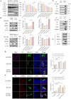
SFTSV NSs induces autophagy through the complex of the BECN1-PIK3C3. (A) BECN1 knockout cells or WT HeLa cells were transfected with pCAGGS and HA-NSs for 24 h. LC3-II and SQSTM1 protein levels were analyzed with western blot (mean ± SEM, n = 3). (B-C) HEK293T cells were transfected with HA-NSs or co-transfected with HA-NSs and Flag-BECN1 for 24 h. The interaction between endogenous and exogenous BECN1 and NSs was analyzed with co-IP using IgG as control (mean ± SEM, n = 3). (D) HEK293T cells were transfected with HA-NSs for 24 h. HEK293T cells were infected with SFTSV for 2 h and then transfected with HA-NSs for 24 h for simulating the infection status. HA-NSs (red), endogenous BECN1 (green), and DAPI (blue) were analyzed with confocal microscopy. Scale bar: 20 μm (mean ± SEM, n = 3, ****p < 0.00001). (E) the schematic diagram of BECN1 truncation mutants and the BECN1-NSs interacting domain. (F) HEK293T cells were co-transfected with HA-NSs and Flag-BECN1, Flag-BECN1-BD, Flag-BECN1-BD+CCD, Flag-BECN1-BD+CCD+ECD, Flag-BECN1-CCD+ECD, or Flag-BECN1-ECD. The interaction between truncations and HA-NSs was analyzed with co-IP using IgG as control (mean ± SEM, n = 3). (G) HEK293T cells were transfected with pCAGGS or HA-NSs for 24 h. Co-IP with HA antibodies and evaluation of BECN1, PIK3C3 and ATG14 with western blot using IgG as control. Co-IP with BECN1 antibodies and evaluation of HA-NSs, PIK3C3 and ATG14 with western blot using IgG as control (mean ± SEM, n = 3).
Figure 4.
Figure 4.
The 8A mutant of NSs displays a reduced ability to stimulate autophagy. (A-B) HEK293T cells were transfected with HA-NSs or co-transfected with HA-NSs and GFP-LC3 for 24 h. The interaction between HA-NSs and LC3 was analyzed with co-IP using IgG as control (mean ± SEM, n = 3). (C) The motif diagram of the WT NSs and mutants. (D) HEK293T cells were co-transfected with HA-NSs and HA-8A, HA-PxxP, HA-DWP, or HA-WxxL for 24 h. The interaction between mutants and NSs was analyzed with co-IP using IgG as control. Western blot data were semi-quantified and normalized against IP group loading control (mean ± SEM, n = 3, ****p < 0.00001). (E-F) HEK293T and HeLa cells were transfected with pCAGGS, HA-8A or HA-NSs at doses of 1 μg and 2 μg for 24 h. LC3-II protein level was analyzed with western blot. Western blot data were semi-quantified and normalized against ACTB protein loading control (mean ± SEM, n = 3, *p < 0.05, **p <0.001). (G) HEK293T cells were co-transfected with GFP-LC3 and pCAGGS, HA-8A or HA-NSs for 24 h. HA-NSs (red) and DAPI (blue) were analyzed with confocal microscopy. Scale bar: 20 μm (mean ± SEM, n = 3, ***p < 0.0001, ns, not significant). (H) HEK293T cells were transfected with HA-8A or HA-NSs at a dose of 2 μg for 24 h. Co-IP with HA antibodies and evaluation of BECN1, PIK3C3 and ATG14 with western blot using IgG as control. Western blot data were semi-quantified and normalized against IP group loading control (mean ± SEM, n = 3, *p < 0.05, ****p < 0.00001).
Figure 5.
Figure 5.
SFTSV NSs-induced immune escape depends on autophagy. (A) HEK293T cells were transfected with pCAGGS or HA-NSs at doses of 0.5 μg, 1 μg, 2 μg for 24 h. TBK1, RIGI, TRIM25 and NFKB protein levels were analyzed with western blot. Western blot data were semi-quantified and normalized against ACTB protein loading control (mean ± SEM, n = 3, *p < 0.05, ***p < 0.0001). (B) HEK293T and HeLa cells were transfected with pCAGGS or HA-NSs for 24 h and then treated with CQ or Baf-A1 for 6 h. TBK1 and TRIM25 protein levels were analyzed with western blot (mean ± SEM, n = 3, *p < 0.05, ***p < 0.0001, ****p < 0.00001). (C) HEK293T cells were co-transfected with pCAGGS or HA-NSs for 24 h. Co-IP with HA antibodies and evaluation of BECN1, PIK3C3 and ATG14 with western blot using IgG as control. (mean ± SEM, n = 3). Co-IP with LC3 antibodies and evaluation of HA-NSs, TBK1 and TRIM25 with western blot using IgG as control (mean ± SEM, n = 3). (D-E) HEK293T cells were co-transfected with HA-NSs and GFP-LC3 for 24 h, HA-NSs (red), TBK1 (purple), and DAPI (blue) were analyzed with confocal microscopy (above). HA-NSs (purple), TRIM25 (red), and DAPI (blue) were analyzed with confocal microscopy (below). Scale bar: 20 μm (mean ± SEM, n = 3, ****p < 0.00001).
Figure 6.
Figure 6.
The 8A mutant of NSs loses the ability of immune escape. (A-B) HeLa cells were transfected with HA-NSs for 24 h, 4-nm gold particles were used to label endogenous LC3 or LAMP1 and 12-nm gold particles were used to label HA-NSs. The orange arrows refer to LC3 or LAMP1, the black arrows refer to the HA-NSs, and the red arrows refer to autophagy vesicles. Scale bar: 250 nm (mean ± SEM, n = 3). (C) HeLa cells were transfected with HA-NSs for 24 h, 12-nm gold particles were used to label HA-NSs. The black arrows refer to the NSs, and the red arrows refer to autophagy vesicles. Scale bar: 250 nm (mean ± SEM, n = 3). (D) HEK293T cells were transfected with pCAGGS, HA-NSs and HA-8A, HA-PxxP, HA-DWP, or HA-WxxL at a dose of 2 μg for 24 h. TBK1 protein level was analyzed with western blot. Western blot data were semi-quantified and normalized against ACTB protein loading control (mean ± SEM, n = 3, **p < 0.001, ****p < 0.00001, ns, not significant). (E) HEK293T cells were transfected with HA-8A or HA-NSs at a dose of 2 μg for 24 h, and then transfected with poly(I:C) for 24 h. Co-IP with HA antibodies and evaluation of LC3, TBK1 and TRIM25 with western blot using IgG as control. Western blot data were semi-quantified and normalized against IP group loading control (mean ± SEM, n = 3, **p < 0.001, ****p < 0.00001). (F) HEK293T cells were transfected with pCAGGS, HA-NSs and HA-8A, HA-PxxP, HA-DWP, or HA-WxxL at a dose of 2 μg for 24 h, and then transfected with poly(I:C) for 24 h. IFNB and IFIT1 mRNA levels were analyzed with RT-qPCR (mean ± SEM, n = 3, *p < 0.05, ****p < 0.00001). (G) HeLa cells were transfected with pCAGGS, HA-8A or HA-NSs at a dose of 2 μg for 24 h, and then transfected with poly(I:C) for 24 h. IFNB, IFIT1 and CXCL10 mRNA levels were analyzed with RT-qPCR (mean ± SEM, n = 3, *p < 0.05, **p < 0.001, ***p < 0.0001, ****p < 0.00001). (H) BECN1 knockout HeLa cells were transfected with pCAGGS, HA-8A or HA-NSs for 24 h, and then transfected with poly(I:C) for 24 h. IFNB, IFIT1 and CXCL10 mRNA levels were analyzed with RT-qPCR (mean ± SEM, n = 3, ns, not significant).
Figure 7.
Figure 7.
Schematic model illustrating the role of the NSs in autophagy and immune escape. SFTSV NSs induces complete autophagy by interacting with BECN1, thus promoting the formation of BECN1-PIK3C3 autophagy initiation complex. SFTSV NSs sequesters TBK1 and other antiviral proteins into autophagy vesicles, leading to their degradation and ultimately mediating immune escape.

Similar articles

Cited by

References

    1. Yu XJ, Liang MF, Zhang SY, et al. Fever with thrombocytopenia associated with a novel bunyavirus in China. N Engl J Med. 2011;364(16):1523–1532. doi: 10.1056/NEJMoa1010095 - DOI - PMC - PubMed
    1. Li J, Li S, Yang L, et al. Severe fever with thrombocytopenia syndrome virus: a highly lethal bunyavirus. Crit Rev Microbiol. 2021;47(1):112–125. doi: 10.1080/1040841X.2020.1847037 - DOI - PubMed
    1. Li J-C, Zhao J, Li H, et al. Epidemiology, clinical characteristics, and treatment of severe fever with thrombocytopenia syndrome. Infect Med. 2022;1(1):40–49. doi: 10.1016/j.imj.2021.10.001 - DOI - PMC - PubMed
    1. Kim YR, Yun Y, Bae SG, et al. Severe fever with thrombocytopenia syndrome virus infection, South Korea, 2010. Emerg Infect Dis. 2018;24(11):2103–2105. doi: 10.3201/eid2411.170756 - DOI - PMC - PubMed
    1. Takahashi T, Maeda K, Suzuki T, et al. The first identification and retrospective study of severe fever with thrombocytopenia syndrome in Japan. J Infect Dis. 2014;209(6):816–827. doi: 10.1093/infdis/jit603 - DOI - PMC - PubMed

Publication types

LinkOut - more resources