

Juvenile peripheral LPS exposure overrides female resilience to prenatal VPA effects on adult sociability in mice
- PMID: 38763939
- PMCID: PMC11102908
- DOI: 10.1038/s41598-024-62217-6
Juvenile peripheral LPS exposure overrides female resilience to prenatal VPA effects on adult sociability in mice
Abstract
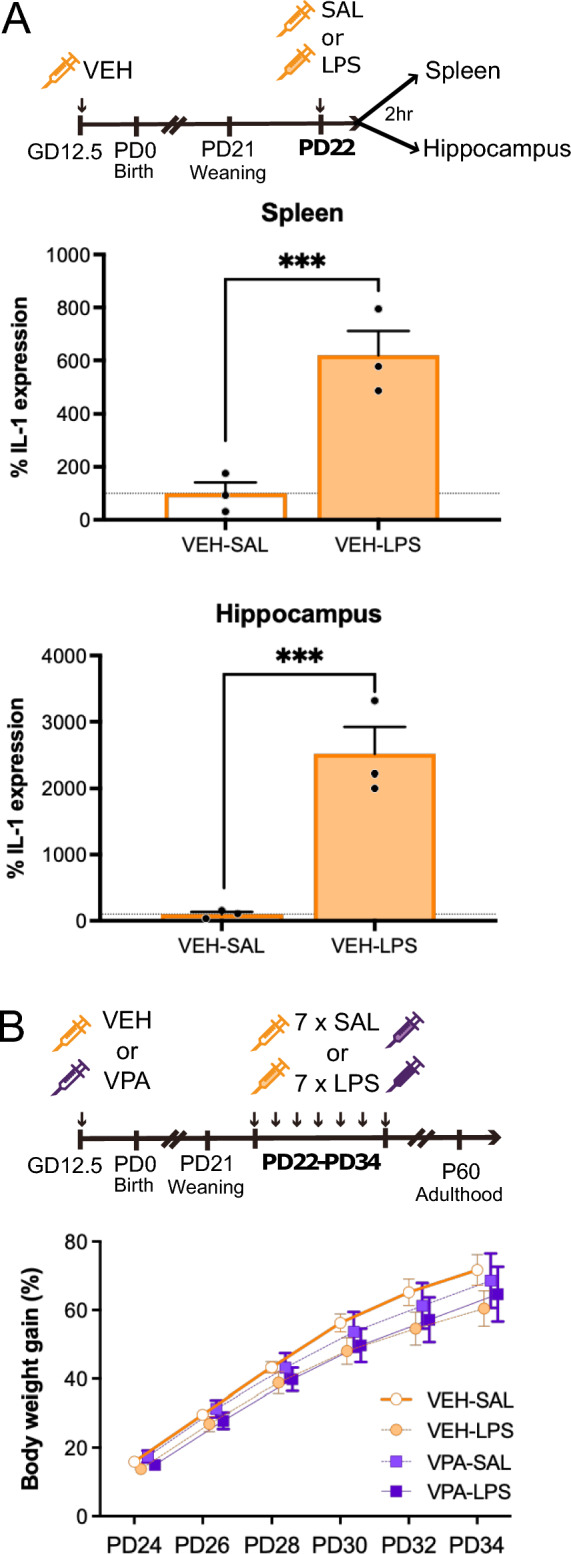
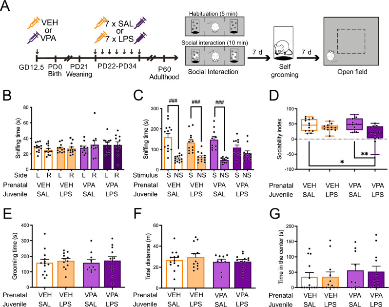
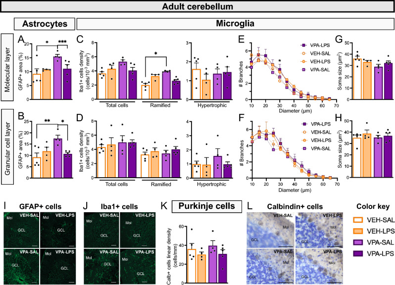
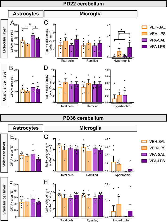
Autism spectrum disorder (ASD) exhibits a gender bias, with boys more frequently affected than girls. Similarly, in mouse models induced by prenatal exposure to valproic acid (VPA), males typically display reduced sociability, while females are less affected. Although both males and females exhibit VPA effects on neuroinflammatory parameters, these effects are sex-specific. Notably, females exposed to VPA show increased microglia and astrocyte density during the juvenile period. We hypothesized that these distinct neuroinflammatory patterns contribute to the resilience of females to VPA. To investigate this hypothesis, we treated juvenile animals with intraperitoneal bacterial lipopolysaccharides (LPS), a treatment known to elicit brain neuroinflammation. We thus evaluated the impact of juvenile LPS-induced inflammation on adult sociability and neuroinflammation in female mice prenatally exposed to VPA. Our results demonstrate that VPA-LPS females exhibit social deficits in adulthood, overriding the resilience observed in VPA-saline littermates. Repetitive behavior and anxiety levels were not affected by either treatment. We also evaluated whether the effect on sociability was accompanied by heightened neuroinflammation in the cerebellum and hippocampus. Surprisingly, we observed reduced astrocyte and microglia density in the cerebellum of VPA-LPS animals. These findings shed light on the complex interactions between prenatal insults, juvenile inflammatory stimuli, and sex-specific vulnerability in ASD-related social deficits, providing insights into potential therapeutic interventions for ASD.
Keywords: Autism spectrum disorder; Inflammation.
© 2024. The Author(s).
Conflict of interest statement
The authors declare no competing interests.
Figures
Similar articles
-
Sex-specific effects of prenatal valproic acid exposure on sociability and neuroinflammation: Relevance for susceptibility and resilience in autism.Psychoneuroendocrinology. 2019 Dec;110:104441. doi: 10.1016/j.psyneuen.2019.104441. Epub 2019 Sep 11. Psychoneuroendocrinology. 2019. PMID: 31541913
-
Exploring the role of Cav3.2 calcium channels in autism-like cognitive behavior induced by prenatal valproic acid exposure.Neuroscience. 2025 Jun 21;577:71-79. doi: 10.1016/j.neuroscience.2025.05.011. Epub 2025 May 6. Neuroscience. 2025. PMID: 40339900
-
Prenatal Exposure to Valproic Acid Affects Microglia and Synaptic Ultrastructure in a Brain-Region-Specific Manner in Young-Adult Male Rats: Relevance to Autism Spectrum Disorders.Int J Mol Sci. 2020 May 18;21(10):3576. doi: 10.3390/ijms21103576. Int J Mol Sci. 2020. PMID: 32443651 Free PMC article.
-
Prenatal valproate in rodents as a tool to understand the neural underpinnings of social dysfunctions in autism spectrum disorder.Neuropharmacology. 2019 Nov 15;159:107477. doi: 10.1016/j.neuropharm.2018.12.024. Epub 2019 Jan 9. Neuropharmacology. 2019. PMID: 30639388 Review.
-
The valproic acid-induced rodent model of autism.Exp Neurol. 2018 Jan;299(Pt A):217-227. doi: 10.1016/j.expneurol.2017.04.017. Epub 2017 May 2. Exp Neurol. 2018. PMID: 28472621 Review.
References
-
- American Psychiatric Association . Diagnostic and Statistical Manual of Mental Disorders. Diagnostic and Statistical Manual of Mental Disorders. 5. American Psychiatric Association; 2013.
-
- Hull L, Petrides KV, Mandy W. The female autism phenotype and camouflaging: A narrative review. Rev. J. Autism Dev. Disord. 2020;7:306–317. doi: 10.1007/s40489-020-00197-9. - DOI
