

TCL1A-expressing B cells are critical for tertiary lymphoid structure formation and the prognosis of oral squamous cell carcinoma
- PMID: 38764038
- PMCID: PMC11103841
- DOI: 10.1186/s12967-024-05292-7
TCL1A-expressing B cells are critical for tertiary lymphoid structure formation and the prognosis of oral squamous cell carcinoma
Abstract
Background: Oral squamous cell carcinoma (OSCC) is a malignant tumor with a poor prognosis. Traditional treatments have limited effectiveness. Regulation of the immune response represents a promising new approach for OSCC treatment. B cells are among the most abundant immune cells in OSCC. However, the role of B cells in OSCC treatment has not been fully elucidated.
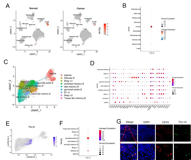
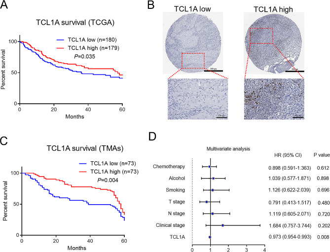
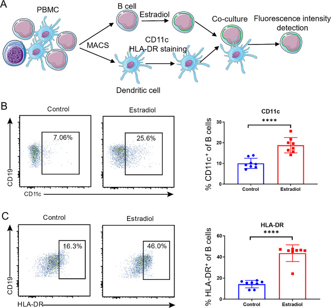
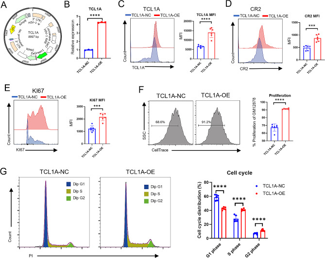
Methods: Single-cell RNA sequencing analysis of 13 tissues and 8 adjacent normal tissues from OSCC patients was performed to explore differences in B-cell gene expression between OSCC tissues and normal tissues. We further investigated the relationship between differentially expressed genes and the immune response to OSCC. We utilized tissue microarray data for 146 OSCC clinical samples and RNA sequencing data of 359 OSCC samples from The Cancer Genome Atlas (TCGA) to investigate the role of T-cell leukemia 1 A (TCL1A) in OSCC prognosis. Multiplex immunohistochemistry (mIHC) was employed to investigate the spatial distribution of TCL1A in OSCC tissues. We then investigated the effect of TCL1A on B-cell proliferation and trogocytosis. Finally, lentiviral transduction was performed to induce TCL1A overexpression in B lymphoblastoid cell lines (BLCLs) to verify the function of TCL1A.
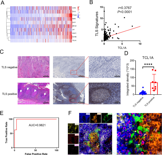
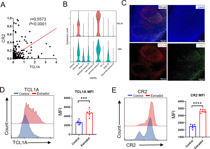
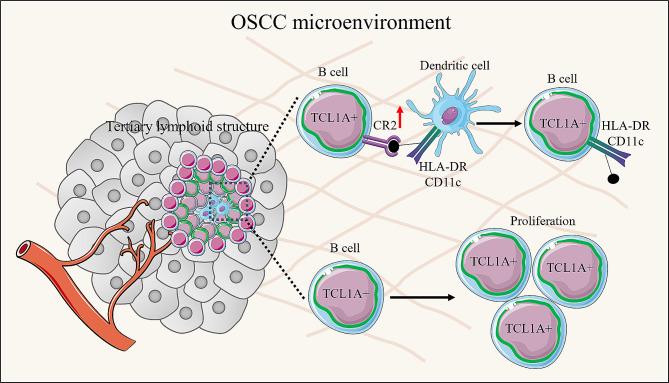
Results: Our findings revealed that TCL1A was predominantly expressed in B cells and was associated with a better prognosis in OSCC patients. Additionally, we found that TCL1A-expressing B cells are located at the periphery of lymphatic follicles and are associated with tertiary lymphoid structures (TLS) formation in OSCC. Mechanistically, upregulation of TCL1A promoted the trogocytosis of B cells on dendritic cells by mediating the upregulation of CR2, thereby improving antigen-presenting ability. Moreover, the upregulation of TCL1A expression promoted the proliferation of B cells.
Conclusion: This study revealed the role of B-cell TCL1A expression in TLS formation and its effect on OSCC prognosis. These findings highlight TCL1A as a novel target for OSCC immunotherapy.
Keywords: B cells; Oral squamous cell carcinoma; TCL1A; Tertiary lymphoid structures.
© 2024. The Author(s).
Conflict of interest statement
The authors declare no competing interests.
Figures
Similar articles
-
Impact of Non-SMC Condensin I Complex Subunit D2 Upregulation on Oral Squamous Cell Carcinoma Prognosis.Int Dent J. 2025 Jun;75(3):1818-1827. doi: 10.1016/j.identj.2024.03.015. Epub 2025 Apr 16. Int Dent J. 2025. PMID: 40245749 Free PMC article.
-
Role of hyaluronan mediated motility receptor gene in oral squamous cell carcinoma and clinical prognosis.Zhong Nan Da Xue Xue Bao Yi Xue Ban. 2021 Dec 28;46(12):1315-1324. doi: 10.11817/j.issn.1672-7347.2021.200955. Zhong Nan Da Xue Xue Bao Yi Xue Ban. 2021. PMID: 35232899 Free PMC article. Chinese, English.
-
Genome-wide DNA methylation profile identified a unique set of differentially methylated immune genes in oral squamous cell carcinoma patients in India.Clin Epigenetics. 2017 Feb 3;9:13. doi: 10.1186/s13148-017-0314-x. eCollection 2017. Clin Epigenetics. 2017. PMID: 28174608 Free PMC article.
-
Mediator complex subunit 1 promotes oral squamous cell carcinoma progression by activating MMP9 transcription and suppressing CD8+ T cell antitumor immunity.J Exp Clin Cancer Res. 2024 Sep 30;43(1):270. doi: 10.1186/s13046-024-03191-9. J Exp Clin Cancer Res. 2024. PMID: 39343952 Free PMC article.
-
Revelation of comprehensive cell profiling of primary and metastatic tumour ecosystems in oral squamous cell carcinoma by single-cell transcriptomic analysis.Int J Med Sci. 2024 Aug 26;21(12):2293-2304. doi: 10.7150/ijms.97404. eCollection 2024. Int J Med Sci. 2024. PMID: 39310253 Free PMC article.
Cited by
-
Multi-omics analysis unveils a four-gene prognostic signature in esophageal squamous carcinoma and the therapeutic potential of PKP1.BMC Cancer. 2025 Apr 25;25(1):777. doi: 10.1186/s12885-025-14150-8. BMC Cancer. 2025. PMID: 40281492 Free PMC article.
-
Review: radiotherapy-mediated B cells within the TLS influence the tumor microenvironment.J Immunother Cancer. 2025 Jul 15;13(7):e011617. doi: 10.1136/jitc-2025-011617. J Immunother Cancer. 2025. PMID: 40664446 Free PMC article. Review.
-
[Chinese Expert Consensus on Assessment and Clinical Application of Tertiary Lymphoid Structure for Non-small Cell Lung Cancer (2025 Version)].Zhongguo Fei Ai Za Zhi. 2025 Feb 20;28(2):95-104. doi: 10.3779/j.issn.1009-3419.2025.102.03. Zhongguo Fei Ai Za Zhi. 2025. PMID: 40114486 Free PMC article. Review. Chinese.
-
Global trends in tertiary lymphoid structures: a bibliometric analysis from 2014 to 2023.Front Immunol. 2024 Nov 15;15:1475062. doi: 10.3389/fimmu.2024.1475062. eCollection 2024. Front Immunol. 2024. PMID: 39620224 Free PMC article.
References
-
- 1. Hosainzadegan M, Aziz E, Rovshan K, Nasibova A, Amir H, Parviz V, et al. Are microbial infections and some antibiotics causes cancer. Adv Biol Earth Sci. 2020; 5: 58–61.
-
- 2. Ali I, Saleem K, Aboul-Enein HY, Rather A. Social Aspects of Cancer Genesis. Cancer Therapy. 2011; 8: 6–14.
-
- 3. Ali I, Wani WA, Khan A, Haque A, Ahmad A, Saleem K, et al. Synthesis and synergistic antifungal activities of a pyrazoline based ligand and its copper (II) and nickel (II) complexes with conventional antifungals. Microb Pathogenesis. 2012; 53: 66–73. - PubMed
-
- 4. Ali I, Wani WA, Haque A, Saleem K. Glutamic acid and its derivatives: candidates for rational design of anticancer drugs. Future Med Chem. 2013; 5: 961–978. - PubMed
-
- 5. Ali I, Wani WA, Saleem K, Hsieh M. Anticancer metallodrugs of glutamic acid sulphonamides: in silico, DNA binding, hemolysis and anticancer studies. Rsc Adv. 2014; 4: 29629–29641.
Publication types
MeSH terms
Substances
Grants and funding
LinkOut - more resources
Full Text Sources
Medical
