. 2024 May 20;20(1):212.
doi: 10.1186/s12917-024-04015-w.

Effect of baicalin on eradicating biofilms of bovine milk derived Acinetobacter lwoffii

Affiliations

Effect of baicalin on eradicating biofilms of bovine milk derived Acinetobacter lwoffii

Chengjun Ma et al. BMC Vet Res. .

Abstract

Background: Acinetobacter lwoffii (A.lwoffii) is a serious zoonotic pathogen that has been identified as a cause of infections such as meningitis, bacteremia and pneumonia. In recent years, the infection rate and detection rate of A.lwoffii is increasing, especially in the breeding industry. Due to the presence of biofilms, it is difficult to eradicate and has become a potential super drug-resistant bacteria. Therefore, eradication of preformed biofilm is an alternative therapeutic action to control A.lwoffii infection. The present study aimed to clarify that baicalin could eradicate A.lwoffii biofilm in dairy cows, and to explore the mechanism of baicalin eradicating A.lwoffii.

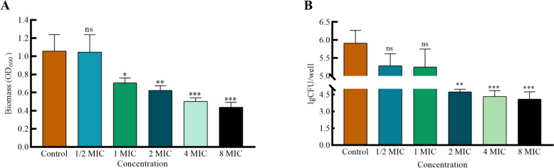
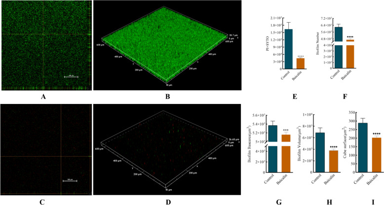
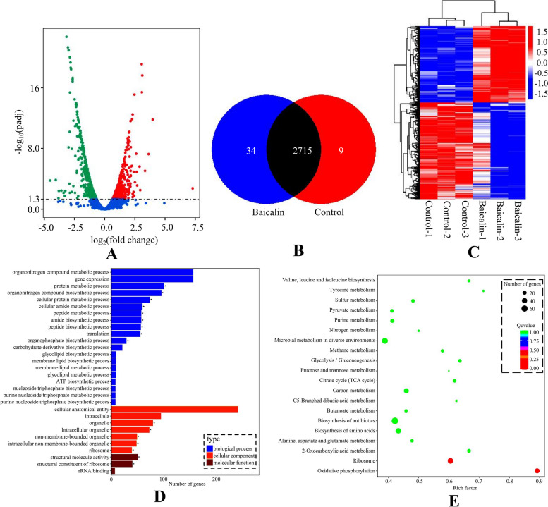
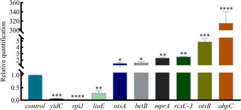
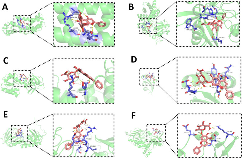
Results: The results showed that compared to the control group, the 4 MIC of baicalin significantly eradicated the preformed biofilm, and the effect was stable at this concentration, the number of viable bacteria in the biofilm was decreased by 0.67 Log10CFU/mL. The total fluorescence intensity of biofilm bacteria decreased significantly, with a reduction rate of 67.0%. There were 833 differentially expressed genes (367 up-regulated and 466 down-regulated), whose functions mainly focused on oxidative phosphorylation, biofilm regulation system and trehalose synthesis. Molecular docking analysis predicted 11 groups of target proteins that were well combined with baicalin, and the content of trehalose decreased significantly after the biofilm of A.lwoffii was treated with baicalin.

Conclusions: The present study evaluated the antibiofilm potential of baicalin against A.lwoffii. Baicalin revealed strong antibiofilm potential against A.lwoffii. Baicalin induced biofilm eradication may be related to oxidative phosphorylation and TCSs. Moreover, the decrease of trehalose content may be related to biofilm eradication.

Keywords: Acinetobacter lwoffii; Baicalin; Biofilm; Eradication; Trehalose.


Conflict of interest statement

The authors declare no competing interests.

Figures

Fig. 1
Fig. 1
Effect of baicalin on A.Lwoffii biofilm. A.Lwoffii biofilms were cultured for 24 h at 37 °C in 96-well plates. Post-washing, these biofilms were exposed to baicalin for 3 h. Biomass quantification was conducted using crystal violet (CV) staining (A) and viable bacteria of biofilm by colony counting (B). The significance of results was analyzed using an unpaired two-tailed t-test. Notably, * P < 0.05 and ** P < 0.01 when compared to the control group
Fig. 2
Fig. 2
CLSM analysis of baicalin treated A.lwoffii pre-biofilms. A.Lwoffii biofilm was formed for 24 h at 37 °C on chambered coverglass slides. After washing, biofilms were treated with baicalin for 3 h at 37 °C as described and subsequently stained with SYTO 9 and PI for 20 min in the dark. Images were acquired by CLSM. Using BiofilmQ software for image processing.. A and B 3D and orthogonal views biofilm representation in the objective of 20X in the control group. C and D 3D and orthogonal views biofilm representation of baicalin in the objective of 20X. E total fluorescence intensity of biofilms (PI + SYTO9). F represents the number of biofilms. G bottom area of biofilms. H represents the volume of biofilms. I represents the surface area of biofilms. Unpaired t-test (two-tailed) was used to measure statistical significance. * P < 0.05, ** P < 0.01 compared with the control group
Fig. 3
Fig. 3
Analysis of differential gene expression. The preformed biofilm was treated with 4 MIC baicalin for 3 h, and the upper culture medium in cell culture flasks was collected and centrifuged. The supernatant was taken and frozen in liquid nitrogen for storage. There were 6 samples in both the baicalin group and the control group. The samples were analyzed by LC–MS, and the data were extracted and preprocessed using Mas-terView (SCIEX). A Volcano plots showed the fold change of differential gene in baicalin group vs control group. The green dots represent significantly down-regulated genes (466), the red dots represent significantly up-regulated genes (367), and grey dots represent non-significantly changed differential genes. B Venn diagram. C Cluster analysis diagram of differentially expressed genes, the twenty-four hours A.Lwoffii pre-biofilms was treated with baicalin (4 MIC) for 3h, and the untreated biofilms was used as the control. All experiments were performed at least in triplicate, and there were 6 samples in both the baicalin group and the control group. D GO annotation enrichmentan analysis diagram and (E) KEGG enrichment analysis diagram. The p value is the significance of enrichment of this metabolic pathway. The ordinate is the name of the metabolic pathway, the abscissa is the rich factor. The larger the rich factor, the more metabolites enriched in the pathway. The size of the dots represents the number of differential metabolites enriched into the pathway
Fig. 4
Fig. 4
Fluorescence quantitative PCR results of differential genes. Effects of 4 MIC baicalin on the regulated genes ahpC, otsB, rcsC-3, mpaA, betB, otsA, liuE, rpiJ and yidC in A.Lwoffii biofilms
Fig. 5
Fig. 5
Three-dimensional interaction between baicalin and target proteins. AutoDockTools 1.5.7 software was applied to process proteins. PyMOL analyze the binding situation of receptor proteins and ligands in molecular docking, and it can mark hydrogen bonds. The top 6 binding force compounds were imported into PyMOL for further visualization. A Baicalin-betI_3, (B) Baicalin-hmp-1, (C) Baicalin-mprA, (D) Baicalin-ppiC, (E) Baicalin-mutM, (F). Baicalin-mymA. The yellow dotted line indicates the hydrogen bond distance or π-stacking distance, while the active ingredient is shown in red
Fig. 6
Fig. 6
Standard curve and content determination of trehalose. Sodium alginate standard product was diluted with ddH2O water at a ratio, with concentrations of 0.1, 0.05, 0.025, 0.0125, 0.00625, 0.003125 mg/mL respectively. Take 0.25 mL of standard solution and 1 mL of working solution to an EP tube, water bath at 95 °C for 10 min, naturally cool to room temperature, take the solution to the colorimetric dish, measure OD620nm. Calculate the concentration of ΔA standard (A) and using the formula obtained from the drawn standard curve to calculate X value can measure the trehalose content of each group (B). Unpaired t-test (two-tailed) was used to measure statistical significance. * P < 0.05, ** P < 0.01 compared with the control group

Similar articles

Cited by

References

    1. Fu S, Wen C, Wang Z, Qiu Y, Zhang Y, Zuo J, Xu Y, Han X, Luo Z, Chen W, et al. Molecular Epidemiology and Antimicrobial Resistance of Outbreaks of Klebsiella pneumoniae Clinical Mastitis in Chinese Dairy Farms. Microbiol Spectr. 2022;10:e299722. doi: 10.1128/spectrum.02997-22. - DOI - PMC - PubMed
    1. Avery TM, Boone RL, Lu J, Spicer SK, Guevara MA, Moore RE, Chambers SA, Manning SD, Dent L, Marshall D, et al. Analysis of Antimicrobial and Antibiofilm Activity of Human Milk Lactoferrin Compared to Bovine Lactoferrin against Multidrug Resistant and Susceptible Acinetobacter baumannii Clinical Isolates. ACS Infect Dis. 2021;7:2116–2126. doi: 10.1021/acsinfecdis.1c00087. - DOI - PMC - PubMed
    1. Xu N, Qiu C, Yang Q, Zhang Y, Wang M, Ye C, Guo M. Analysis of Phenol Biodegradation in Antibiotic and Heavy Metal Resistant Acinetobacter lwoffii NL1. Front Microbiol. 2021;12:725755. doi: 10.3389/fmicb.2021.725755. - DOI - PMC - PubMed
    1. Rumbaugh KP, Sauer K. Biofilm dispersion. Nat Rev Microbiol. 2020;18:571–586. doi: 10.1038/s41579-020-0385-0. - DOI - PMC - PubMed
    1. Ju X, Chen J, Zhou M, Zhu M, Li Z, Gao S, Ou J, Xu D, Wu M, Jiang S, et al. Combating Pseudomonas aeruginosa Biofilms by a Chitosan-PEG-Peptide Conjugate via Changes in Assembled Structure. ACS Appl Mater Interfaces. 2020;12:13731–13738. doi: 10.1021/acsami.0c02034. - DOI - PubMed