. 2024 May 20;15(1):143.
doi: 10.1186/s13287-024-03758-5.

Stem cell exosome-loaded Gelfoam improves locomotor dysfunction and neuropathic pain in a rat model of spinal cord injury

Affiliations

Stem cell exosome-loaded Gelfoam improves locomotor dysfunction and neuropathic pain in a rat model of spinal cord injury

Raju Poongodi et al. Stem Cell Res Ther. .

Abstract

Background: Spinal cord injury (SCI) is a debilitating illness in humans that causes permanent loss of movement or sensation. To treat SCI, exosomes, with their unique benefits, can circumvent limitations through direct stem cell transplantation. Therefore, we utilized Gelfoam encapsulated with exosomes derived from human umbilical cord mesenchymal stem cells (HucMSC-EX) in a rat SCI model.

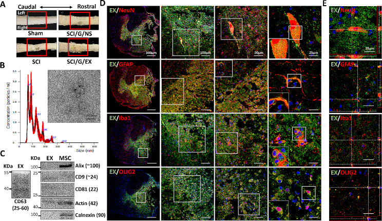
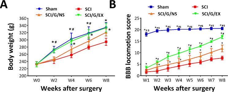
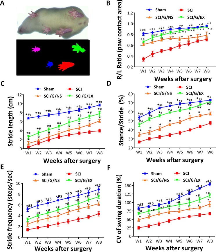
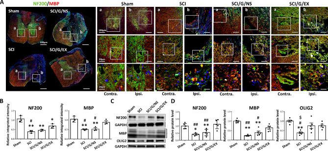
Methods: SCI model was established through hemisection surgery in T9 spinal cord of female Sprague-Dawley rats. Exosome-loaded Gelfoam was implanted into the lesion site. An in vivo uptake assay using labeled exosomes was conducted on day 3 post-implantation. Locomotor functions and gait analyses were assessed using Basso-Beattie-Bresnahan (BBB) locomotor rating scale and DigiGait Imaging System from weeks 1 to 8. Nociceptive responses were evaluated through von Frey filament and noxious radiant heat tests. The therapeutic effects and potential mechanisms were analyzed using Western blotting and immunofluorescence staining at week 8 post-SCI.

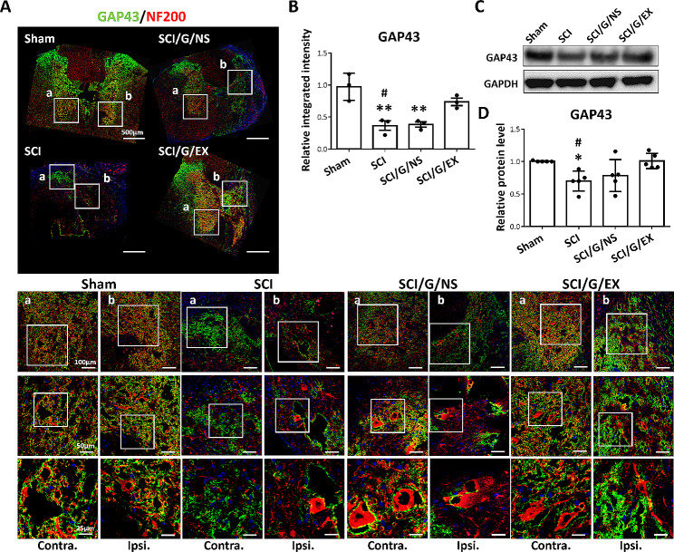
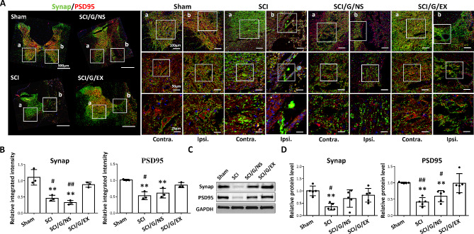
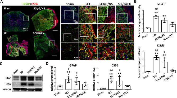
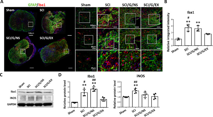
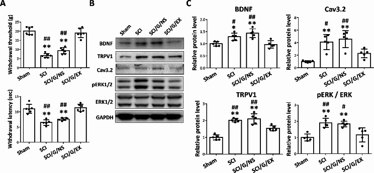
Results: For the in vivo exosome uptake assay, we observed the uptake of labeled exosomes by NeuN+, Iba1+, GFAP+, and OLIG2+ cells around the injured area. Exosome treatment consistently increased the BBB score from 1 to 8 weeks compared with the Gelfoam-saline and SCI control groups. Additionally, exosome treatment significantly improved gait abnormalities including right-to-left hind paw contact area ratio, stance/stride, stride length, stride frequency, and swing duration, validating motor function recovery. Immunostaining and Western blotting revealed high expression of NF200, MBP, GAP43, synaptophysin, and PSD95 in exosome treatment group, indicating the promotion of nerve regeneration, remyelination, and synapse formation. Interestingly, exosome treatment reduced SCI-induced upregulation of GFAP and CSPG. Furthermore, levels of Bax, p75NTR, Iba1, and iNOS were reduced around the injured area, suggesting anti-inflammatory and anti-apoptotic effects. Moreover, exosome treatment alleviated SCI-induced pain behaviors and reduced pain-associated proteins (BDNF, TRPV1, and Cav3.2). Exosomal miRNA analysis revealed several promising therapeutic miRNAs. The cell culture study also confirmed the neurotrophic effect of HucMSCs-EX.

Conclusion: Implantation of HucMSCs-EX-encapsulated Gelfoam improves SCI-induced motor dysfunction and neuropathic pain, possibly through its capabilities in nerve regeneration, remyelination, anti-inflammation, and anti-apoptosis. Overall, exosomes could serve as a promising therapeutic alternative for SCI treatment.

Keywords: And neuropathic pain; Exosomes; Glial scar; Locomotory function; Nerve regeneration; Spinal cord injury; Synapse formation.


Conflict of interest statement

The authors declare that they have no competing interests.

Figures

Fig. 1
Fig. 1
Treatment of spinal cord injury, characterization of HucMSC-EX, and in vivo exosome uptake by neurons and glial cells around the lesion site. (A) Overview of the lesion site in the spinal cord at week 8 post-surgery. The red box reflects the lesion area. (B). Transmission electron microscope image of HucMSC-EX and analysis of exosome size distribution by NanoSight. (C) Western blot analysis of exosome markers. (D, E) Confocal microscopy images and orthogonal views (Z-stack projection) of internalized Exo-fluorescent green-labeled HucMSC-EX by neuronal nuclei (NeuN+), glial fibrillary acidic protein (GFAP+), ionized calcium-binding adaptor molecule 1 (Iba1+), and oligodendrocyte transcription factor 2 (OLIG2+) cells in the lesion site on day 3 post-surgery. The yellow spots indicate exosome uptake. The white box shows the magnification of the specific area. Scale bar: 500, 100, 50, and 25 μm as indicated. HucMSC-EX: human umbilical cord mesenchymal stem cell-derived exosome; SCI: spinal cord injury; G: Gelfoam; NS: normal saline; EX: exosome
Fig. 2
Fig. 2
Temporal changes in the distribution of HucMSC-EX in the lesion site downstream area on days 1 and 3 post-SCI surgery. Orthogonal views (Z-stack projection) showed the uptake of Exo-fluorescent green-labeled exosome by neuronal nuclei (NeuN+) cells in the dorsal and ventral areas of the thoracic (T) and lumbar (L) regions. The yellow spots reflect the uptake of exosomes. Scale bar: 25 μm. HucMSC-EX: human umbilical cord mesenchymal stem cell-derived exosome; EX: exosome
Fig. 3
Fig. 3
Locomotor function recovery in rats from week 1 to 8 after spinal cord injury. Temporal changes in animal’s body weight (A) and Basso-Beattie-Bresnahan (BBB) locomotion score (B). Each data point represents the mean ± standard deviation. Group effect: significant, *p < 0.05 compared with the SCI group, #p < 0.05 compared with the SCI/G/NS group, and $p < 0.05 compared with the SCI/G/EX group using two-way analysis of variance (ANOVA) with post hoc Tukey’s multiple comparisons test (n = 6 in each group). SCI: spinal cord injury; G: Gelfoam; NS; normal saline; EX: exosome
Fig. 4
Fig. 4
DigiGait walking analysis of rats from week 1 to 8 after spinal cord injury. (A) Representative image of the paw contact area. Temporal changes in right-to-left (R/L) hind paw ratio of the maximal paw contact area (B), stride length (C), stance/stride (time) ratio (D), stride frequency (E), and coefficient of variation (CV) of the swing duration (F) of the right hind paw in four groups. Each data point represents the mean ± standard deviation. Group effect: significant, *p < 0.05 compared with the SCI group, #p < 0.05 compared with the SCI/G/NS group, and $p < 0.05 compared with the SCI/G/EX group using two-way ANOVA with post hoc Tukey’s multiple comparisons test (n = 6–8 in each group). SCI: spinal cord injury; G: Gelfoam; NS: normal saline; EX: exosome
Fig. 5
Fig. 5
Implantation of HucMSC-EX-loaded Gelfoam promoted nerve regeneration and remyelination around the lesion site at week 8 after spinal cord injury. (A, B) Representative immunofluorescence images and relative integrated intensities of neuronal filament 200 (NF200, green) and myelin basic protein (MBP, red) around the T9 lesion site in four groups. The white box indicates the magnification of the specific area. Scale bar: 500, 100, 50, and 25 μm as indicated. n = 3 in each group. (C, D) Representative Western blots and relative protein levels of NF200, MBP, and oligodendrocyte transcription factor 2 (OLIG2) around the T9 lesion site. GAPDH was used as an internal control. Data are presented as mean ± standard deviation, taking the Sham group as 100%. *p < 0.05, **p < 0.01 compared with the Sham group, #p < 0.05, ##p < 0.01 compared with the SCI/G/EX group, and $p < 0.05 compared with the SCI/G/NS group through one-way ANOVA with post hoc Tukey’s multiple comparisons test, n = 5 in each group. HucMSC-EX: human umbilical cord mesenchymal stem cell-derived exosome; SCI: spinal cord injury; G: Gelfoam; NS: normal saline; Ipsi: ipsilateral; Contra: contralateral
Fig. 6
Fig. 6
HucMSC-EX treatment decreased the spinal cord injury-induced growth-associated protein 43 (GAP43) loss around the lesion site at week 8 after surgery. (A, B) Representative immunofluorescence images of GAP43+ (green) and neuronal filament 200 (NF200, red) around the T9 lesion site in four groups. The white box reflects the magnification of a specific area. Scale bar: 500, 100, 50, and 25 μm as indicated. n = 3 in each group. (C, D) Representative Western blots and relative protein levels of GAP43 of the spinal cord around the T9 lesion site. GAPDH was used as an internal control. Data are presented as mean ± standard deviation, taking the Sham group as 100%. *p < 0.05, **p < 0.01 compared with the Sham group, and #p < 0.05 compared with the SCI/G/EX group by one-way ANOVA with post hoc Tukey’s multiple comparisons test, n = 5 in each group. HucMSC-EX: human umbilical cord mesenchymal stem cell-derived exosome; SCI: spinal cord injury; G: Gelfoam; NS; normal saline; Ipsi: ipsilateral; Contra: contralateral
Fig. 7
Fig. 7
HucMSC-EX treatment attenuated the spinal cord injury-induced synapse loss at week 8 after surgery. (A, B) Representative Western blots and relative protein levels of synaptophysin (Synap, pre-synaptic marker, green) and postsynaptic density 95 (PSD95, post synaptic marker, red) around the T9 lesion site in four groups. The white box indicates the magnification of the specific area. Scale bar: 500, 100, 50, and 25 μm as indicated. n = 3 in each group. (C, D) Representative Western blots and relative protein levels of Synap and PSD95 around the T9 lesion site. GAPDH was used as the internal control. Data are presented as mean ± standard deviation, taking the Sham group as 100%. **p < 0.01 compared with the Sham group, #p < 0.05, and ##p < 0.01 compared with the SCI/G/EX group through one-way ANOVA with post hoc Tukey’s multiple comparisons test, n = 5 in each group. HucMSC-EX: human umbilical cord mesenchymal stem cell-derived exosome. SCI: spinal cord injury; G: Gelfoam; NS; normal saline; Ipsi: ipsilateral; Contra: contralateral
Fig. 8
Fig. 8
HucMSC-EX treatment decreased the spinal cord injury-induced astrocyte activation and CS56 production at week 8 after surgery. (A, B) Representative immunofluorescence images and relative integrated intensities of glial fibrillary acidic protein (GFAP, green) and chondroitin sulfate 56 (CS56, red) around the T9 lesion site in four groups. The white box indicates the magnification of a specific area. Scale bar: 500, 100, 50, and 25 μm as indicated. n = 3 in each group. (C, D) Representative Western blots and relative protein levels of GFAP and CS56 around the T9 lesion site. GAPDH was used as the internal control. Data are presented as mean ± standard deviation, taking the Sham group as 100%. *p < 0.05, **p < 0.01 compared with the Sham group, #p < 0.05, and ##p < 0.01 compared with the SCI/G/EX group by one-way ANOVA with post hoc Tukey’s multiple comparisons test, n = 5 in each group. HucMSC-EX: human umbilical cord mesenchymal stem cell-derived exosome. SCI: spinal cord injury; G: Gelfoam; NS; normal saline
Fig. 9
Fig. 9
HucMSC-EX-loaded Gelfoam implantation reduced spinal cord injury-induced neuroinflammation at week 8 after surgery. (A) Representative immunofluorescence images of glial fibrillary acidic protein (GFAP, green) and ionized calcium-binding adaptor molecule 1 (Iba1, red) around the T9 lesion site in four groups. The white box indicates the magnification of the specific area. Scale bar: 500, 100, 50, and 25 μm as indicated. (B) Relative integrated intensity of Iba1+ staining in four groups. n = 3 in each group. (C, D) Representative Western blots and relative protein levels of Iba1 and inducible nitric oxide synthase (iNOS) around the T9 lesion site. GAPDH was used as an internal control. Data are presented as mean ± standard deviation, taking the Sham group as 100%. **p < 0.01 compared with the Sham group, #p < 0.05, and ##p < 0.01 compared with the SCI/G/EX group through one-way ANOVA with post hoc Tukey’s multiple comparisons test, n = 5 in each group. HucMSC-EX: human umbilical cord mesenchymal stem cell-derived exosome; SCI: spinal cord injury; G: Gelfoam; NS: normal saline
Fig. 10
Fig. 10
Implantation of HucMSC-EX-loaded Gelfoam alleviated spinal cord injury-induced neuropathic pain and pain-related protein upregulation at week 8 after surgery. (A) The withdrawal threshold and latency of the right hind paw in four groups. n = 6 in each group. (B, C) Representative Western blots and relative protein levels of brain-derived neurotrophic factor (BDNF), transient receptor potential vanilloid type-1 (TRPV1), Cav3.2 T-type calcium channel (Cav3.2), and phosphor-ERK1/2 (pERK1/2) around the T9 lesion site. GAPDH was used as an internal control. Data are presented as mean ± standard deviation, taking the Sham group as 100%. n = 5 in each group. *p < 0.05, **p < 0.01 compared with the Sham group, #p < 0.05, and ##p < 0.01 compared with the SCI/G/EX group through one-way ANOVA with post hoc Tukey’s multiple comparisons test. HucMSC-EX: human umbilical cord mesenchymal stem cell-derived exosome; SCI: spinal cord injury; G: Gelfoam; NS: normal saline

References

    1. La Spada A, Ranum LPW. Molecular genetic advances in neurological disease: special review issue. Hum Mol Genet. 2010;19(R1):R1–3. doi: 10.1093/hmg/ddq193. - DOI - PMC - PubMed
    1. Ahuja CS, Wilson JR, Nori S, Kotter MRN, Druschel C, Curt A, Fehlings MG. Traumatic spinal cord injury. Nat Reviews Disease Primers. 2017;3(1):17018. doi: 10.1038/nrdp.2017.18. - DOI - PubMed
    1. Ramer LM, Ramer MS, Bradbury EJ. Restoring function after spinal cord injury: towards clinical translation of experimental strategies. Lancet Neurol. 2014;13(12):1241–56. doi: 10.1016/S1474-4422(14)70144-9. - DOI - PubMed
    1. Thuret S, Moon LDF, Gage FH. Therapeutic interventions after spinal cord injury. Nat Rev Neurosci. 2006;7(8):628–43. doi: 10.1038/nrn1955. - DOI - PubMed
    1. Assinck P, Duncan GJ, Hilton BJ, Plemel JR, Tetzlaff W. Cell transplantation therapy for spinal cord injury. Nat Neurosci. 2017;20(5):637–47. doi: 10.1038/nn.4541. - DOI - PubMed

Publication types