. 2024 May 19;16(1):105.
doi: 10.1186/s13098-024-01276-1.

Adipose stem cells-derived small extracellular vesicles transport Thrombospondin 1 cargo to promote insulin resistance in gestational diabetes mellitus

Affiliations

Adipose stem cells-derived small extracellular vesicles transport Thrombospondin 1 cargo to promote insulin resistance in gestational diabetes mellitus

Huaping Li et al. Diabetol Metab Syndr. .

Abstract

Background: Gestational diabetes mellitus (GDM) is a highly prevalent disease and poses a significant risk to the health of pregnant women. Abdominal adipose tissue (AT) contributes to insulin resistance (IR) associated with GDM. However, the underlying mechanisms remain unclear.

Methods: In this study, we developed a mouse model of GDM by subjecting mice to a high-fat diet. We collected adipose-derived stem cells (ADSCs) from the abdominal and inguinal regions and examined their role in inducing IR in normal tissues through the secretion of small extracellular vesicles (sEVs). The sEVs derived from ADSCs isolated from GDM mice (ADSC/GDM) were found to inhibit cell viability and insulin sensitivity in AML12, a normal mouse liver cell line.

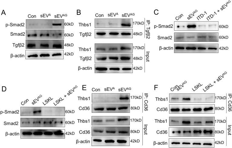
Results: Through proteomic analysis, we identified high levels of the thrombospondin 1 (Thbs1) protein in the sEVs derived from ADSC/GDM. Subsequent overexpression of Thbs1 protein in AML12 cells demonstrated similar IR as observed with ADSC/GDM-derived sEVs. Mechanistically, the Thbs1 protein within the sEVs interacted with CD36 and transforming growth factor (Tgf) β receptors in AML12 cells, leading to the activation of Tgfβ/Smad2 signaling. Furthermore, the administration of LSKL, an antagonistic peptide targeting Thbs1, suppressed Thbs1 expression in ADSC/GDM-derived sEVs, thereby restoring insulin sensitivity in AML12 cells and GDM mice in vivo.

Conclusions: These findings shed light on the intercellular transmission mechanism through which ADSCs influence hepatic insulin sensitivity and underscore the therapeutic potential of targeting the Thbs1 protein within sEVs.

Keywords: Adipose-derived stem cells; Gestational diabetes mellitus; Insulin resistance; Small extracellular vesicles; Thrombospondin 1.


Conflict of interest statement

The authors have no competing interests to declare.

Figures

Fig. 1
Fig. 1
Isolation, culture and purification of GDM mice-derived ADSCs and their sEVs. A Plasma glucose values were quantified in eight normal gestation and GDM mice. B The expression of insulin, leptin, adiponectin and hs-CRP in the plasma of mice was detected by ELISA assays. C ADSCs was obtained by collagenase digestion of abdominal and inguinal adipose tissue from gestational mice and further in vitro culture. D Microscopic images of ADSCs isolated and cultured from normal gestational and GDM mice. Scale bar, 25 μm. E The expression of Cd44 marker (red) was identified by immunofluorescence. DAPI staining (blue) indicated the nucleus. Scale bar, 25 μm. F The sEVA and sEVAG were identified by transmission electron microscope. Scale bar, 100 nm. G The sEVA and sEVAG solution was diluted 500 times and particle size distribution and concentration were determined by nanoparticle tracking analysis. H The sEV markers and stem cell markers were detected by western blotting in sEVA and sEV.AG samples. Data were presented with mean ± standard deviation (SD), *P < 0.05, **P < 0.01, ***P < 0.001
Fig. 2
Fig. 2
ADSC/GDM-derived inhibits hepatocyte proliferation and insulin sensitivity in normal mice. A, B The sEVA and sEVAG with different concentrations were co-cultured with AML12 cells for 48 or 72 h. Cell viability was obtained by CCK8 assays. C, D The sEVA and sEVAG with 108 particles / mL were co-cultured with AML12 cells for 48 or 72 h. Cell apoptosis was detected by flow cytometry. E, F The sEVA and sEVAG with 108 particle number / mL were co-cultured with AML12 cells for 48 or 72 h, then cells were treated with 1 μg/ml insulin for 1 h. Glucose uptake was measured to reflect insulin sensitivity of AML12 cells in each treatment. G The sEVA and sEVAG with 10.8 particle number /mL were co-cultured with AML12 cells for 48 h, and mRNA expression of endoplasmic reticulum (ER)-related genes was detected by RT-PCR. H The expression of ER-related proteins was detected by western blotting. Data were presented with mean ± standard deviation (SD); ns, no significance; *P < 0.05, **P < 0.01, ***P < 0.001
Fig. 3
Fig. 3
Proteomic identification of high Thbs1 expression in ADSC/GDM-derived sEV. A Venn diagram of the number of proteins detected by unlabeled protein profiles for and sEVA (AD in graph) and sEVAG (ADGDM in graph). B The number of proteins with high and low expression in sEVAG. C Volcanic map of differentially expressed protein in sEVAG. D GO signaling pathway analysis of sEVA and sEVAG proteins. E KEGG signal analysis of sEVA and sEVAG proteins. F GSEA analysis revealed enrichment of the ECm-receptor interaction pathway. G Network analysis of interactions between differential ECM regulatory proteins and other differential proteins. H. Heat maps of 10 proteins significantly overexpressed in sEVAG. I, J Mass spectrometry quantification and western blotting of Thbs1 in sEVA and sEV.AG. ***P < 0.001
Fig. 4
Fig. 4
Overexpression of Thbs1 inhibits insulin sensitivity in AML12 cells. A Flag-eGFP-Thbs1 plasmid and control vector were transfected into AML12 cells. The expression of eGFP-thbs1 fusion protein in AML12 was identified by western blotting and fluorescence microscopy. B Plasmid transfected AML12 cells were placed in 96-well plates, and cell viability was detected by CCK8 assay. C. Apoptosis assays of AML12 cells after plasmid transfection were performed. D Plasmid transfected AML12 cells were treated with 1 μg/ml insulin for 1 h, and the culture medium was collected for glucose uptake measurement. EG AML12 cells were treated with 108/mL sEVAG, 10 μM LSKL and 108/mL sEVAG + 10 μM LSKL for 48 h. Cell viability, apoptosis, and insulin-induced glucose uptake were then measured. H The expression of endoplasmic reticulum stress (ERS)-related mRNA in AML12 cells after plasmid transfection was detected by RT-PCR. I The expression of ERS-related mRNA in 108/mL sEVAG and/or 10 μM LSKL treated AML12 cells was detected by RT-PCR. J The expression of ERS-related proteins in AML12 cells was detected by western blotting. Statistical data were presented as mean ± standard deviation (SD); ns, no significance; *P < 0.05, **P < 0.01, ***P < 0.001
Fig. 5
Fig. 5
ADSC/GDM-derived sEV carrying Thbs1 induces Tgfβ and CD36 activation. A sEVAG was co-cultured with AML12 for 48 h, and the expression of Smad2, phosphorylated Smad2 and Tgfβ was detected by western blotting. B sEVAG was co-cultured with AML12 for 48 h, and the cell lysate was subsequently subjected to co-immunoprecipitation (co-IP) with anti-Tgfβ antibodies. Thbs1 levels in co-IP products and input samples were detected. C, D AML12 cells were treated with 108 /mL sEVAG, 2 μM ITD-1, 10 μM LSKL or a combination for 48 h. The expression of Smad2 and phosphorylated Smad2 was detected by western blotting. E Cell lysate was co-immunoprecipitated with anti-CD36 antibody. Thbs1 levels in co-IP products and input samples were detected. F AML12 cells were treated with 108/mL sEVAG, 10 μM LSKL or a combination for 48 h, followed by co-IP with anti-CD36 antibody
Fig. 6
Fig. 6
ADSC/GDM sEV restore insulin sensitivity to liver cells after Thbs1 pharmacological interference. A ADSC-GDM cells were treated with different concentrations of LSKL (0, 5 μM, 10 μM) for 72 h. sEVAG, sEVAG−L5 and sEVAG−L10 were collected and identified by TEM and NTA. B The expression of Thbs1 in sEVAG, sEVAG−L5 and sEVAG−L10 was identified by western blotting. C sEVAG, sEVAG−L5 and sEVAG−L10 were co-cultured with AML12 for 24–96 h, and cell viability was detected by CCK8 assays. D sEVAG, sEVAG−L5 and sEVAG−L10 were co-cultured with AML12 for 48 h, and cell apoptosis was detected by flow cytometry. E Glucose uptake of AML12 cells after insulin treatment was measured. F. sEVAG, sEVAG−L5 and sEVAG−L10 were co-cultured with AML12 for 48 h, and the expression of phosphorylated Smad2 and total Smad2 protein was detected by western blotting. G, H RT-PCR and western blotting were used to detect the expression of ER stress related genes in AML12 cells. Statistical data were presented as mean ± standard deviation (SD); ns, no significance; *P < 0.05, **P < 0.01, ***P < 0.001
Fig. 7
Fig. 7
Targeted Thbs1 pharmacological therapy improves insulin resistance phenotype in GDM mice. A Plasma was extracted from normal, GDM, and LSKL-treated GDM mice, and blood glucose and insulin, leptin, adiponectin and hs-CRP levels were measured by glucose meter and ELISA kit. B HE staining and immunohistochemical detection of adipose tissues from normal, GDM, and LSKL treated mice. C HE staining and immunohistochemical detection of liver tissues from these mice. Statistical data were presented as mean ± standard deviation (SD); ns, no significance; *P < 0.05, **P < 0.01, ***P < 0.001

Similar articles

Cited by

References

    1. Pinto Y, Frishman S, Turjeman S, Eshel A, Nuriel-Ohayon M, Shrossel O, Ziv O, Walters W, Parsonnet J, Ley C, et al. Gestational diabetes is driven by microbiota-induced inflammation months before diagnosis. Gut. 2023;72(5):918–928. doi: 10.1136/gutjnl-2022-328406. - DOI - PMC - PubMed
    1. Ferrara A. Increasing prevalence of gestational diabetes mellitus: a public health perspective. Diabetes Care. 2007;30(Suppl 2):S141–146. doi: 10.2337/dc07-s206. - DOI - PubMed
    1. McIntyre HD, Catalano P, Zhang C, Desoye G, Mathiesen ER, Damm P. Gestational diabetes mellitus. Nat Rev Dis Primers. 2019;5(1):47. doi: 10.1038/s41572-019-0098-8. - DOI - PubMed
    1. Gao C, Sun X, Lu L, Liu F, Yuan J. Prevalence of gestational diabetes mellitus in mainland China: a systematic review and meta-analysis. J Diabetes Investig. 2019;10(1):154–162. doi: 10.1111/jdi.12854. - DOI - PMC - PubMed
    1. Geach T. Diabetes: a metabolomic signature to predict the transition from GDM to T2DM. Nat Rev Endocrinol. 2016;12(9):498. - PubMed

LinkOut - more resources