

A Pilot Study on Cerebral Blood Flow and Mini-Mental State Examination to Predict Amyloid Deposition in Preclinical Alzheimer's Disease
- PMID: 38764533
- PMCID: PMC11082584
- DOI: 10.5152/pcp.2023.22524
A Pilot Study on Cerebral Blood Flow and Mini-Mental State Examination to Predict Amyloid Deposition in Preclinical Alzheimer's Disease
Abstract
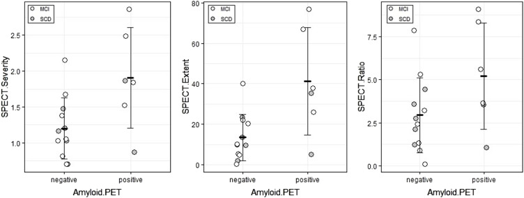
Background: Earlier differential diagnosis of dementia remains a major challenge. Although amyloid deposition by positron emission tomography is an emerging standard for the diagnosis of Alzheimer's disease, it is too expensive for routine use in clinical settings. We conducted a pilot study on the potential usefulness of single-photon emission computed tomography and the Mini-Mental State Examination to predict amyloid positron emission tomography positivity in preclinical Alzheimer's disease.
Methods: Eighteen subjects, including 11 with mild cognitive impairment and 7 with subjective cognitive decline, underwent 18F-florbetapir positron emission tomography, 99mTc-ethylcysteinate dimer cerebral perfusion single-photon emission computed tomography, and the Mini-Mental State Examination. For the assessment of amyloid deposition, visual judgment as a qualitative method and a semiautomatic software analysis as a quantitative method were used.
Results: Six subjects were judged as amyloid positive, including 4 mild cognitive impairment and 2 subjective cognitive decline subjects. Compared to the amyloid positron emission tomography-negative group, this group showed a statistically significant difference in the Mini-Mental State Examination recall score [2 (1 : 3) vs. 3 (2 : 3), P = .041] and single-photon emission computed tomography findings from the amyloid-negative group. In the mild cognitive impairment subgroup, correlations were found between amyloid deposition and single-photon emission computed tomography indicators, while in the subjective cognitive decline subgroup, only the Mini-Mental State Examination recall score correlated with amyloid deposition.
Conclusion: The Mini-Mental State Examination recall score and single-photon emission computed tomography indicators may be worthwhile for further evaluation as predictors of amyloid deposition in the preclinical stage.
2023 authors.
Figures
References
-
- Sperling RA, Aisen PS, Beckett LA, et al. Toward defining the preclinical stages of Alzheimer's disease: recommendations from the National Institute on Aging-Alzheimer's Association workgroups on diagnostic guidelines for Alzheimer's disease. Alzheimers Dement. 2011;7(3):280 292. (10.1016/j.jalz.2011.03.003) - DOI - PMC - PubMed
LinkOut - more resources
Full Text Sources