[Preprint]. 2024 May 6:arXiv:2405.03120v1.

A Curated Rotamer Library for Common Post-Translational Modifications of Proteins

Affiliations

A Curated Rotamer Library for Common Post-Translational Modifications of Proteins

Oufan Zhang et al. ArXiv. .

Update in

Abstract

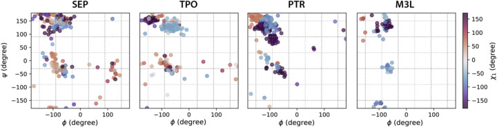
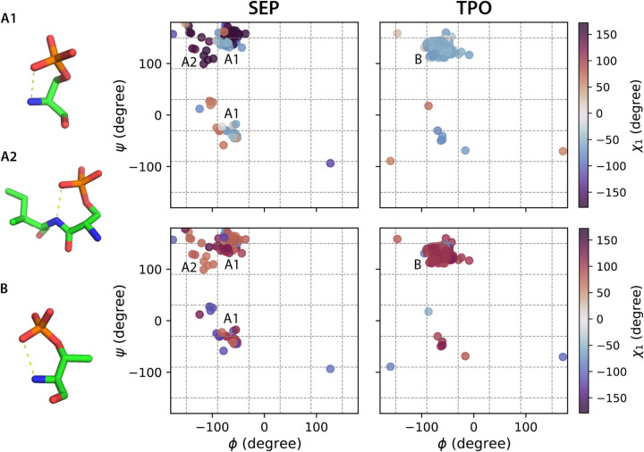
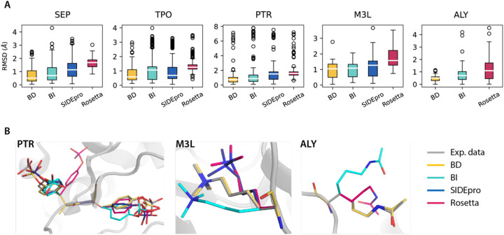
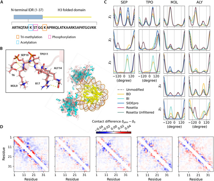
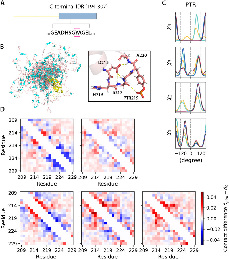
Sidechain rotamer libraries of the common amino acids of a protein are useful for folded protein structure determination and for generating ensembles of intrinsically disordered proteins (IDPs). However much of protein function is modulated beyond the translated sequence through thFiguree introduction of post-translational modifications (PTMs). In this work we have provided a curated set of side chain rotamers for the most common PTMs derived from the RCSB PDB database, including phosphorylated, methylated, and acetylated sidechains. Our rotamer libraries improve upon existing methods such as SIDEpro and Rosetta in predicting the experimental structures for PTMs in folded proteins. In addition, we showcase our PTM libraries in full use by generating ensembles with the Monte Carlo Side Chain Entropy (MCSCE) for folded proteins, and combining MCSCE with the Local Disordered Region Sampling algorithms within IDPConformerGenerator for proteins with intrinsically disordered regions.


Figures

Figure 1.
Figure 1.. Ramachandran plots color coded by χ1 angle ranges for PTM-modified amino acids using the backbone-dependent library.
Backbone bins in 60° separation are shown in gray dash lines.
Figure 2.
Figure 2.. Ramachandran plots color coded by χ1 and χ2 angle ranges for SEP and TPO with hydrogen bond formation using the backbone-dependent library.
Backbone bins in 60° separation are shown in gray dash lines. Hydrogen bonds are defined by within a donor-acceptor distance cutoff of 3.5 Å. A1, A2 and B show representative configurations for SEP (A1, A2) and TPO (B) associated with the annotated backbone regions.
Figure 3.
Figure 3.. RMSD distributions of repacked PTM-modified residues using different rotamer libraries to experimental structures.
SIDEpro does not support ALY packing. A). Boxplots for the RMSD distributions. Medians are highlighted in white and each box extends from the first quartile (Q1) to the third quartile (Q3). Outliers in circle are defined as points outside of 1.5 times the interquartile range below Q1 or above Q3. B). Repacked PTM-containing structures using different rotamer libraries compared to the experimental PDB structures. Examples are taken from PDB ID 3CLY (PTR), 4EZH (M3L) and 4QUT (ALY).
Figure 4.
Figure 4.. Histone H3 conformers generated with different PTMs libraries.
A). Modifications on the histone H3 N-terminus on chains C/G of the nucleosome. B). Ensembles of 30 all-atom H3-modified nucleosome conformers (folded domains and DNA are taken from PDB ID 8SIY). The N-terminal IDRs on chain C and G are shown in salmon (loop) and cyan (helices), and folded domains (yellow). PTM-modified residues are highlighted with stick representations. C). Comparison of torsion angle probability distributions for PTM-modified residues in the H3 conformers with different libraries. D). Fractional inter-residue contacts (Cα-Cα distances within 8 Å) of PTMs containing ensembles (δptm) subtracted by the ensemble without modifications (δ0) for the IDR regions. The maps were calculated with 500 randomly sampled conformers (based on convergence of Rgand averaged over 10 trials (blue-less, red-more). From left to right: BD-rotamer, BI-rotamer, SIDEpro, Rosetta and Rosetta without clash filtering, with contacts averaged from chain C and G.
Figure 5.
Figure 5.. UDF1 conformers generated with different PTM libraries
. A). Modifications on the C-terminal IDR of UDF1 at Y219. B). Cartoon representations of 30 all-atom UDF1 conformers (structure of the folded domain taken from PDB ID 2YUJ model). The C-terminal IDR is shown in salmon red (loop) and cyan (helices), and the folded domains in yellow. C). Comparison of torsion angle probability distributions for PTM in the UDF1 conformers with different libraries. D). Fractional inter-residue contacts (Cα-Cα distances within 8 Å) of PTMs containing ensembles (δptm) subtracted by the ensemble without modifications (δ0) for the IDR region around the PTM site. The maps were calculated with ensembles of 500 randomly sampled conformers and averaged over 10 trials. Top: BD-rotamer, BI-rotamer; Bottom (L to R): SIDEpro, Rosetta and Rosetta without clash filtering.

Similar articles

References

    1. Ramazi S. & Zahiri J. Post-translational modifications in proteins: resources, tools and prediction methods. Database 2021, baab012, doi:10.1093/database/baab012 (2021). - DOI - PMC - PubMed
    1. Huang K.-Y. et al. dbPTM in 2019: exploring disease association and cross-talk of post-translational modifications. Nucleic Acids Research 47, D298–D308, doi:10.1093/nar/gky1074 (2019). - DOI - PMC - PubMed
    1. Jumper J. et al. Highly accurate protein structure prediction with AlphaFold. Nature 596, 583–589, doi:10.1038/s41586-021-03819-2 (2021). - DOI - PMC - PubMed
    1. Baek M. et al. Accurate prediction of protein structures and interactions using a three-track neural network. Science 373, 871–876, doi:10.1126/science.abj8754 (2021). - DOI - PMC - PubMed
    1. Lane T. J. Protein structure prediction has reached the single-structure frontier. Nature Methods 20, 170–173, doi:10.1038/s41592-022-01760-4 (2023). - DOI - PMC - PubMed

Publication types

LinkOut - more resources