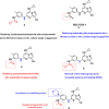
Review
. 2024 May 3;9(19):20702-20719.
doi: 10.1021/acsomega.3c10478. eCollection 2024 May 14.

Recent Advances in the Discovery of CK2 Inhibitors

Affiliations
Review

Recent Advances in the Discovery of CK2 Inhibitors

Mustafa A Al-Qadhi et al. ACS Omega. .

Abstract

CK2 is a vital enzyme that phosphorylates a large number of substrates and thereby controls many processes in the body. Its upregulation was reported in many cancer types. Inhibitors of CK2 might have anticancer activity, and two compounds are currently under clinical trials. However, both compounds are ATP-competitive inhibitors that may have off-target side effects. The development of allosteric and dual inhibitors can overcome this drawback. These inhibitors showed higher selectivity and specificity for the CK2 enzyme compared to the ATP-competitive inhibitors. The present review summarizes the efforts exerted in the last five years in the design of CK2 inhibitors.


Conflict of interest statement

The authors declare no competing financial interest.

Figures

Figure 1
Figure 1
Structures of reported CK2 inhibitors.
Figure 2
Figure 2
X-ray cocrystal structure of CX-4945 with CK2 (PDB ID 3PE1; 1.60 Å). The figure was generated using PyMOL 2.4.0 with the CK2α protein shown as a cartoon and CX-4945 shown as sticks colored by the atom type: C, yellow; O, red; and N, blue. The H-bonds are illustrated in yellow dashed lines. The bound water molecules are shown as red spheres.
Figure 3
Figure 3
Structure modification of CX-4945 to afford compound 1.
Figure 4
Figure 4
X-ray cocrystal structure of compound 1 with CK2 (PDB ID 8BGC; 2.80 Å). The figure was generated using PyMOL 2.4.0 with the CK2α protein shown as a cartoon and compound 1 shown as sticks colored by the atom type: C, yellow; O, red; and N, blue. The H-bonds are illustrated in yellow dashed lines.
Figure 5
Figure 5
Design of compounds 26.
Figure 6
Figure 6
X-ray cocrystal structure of compound 3 with CK2α (PDB ID 6Z83; 2.17 Å). The figure was generated using PyMOL 2.4.0 with the CK2α protein shown as a cartoon and compound 3 shown as sticks colored by the atom type: C, yellow; O, red; and N, blue. The H-bonds are illustrated in yellow dashed lines. The bound water molecules are shown as red spheres.
Figure 7
Figure 7
Structures of compounds 7 and 8.
Figure 8
Figure 8
Key interactions between compound 7 and CK2 residue inside active site.
Figure 9
Figure 9
Design of compounds 9 and 10.
Figure 10
Figure 10
Design of compounds 1116.
Figure 11
Figure 11
Key interactions of tetrabromo-1H-benzimidazole (TBBi) and tetrabromo-1H-benzotriazole (TBBt) within CK2 pocket.
Figure 12
Figure 12
Structures of compounds 17 and 18.
Figure 13
Figure 13
Design of compound 21.
Figure 14
Figure 14
Main interaction between compound 21 and CK2 pocket.
Figure 15
Figure 15
Structures of compounds 22 and 23.
Figure 16
Figure 16
Main interaction between compound 23 and CK2 pocket.
Figure 17
Figure 17
Structures of emodin, endocrocin, and compound 24.
Figure 18
Figure 18
Design of compounds 2629.
Figure 19
Figure 19
X-ray cocrystal structure of compound 29 with CK2α (PDB ID 6YPG; 1.51 Å). The figure was generated using PyMOL 2.4.0 with the CK2α protein shown as a cartoon and compound 29 shown as sticks colored by the atom type: C, yellow; O, red; and N, blue. The H-bonds are illustrated in yellow dashed lines, π–π interaction in red color. The bound water molecules are shown as red spheres.
Figure 20
Figure 20
SAR study of compound 30.
Figure 21
Figure 21
X-ray cocrystal structure of compound 30 with CK2α (PDB ID 6A1C; 1.68 Å). The figure was generated using PyMOL 2.4.0 with the CK2α protein shown as a cartoon and compound 30 shown as sticks colored by the atom type: C, yellow; O, red; N, blue; and Br, magenta. The H-bonds are illustrated in yellow dashed lines.
Figure 22
Figure 22
Structures of CK2 allosteric inhibitors.
Figure 23
Figure 23
Interactions of compound 31 with residues in CK2α active site.
Figure 24
Figure 24
Design of compound 32.
Figure 25
Figure 25
Main interactions of compound 32 in CK2 pocket.
Figure 26
Figure 26
Structure of compounds 33 and 34.
Figure 27
Figure 27
Design of compounds 36 and 37.
Figure 28
Figure 28
X-ray cocrystal structure of compound 37 with CK2α (PDB ID 6FVF; 1.47 Å). The figure was generated using PyMOL 2.4.0 with the CK2α protein shown as a cartoon and compound 37 shown as sticks colored by the atom type: C, yellow; O, red; and N, blue. The H-bonds are illustrated in yellow dashed lines. The bound water molecules are shown as red spheres.
Figure 29
Figure 29
Design of dual HDAC/CK2 inhibitor 38.
Figure 30
Figure 30
Design of dual HDAC/CK2 inhibitor 39.
Figure 31
Figure 31
Design of cisplatin and CK2 inhibitor conjugate.
Figure 32
Figure 32
Design of cisplatin and CK2 inhibitor conjugate 40.
Figure 33
Figure 33
Design of dual Clk2/CK2 inhibitor 41.
Figure 34
Figure 34
(a) The main interactions of compound 41 in CK2 pocket. (b) The main interactions of compound 41 in Clk2 pocket.
Figure 35
Figure 35
Design of dual BRD4/CK2 inhibitor 43.
Figure 36
Figure 36
Design of dual PIM1/CK2α inhibitors 44 and 45.
Figure 37
Figure 37
Design of dual PIM1/CK2α inhibitors 46 and 47.
Figure 38
Figure 38
Structures of multikinase inhibitor 48 and the dual CDK2/CK2 inhibitor 49.

References

    1. Meggio F.; Pinna L. A. One-thousand-and-one Substrates of Protein Kinase CK2?. FASEB J. 2003, 17 (3), 349–368. 10.1096/fj.02-0473rev. - DOI - PubMed
    1. Franchin C.; Borgo C.; Zaramella S.; Cesaro L.; Arrigoni G.; Salvi M.; Pinna L. A. Exploring the CK2 Paradox: Restless, Dangerous, Dispensable. Pharmaceuticals 2017, 10 (1), 11.10.3390/ph10010011. - DOI - PMC - PubMed
    1. Niefind K.; Guerra B.; Pinna L. A.; Issinger O. G.; Schomburg D. Crystal Structure of the Catalytic Subunit of Protein Kinase CK2 from Zea Mays at 2.1 Å Resolution. EMBO J. 1998, 17 (9), 2451–2462. 10.1093/emboj/17.9.2451. - DOI - PMC - PubMed
    1. Poletto G.; Vilardell J.; Marin O.; Pagano M. A.; Cozza G.; Sarno S.; Itarte E.; Pinna L. A.; Meggio F.; Falques A. The Regulatory Subunit of Protein Kinase CK2 Contributes to the Recognition of the Substrate Consensus Sequence. A Study with an EIF2 -Derived Peptide. Biochemistry 2008, 47, 8317–8325. 10.1021/bi800216d. - DOI - PubMed
    1. Franchin C.; Borgo C.; Cesaro L.; Zaramella S.; Vilardell J.; Salvi M.; Arrigoni G.; Pinna L. A. Re-Evaluation of Protein Kinase CK2 Pleiotropy: New Insights Provided by a Phosphoproteomics Analysis of CK2 Knockout Cells. Cell. Mol. Life Sci. 2018, 75 (11), 2011–2026. 10.1007/s00018-017-2705-8. - DOI - PMC - PubMed

LinkOut - more resources