. 2024 May 4;10(10):e30738.
doi: 10.1016/j.heliyon.2024.e30738. eCollection 2024 May 30.

Kinetics of inactivation of bacteria responsible for infections in hospitals using UV-LED

Affiliations

Kinetics of inactivation of bacteria responsible for infections in hospitals using UV-LED

Beatriz Rito et al. Heliyon. .

Abstract

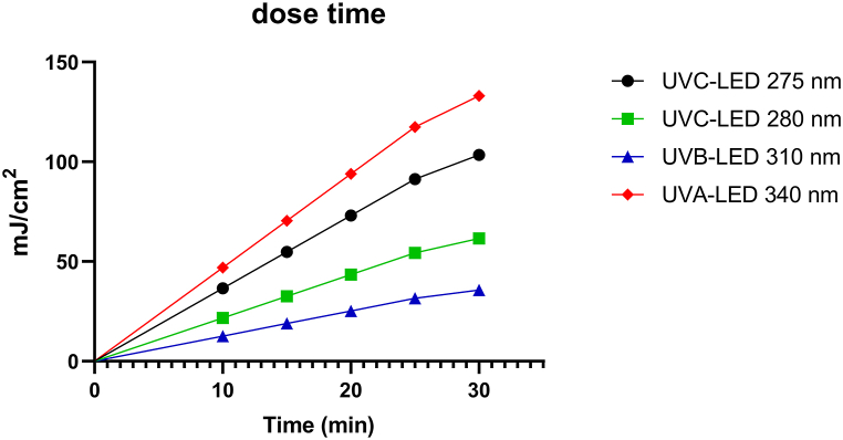
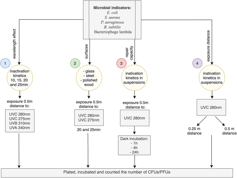
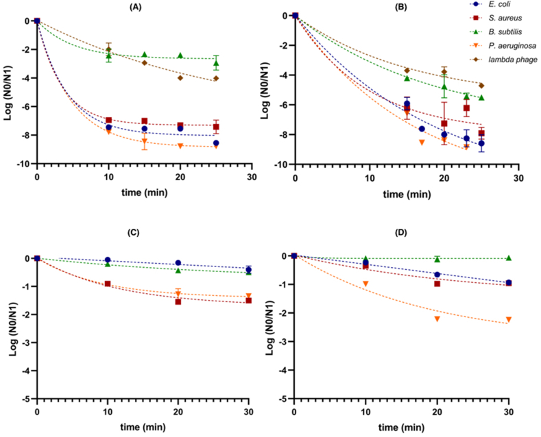
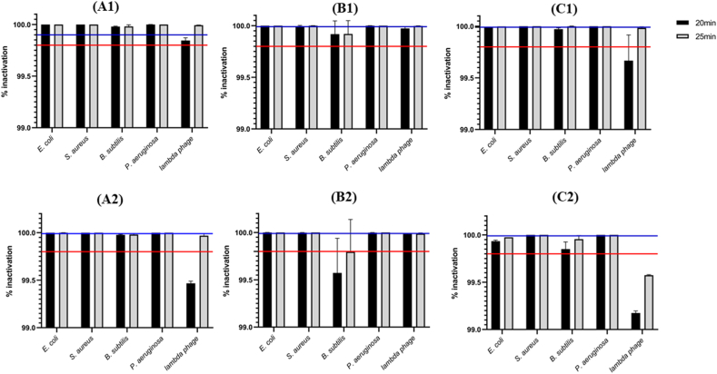
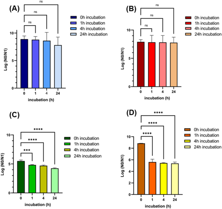
Controlling the microbial load in the environment is crucial to prevent the spread of organisms. The continuous spread of nosocomial infections in hospital facilities and the emergence of the coronavirus (COVID-19) highlighted the importance of disinfection processes in health safety. This work aimed to evaluate the effectiveness of LED-based disinfection lamps on bacteria from the ESKAPEE group and virus phage in vitro inactivation to be applied in hospital environments and health facilities disinfection. This study evaluated the effect of different UV wavelengths (275 nm, 280 nm (UVC), 310 nm (UVB) and 340 nm (UVA)) on the disinfection process of various microbial indicators including E. coli, S. aureus, P. aeruginosa, B. subtilis and Bacteriophage lambda DSM 4499. Exposure time (5 min-30 min), exposure distance (0.25 m and 0.5 m) and surface materials (glass, steel, and polished wood) were evaluated on the disinfection efficiency. Furthermore, the study determined the recovery capacity of each species after UV damage. UVC-LED lamps could inactivate 99.99 % of microbial indicators after 20 min exposures at a 0.5 m distance. The exposure time needed to completely inactivate E. coli, S. aureus, P. aeruginosa, B. subtilis and Bacteriophage lambda DSM 4499 can be decreased by reducing the exposure distance. UVB-LED and UVA-LED lamps were not able to promote a log reduction of 4 and were not effective on B. subtilis or bacteriophage lambda DSM 4499 inactivation. Thus, only UVC-LED lamps were tested on the decontamination of different surface materials, which was successful. P. aeruginosa showed the ability to recover from UV damage, but its inactivation rate remains 99.99 %, and spores from B. subtilis were not completely inactivated. Nevertheless, the inactivation rate of these indicators remained at 99.99 % with 24 h incubation after UVC irradiation. UVC-LED lamps emitting 280 nm were the most indicated to disinfect surfaces from microorganisms usually found in hospital environments.

Keywords: Disinfection; Health facilities; Nosocomial infections; Surfaces; UV-LED.


Conflict of interest statement

The authors declare the following financial interests/personal relationships which may be considered as potential competing interests:Paula V. Morais reports financial support was provided by Foundation for Science and Technology. If there are other authors, they declare that they have no known competing financial interests or personal relationships that could have appeared to influence the work reported in this paper.

Figures

Fig. 1
Fig. 1
Cumulative fluence of UVC-LED 275 nm (red), UVC-LED 280 nm (black), UVB-LED 310 nm (green) and UVA-LED 340 nm (blue) lamps depending on exposure time. (For interpretation of the references to colour in this figure legend, the reader is referred to the Web version of this article.)
Fig. 2
Fig. 2
Diagram of the experimental design performed with the tested UV-LED lamps.
Fig. 3
Fig. 3
Inactivation kinetics of the microbial indicators E. coli DSM 30083, S. aureus DSM 20231, B. subtilis DSM 10, P. aeruginosa DSM 1117 and Bacteriophage Lambda DSM 4499 by irradiation with (A) UVC-LED 275 nm, (B) UVC-LED 280 nm, (C) UVB-LED 310 nm and (D) UVA-LED 340 nm, with an exposure distance of 0.5 m. The error bars represent the standard deviation of the mean of three replicates (n = 3). The trend line on inactivation patterns was determined with a 95 % confidence interval based on best least squares fit.
Fig. 4
Fig. 4
Inactivation rate of the microbial indicators E. coli DSM 30083, S. aureus DSM 20231, B. subtilis DSM 10, P. aeruginosa DSM 1117 and Bacteriophage Lambda DSM 4499, inoculated in glass (A), steel (B) and polished wood (C), after 20 and 25 min irradiation with (A1), (B1), (C1) UVC-LED 275 nm and (A2), (B2), (C2) UVC-LED 280 nm. The error bars in the graph represent the standard deviation of the mean of three replicates (n = 3). The red lines indicate the standard effectiveness established by U.S. Pharmacopeia (corresponding to a reduction of three logs) and the blue lines correspond to the criteria established by WHO and ISO 14937 (corresponding to a six-log reduction). (For interpretation of the references to colour in this figure legend, the reader is referred to the Web version of this article.)
Fig. 5
Fig. 5
Inactivation rate of the microbial indicators (A) E. coli DSM 30083, (B) S. aureus DSM 20231, (C) B. subtilis DSM 10 and (D) P. aeruginosa DSM 1117 after being exposed to UVC-LED radiation with the wavelength at 280 nm for 25 min at a 0.5 m distance, to determine their ability to recover the viability after incubations of 1 h, 12 h and 24 h. The error bars in the graph represent the standard deviation of the mean of three replicates (n = 3). Statistical difference between groups was evaluated by one-way analysis of variance (ANOVA) with post hoc comparisons made by the Dunnett's test. ns: not significant with p-value >0.05; ****: significant with p-value <0.05.
Fig. 6
Fig. 6
Inactivation kinetics of the microbial indicators (A) E. coli DSM 30083, (B) S. aureus DSM 20231, (C) B. subtilis DSM 10 and (D) P. aeruginosa DSM 1117 with exposure to the UVC-LED 280 nm lamp light at a 0.25 m and 0.5 m distances. The error bars in the graph represent the standard deviation of the mean of three replicates (n = 3). The trend line on inactivation patterns was determined with a 95 % confidence interval based on best least squares fit.

Similar articles

Cited by

References

    1. Rezaie A., Leite G.G., Melmed G.Y., Mathur R., Villanueva-Millan M.J., Parodi G., et al. Ultraviolet A light effectively reduces bacteria and viruses including coronavirus. PLoS One. 2020;15(7) doi: 10.1371/journal.pone.0236199. - DOI - PMC - PubMed
    1. Raoofi S., Pashazadeh Kan F., Rafiei S., Hosseinipalangi Z., Noorani Mejareh Z., Khani S., et al. Global prevalence of nosocomial infection: a systematic review and meta-analysis. PLoS One. 2023;18(1) doi: 10.1371/journal.pone.0274248. - DOI - PMC - PubMed
    1. øiby N. Pandemics: past, present, future: that is like choosing between cholera and plague. Apmis. 2021;129(7):352–371. doi: 10.1111/apm.13098. - DOI - PMC - PubMed
    1. Henriques T.M., Rito B., Proença D.N., Morais P.V. Application of an ultrasonic nebulizer closet in the disinfection of textiles and footwear. Int. J. Environ. Res. Publ. Health. 2022;19(17) doi: 10.3390/ijerph191710472. - DOI - PMC - PubMed
    1. Inglis T.J., Spittle C., Carmichael H., Downes J., Chiari M., McQueen-Mason A., et al. Legionnaires' disease outbreak on a merchant vessel, Indian Ocean, Australia, 2015. Emerg. Infect. Dis. 2018;24(7):1345. doi: 10.3201/eid2407.171978. - DOI - PMC - PubMed

LinkOut - more resources