[Preprint]. 2024 May 10:rs.3.rs-4356493.
doi: 10.21203/rs.3.rs-4356493/v1.

Pharmacological PINK1 activation ameliorates Pathology in Parkinson's Disease models

Affiliations

Pharmacological PINK1 activation ameliorates Pathology in Parkinson's Disease models

Nicholas Hertz et al. Res Sq. .

Abstract

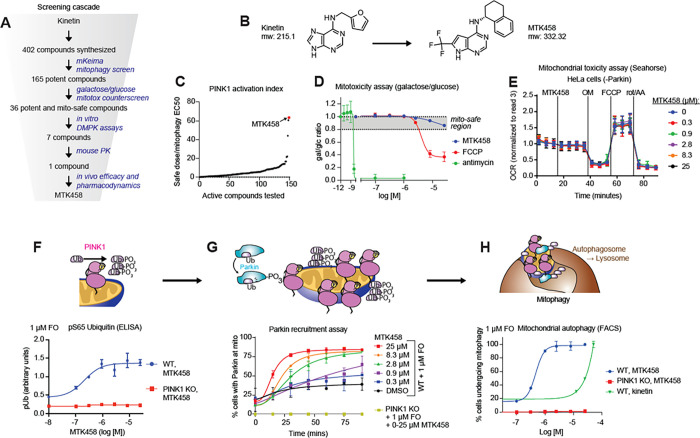
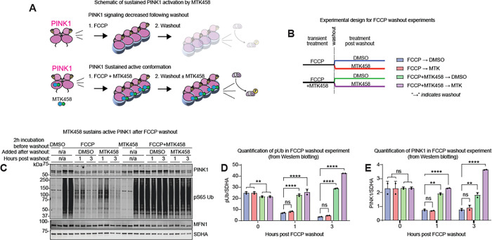
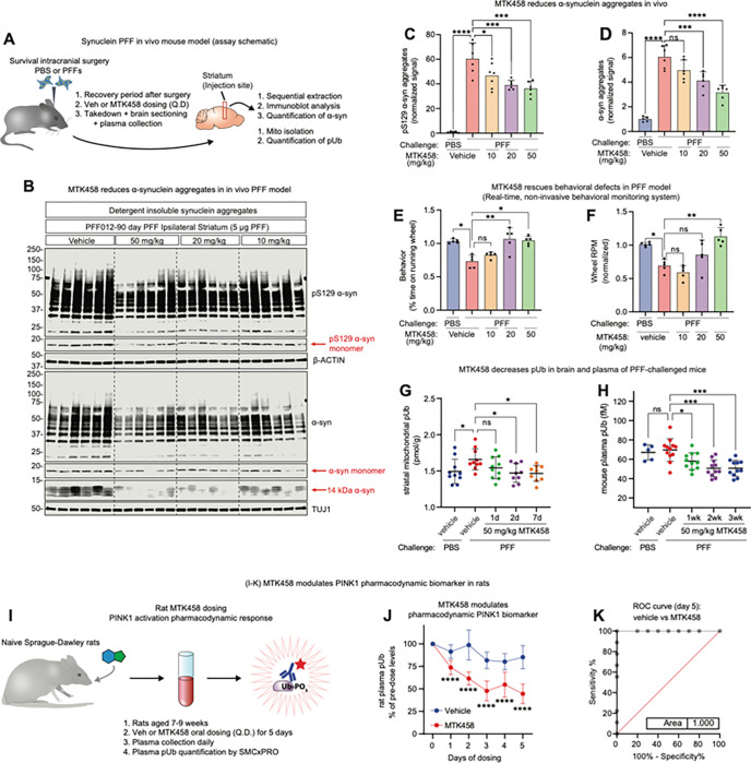
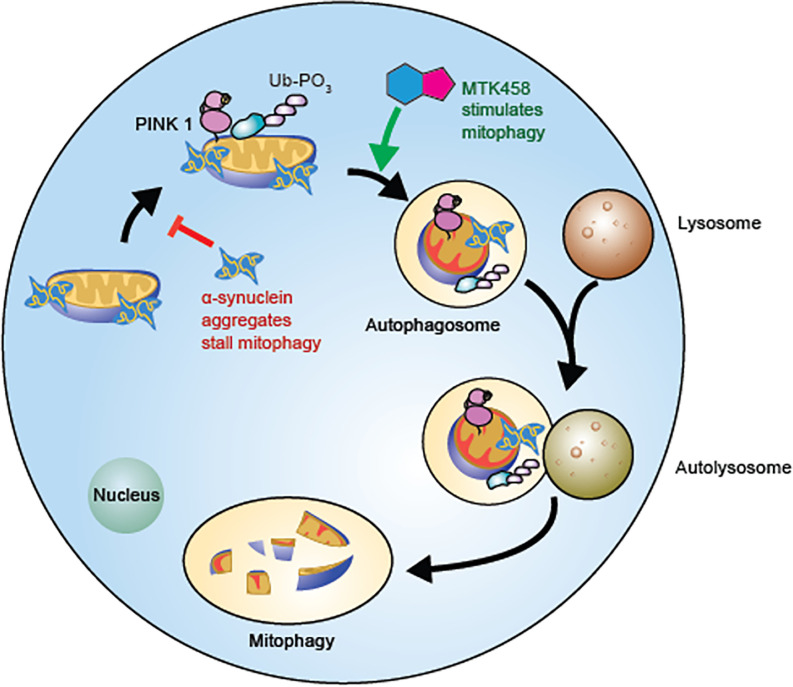
PINK1 loss-of-function mutations and exposure to mitochondrial toxins are causative for Parkinson's disease (PD) and Parkinsonism, respectively. We demonstrate that pathological α-synuclein deposition, the hallmark pathology of idiopathic PD, induces mitochondrial dysfunction, and impairs mitophagy as evidenced by the accumulation of the PINK1 substrate pS65-Ubiquitin (pUb). We discovered MTK458, a brain penetrant small molecule that binds to PINK1 and stabilizes its active complex, resulting in increased rates of mitophagy. Treatment with MTK458 mediates clearance of accumulated pUb and α-synuclein pathology in α-synuclein pathology models in vitro and in vivo. Our findings from preclinical PD models suggest that pharmacological activation of PINK1 warrants further clinical evaluation as a therapeutic strategy for disease modification in PD.


Conflict of interest statement

Competing Interests: The following authors are employees of Mitokinin Inc: R.M.C., R.R., D.D., C.W., S.L., A.M., A.L., A.E., C.T., R.Y.K ., J.L., S.H., V.R., D.H., V.G., J.P., D.d.R., and N.T.H : The authors at Mitokinin Inc are equity holders in Mitokinin Inc. S.K. and J.S. are employees of AbbVie Inc. J.W.H. is a consultant and founder of Caraway Therapeutics and a founding scientific advisory board member of Interline Therapeutics. J.B. and S.J are employees of X-Chem. Patent number related to this work: WO2020206363A1

Figures

Figure 1
Figure 1. Mitochondria-associated α-synuclein is present in PD patient brain tissue and induces mitochondrial dysfunction in primary neurons.
(A) Human brain pieces from HC or PD individuals were separated into mitochondrial or cytoplasmic fractions, or left unfractionated as whole cell lysate, and analyzed by immunoblotting. pS129 α-syn is enriched in the mitochondrial fraction. (B-C) Quantification of (A) is shown. (D-J) M83 primary neuron cultures were challenged with various concentrations of PFFs, and after 21 days, pS129 α-synuclein staining (D-E), mitochondrial respiration (F-I), and mitophagy (J) were assessed. (D-E) pS129 α-synuclein intensity increases dose dependently with PFF challenge. (F-I) PFF challenge dose-dependently decreases basal respiration, maximal respiration, and ATP production rate. (J) Primary neurons challenged for 21 days with 1 μg/ml PFFs or 10 nM CCCP show impaired mitophagy as measured by the mKeima reporter. (K) Primary neurons challenged with PFFs show an accumulation in pUb as measured by the MSD pUb assay. (L) A model of our hypothesis: aggregation of pS129 α-syn occurs at the mitochondria, leading to dysfunctional mitochondria, insufficient mitophagy, and accumulation of pUb. Where applicable, mean and SD are shown; one-way ANOVA was used for statistical analysis. *p < 0.05, **p < 0.01, ***p < 0.001, ****p < 0.0001,n.s., not significant.
Figure 2
Figure 2. Identification of the small molecule PINK1 activator MTK458
(A) Schematic of the screening funnel used to discover MTK458. (B) The structures of kinetin and MTK458 are shown. (C) Compounds were screened for their ability to induce mitophagy in the mKeima assay. MTK458 is one of the best compounds in terms of potency. (D) SK-OV-3 cells grown in galactose or glucose media were treated with MTK458, FCCP, or antimycin. After 24 hours, the ratio of cells remaining in galactose or glucose media (gal/glc ratio) was plotted. A 20% decrease in the gal/glc ratio was designated as the threshold for a mitotoxic compound. FCCP and antimycin are mitotoxic, but MTK458 is “mito-safe.” (E) HeLa cells (which do not express Parkin) were treated with MTK458 and mitochondrial respiration was assessed by the Seahorse Bioanalyzer. MTK458 does not impair mitochondrial respiration in these cells. (F) YPMK (WT or PINK1 KO) cells were treated with a dose range of MTK458 and 1 μM FCCP/oligomycin (FO), and lysed after 2h. pUb levels were measured by ELISA and shown as raw absorbance units. (G) YPMK (WT or PINK1 KO) cells were treated with MTK458 and 1 μM FO, immediately followed by live cell imaging. The percentage of cells with colocalization of YFP-Parkin with mitochondrial mKeima was assessed at each timepoint. (H) YPMK (WT or PINK1 KO) cells were treated with MTK458 (or kinetin) and 1 μM FO for 6h and then analyzed by FACS to measure the percent of cells undergoing mitophagy. Mean ± SD. *p < 0.05, **p < 0.01, ***p < 0.001, n.s., not significant.
Figure 3
Figure 3. MTK458 stabilizes and sustains the active form of PINK1.
(A) Schematic for nanoBRET assay. The nanoBRET assay assesses whether a small molecule (labeled with a BODIPY dye) binds to a particular protein (tagged with NanoLuc). Only when the drug binds to the protein will there be energy transfer between the NanoLuc and the nanoBRET dye, generating a BRET signal. (B) HEK293 PINK1-KO cells were transfected with plasmids encoding PINK1 N-terminally tagged with nanoLuc (NL-PINK1), or GSK-3b C-terminally tagged with NanoLuc (GSK-3b-NL). Cells were then treated with MTK458 labeled with the nanoBRET 590 BODIPY dye (MTK458 tracer). A high nanoBRET signal was observed with NL-PINK1, but not GSK-3b-NL, suggesting specific binding between PINK1 and MTK458. (C) Schematic of PINK1 dimerization (nanoBiT) assay. Plasmids encoding PINK1 tagged with either LgBiT or SmBiT were transfected into YPMK PINK1 KO cells. The dimerization of PINK1-LgBiT and PINK1-SmBiT brings the LgBiT and SmBiT together, forming an active luciferase enzyme capable of generating a luminescent signal. (D) YPMK PINK1 KO cells expressing PINK1-LgBiT and PINK1-SmBiT were treated with 2 μM FO and MTK458, and dimerization (as luminescence) was measured and plotted. (E) Schematic of PINK1 autophosphorylation at S228 generating the active form of PINK1. (F) In EPF1 cells (cells overexpressing PINK1-FLAG) treated with FO and MTK458, there is an increase in the phospho-PINK1 species (red arrow). (G) Schematic of the active, high molecular weight PINK1/TOM complex. (H) EPF1 cells were treated with FO and 2.8 μM MTK458 for 2.5 hours. Whole cell lysates were analyzed by immunoblotting on blue native gels (BNPAGE) or denaturing gels, showing stabilization of the active, high molecular weight PINK1 complex by MTK458. Where applicable, mean and SD are shown and one-way ANOVA was used for statistical analysis. *p < 0.05, **p < 0.01, ***p < 0.001, n.s., not significant.
Figure 4
Figure 4. PINK1 activator MTK458 rescues proteotoxicity and PD pathology in immortalized cells, primary neurons, and iPSC-derived neurons.
(A) A schematic of PINK1 sustained active conformation with MTK458 treatment is shown. (B) A schematic of the FCCP washout experiment in (C-E). SK-OV-3 cells were treated with 10 μM FCCP or 10 μM FCCP + 2.8 μM MTK458 for 2 hours. Cells were washed 3 times to remove FCCP (“washout”), and then media containing either DMSO or MTK458 was added back to the cells. Cells were harvested for analysis just before the washout, or 1 hour or 3 hours after the washout. (C) Immunoblot analysis of cells collected in the FCCP washout experiment, showing sustained PINK1 stability and activity in cells that experienced co-treatment of FCCP with MTK458. (D-E) Band intensities in (C) were quantified and plotted. Where applicable, mean and SD is shown; one-way ANOVA was used for statistical analysis. *p < 0.05, **p < 0.01, ***p < 0.001, ****p < 0.0001, n.s., not significant.
Figure 5
Figure 5. PINK1 activator MTK458 rescues proteotoxicity and PD pathology in immortalized cells, primary neurons, and iPSC-derived neurons.
(A) Schematic describing the inducible cell-based model expressing a deletion mutant of ornithine transcarbamylase (ΔOTC). This mutant yields detergent-insoluble protein aggregates in the mitochondrial matrix. (B-C) In HeLa cells expressing YFP-Parkin and doxycycline-induced ΔOTC, MTK458 treatment (25 μM) results in the clearance of ΔOTC, as assessed by immunofluorescence (B) or immunoblotting (C). (D) Schematic describing the primary neuron culture experiments in (E-G). Mouse primary cultured neurons were challenged with PFFs (0.5 ug/mL) on DIV7, treated with MTK458 on DIV9 and DIV12, and then harvested on DIV14. (E) Following serial extraction, the amount of pS129 α-syn in the NP-40 insoluble fraction was quantified from immunoblots, showing a dose dependent decrease in both pS129 α-syn aggregates and α-syn aggregates (12–250 kDa) by MTK458. (F-G) Quantification of (E) is shown. (H-I) WT or PINK1 KO mouse primary cultured neurons were challenged with PFFs and treated with MTK458 as in (D), but pS129 α-syn was analyzed by immunofluorescence. MTK458 dose-dependently decreases pS129 α-syn signal in WT neurons, but not PINK1 KO neurons. Where applicable, mean and SD is shown; one-way ANOVA was used for statistical analysis. *p < 0.05, **p < 0.01, ***p < 0.001, ****p < 0.0001, n.s., not significant.
Figure 6
Figure 6. PINK1 activator MTK458 rescues PD pathology and normalizes p-S65-Ub levels in vivo
(A) Schematic of in vivo PFF experiments in mice. Mice were challenged with striatal injection of PFFs and dosed (QD, PO) with the indicated dose levels of MTK458. (B) After 90 days, striatum brain pieces were analyzed for pS129 α-syn aggregates or total α-syn aggregates (12–250 kDa). (C-D) Quantification of analysis of the full cohort is shown here. (E-F) Parallel mice were challenged with striatal injection of PBS or PFF, and then dosed with MTK458 for 6 months. Mice were analyzed for time spent on the running wheel using the Vium system. MTK458 treatment rescued the reductions in overall wheel activity and wheel speed (RPM) induced by PFF challenge. (G) Mice were challenged with striatal injection of PFFs and after 3 months, were dosed (QD, PO) with 50 mg/kg MTK458 for the indicated times. Mitochondria was isolated from striatal brain pieces and analyzed for pUb content on the MSD assay. (H) Mice were challenged with PFFs, and after 3 months, were dosed (PO, QD) with 50 mg/kg MTK458 for the indicated durations. Plasma pUb levels were measured by the SMCxPRO pUb assay. (I) Schematic of rat dosing study to evaluate plasma pUb as a target engagement biomarker for PINK1 activator compounds. (J) Naïve Sprague-Dawley rats were dosed (PO, QD) with 50 mg/kg MTK458 for 5 days (6 doses). Plasma pUb levels at the indicated timepoints were determined by the SMCxPRO pUb assay. (K) ROC curve for plasma pUb levels in vehicle vs MTK458 dosed rats (after 5 days of dosing) is shown. Where applicable, mean and SD is shown; one-way ANOVA was used for statistical analysis. *p < 0.05, **p < 0.01, ***p < 0.001, ****p < 0.0001 n.s., not significant.
Figure 7
Figure 7. Schematic of pUb regulation.
Model for the increased pUb induced by α-synuclein aggregates, which is opposed by MTK458. Cells can dispose of dysfunctional mitochondria by PINK1/Parkin mediated mitophagy. α-synuclein aggregates stall mitophagy and result in an increase of pUb inside the cell. MTK458 increases PINK1/Parkin mediated mitophagy, reducing α-synuclein aggregates and pUb.

References

    1. Antico O., Ordureau A., Stevens M., Singh F., Nirujogi R.S., Gierlinski M., Barini E., Rickwood M.L., Prescott A., Toth R., et al. (2021). Global ubiquitylation analysis of mitochondria in primary neurons identifies endogenous Parkin targets following activation of PINK1. Sci. Adv. 7. - PMC - PubMed
    1. Arroyo J.D., Jourdain A.A., Calvo S.E., Ballarano C.A., Doench J.G., Root D.E., and Mootha V.K. (2016). A Genome-wide CRISPR Death Screen Identifies Genes Essential for Oxidative Phosphorylation. Cell Metab. 24, 875–885. - PMC - PubMed
    1. Ball N., Teo W.P., Chandra S., and Chapman J. (2019). Parkinson’s disease and the environment. Front. Neurol. 10, 218. - PMC - PubMed
    1. Bingol B., Tea J.S., Phu L., Reichelt M., Bakalarski C.E., Song Q., Foreman O., Kirkpatrick D.S., and Sheng M. (2014). The mitochondrial deubiquitinase USP30 opposes parkin-mediated mitophagy. Nature 510, 370–375. - PubMed
    1. Bloem B.R., Okun M.S., and Klein C. (2021). Parkinson’s disease. Lancet 397, 2284–2303. - PubMed

Publication types

LinkOut - more resources