[Preprint]. 2024 May 13:2024.05.13.593939.
doi: 10.1101/2024.05.13.593939.

Glycosaminoglycan-mediated lipoprotein uptake protects cancer cells from ferroptosis

Affiliations

Glycosaminoglycan-mediated lipoprotein uptake protects cancer cells from ferroptosis

Dylan Calhoon et al. bioRxiv. .

Update in

  • Glycosaminoglycan-driven lipoprotein uptake protects tumours from ferroptosis.
    Calhoon D, Sang L, Ji F, Bezwada D, Hsu SC, Cai F, Kim N, Basu A, Wu R, Pimentel A, Brooks B, La K, Paulina Serrano A, Cassidy DL, Cai L, Toffessi-Tcheuyap V, Moussa ME, Uritboonthai W, Hoang LT, Kolli M, Jackson B, Margulis V, Siuzdak G, Brugarolas J, Corbin I, Pratt DA, Weiss RJ, DeBerardinis RJ, Birsoy K, Garcia-Bermudez J. Calhoon D, et al. Nature. 2025 Aug;644(8077):799-808. doi: 10.1038/s41586-025-09162-0. Epub 2025 Jun 11. Nature. 2025. PMID: 40500442

Abstract

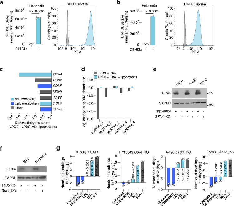
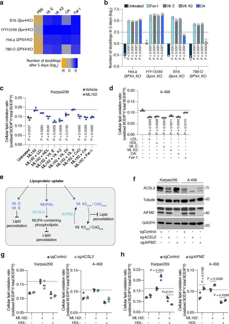
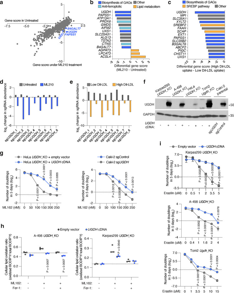
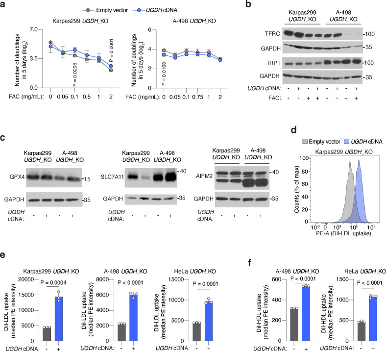
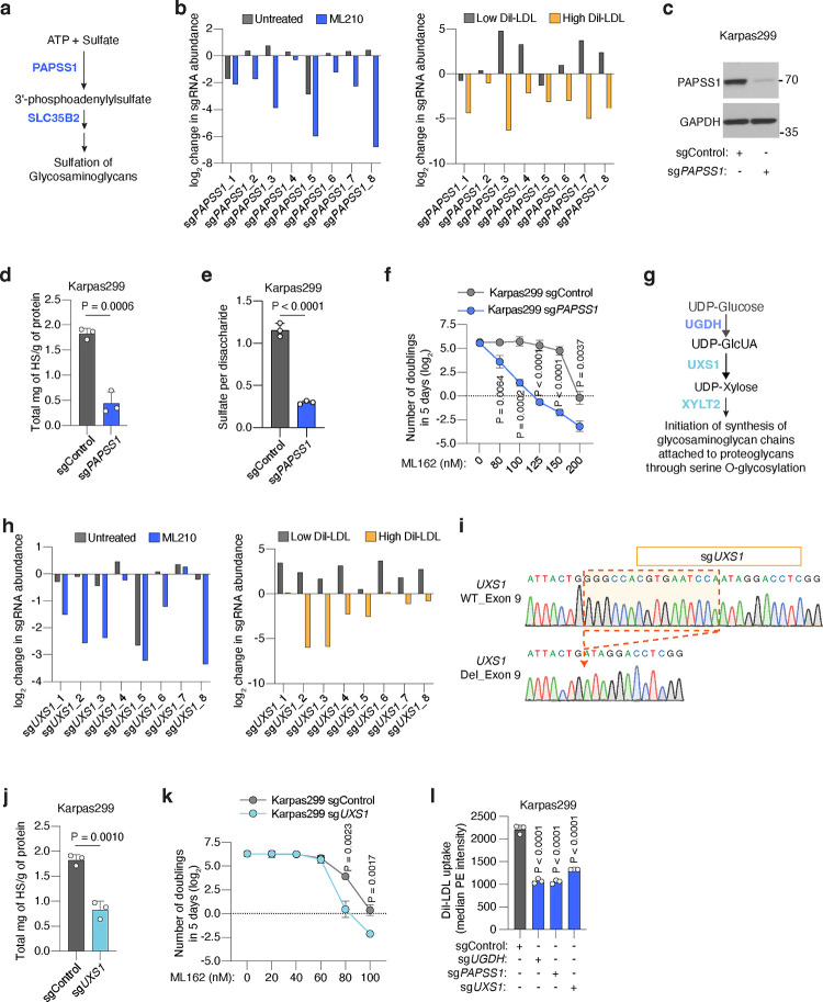
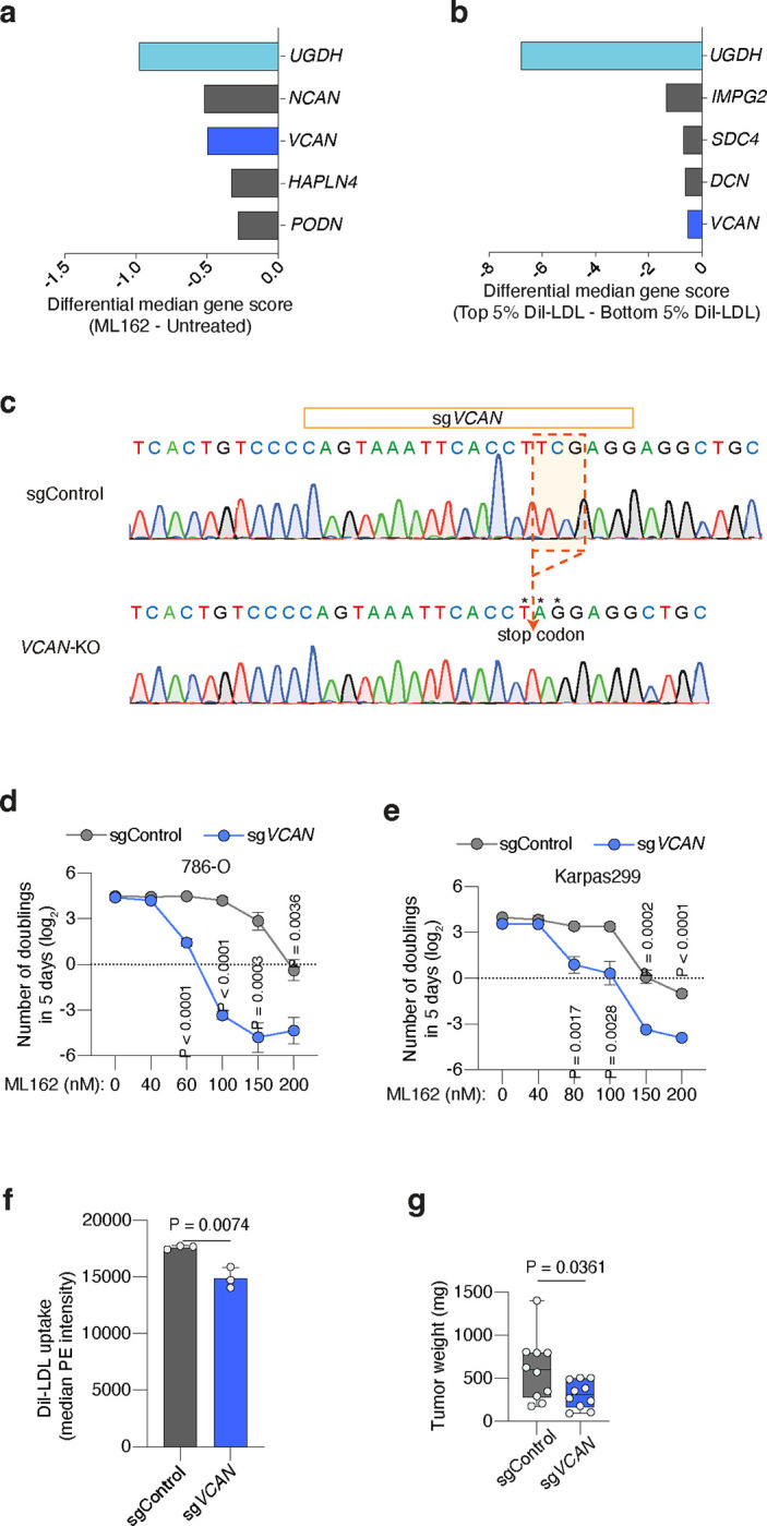
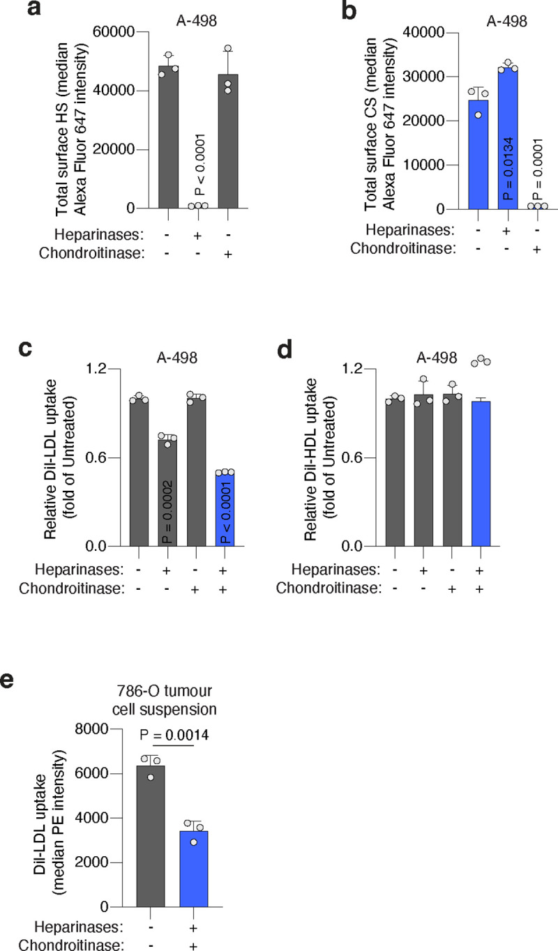
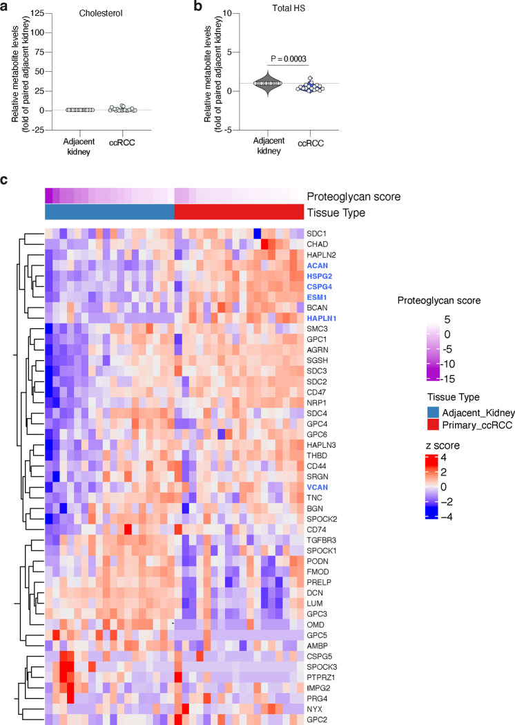
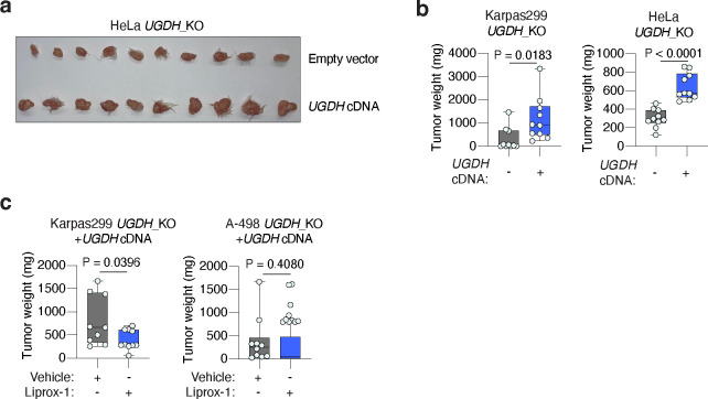
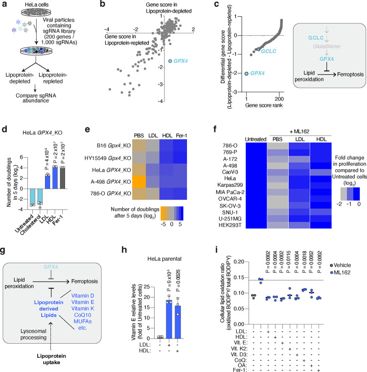
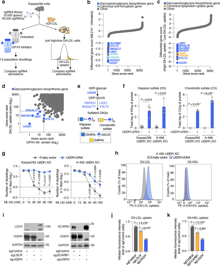
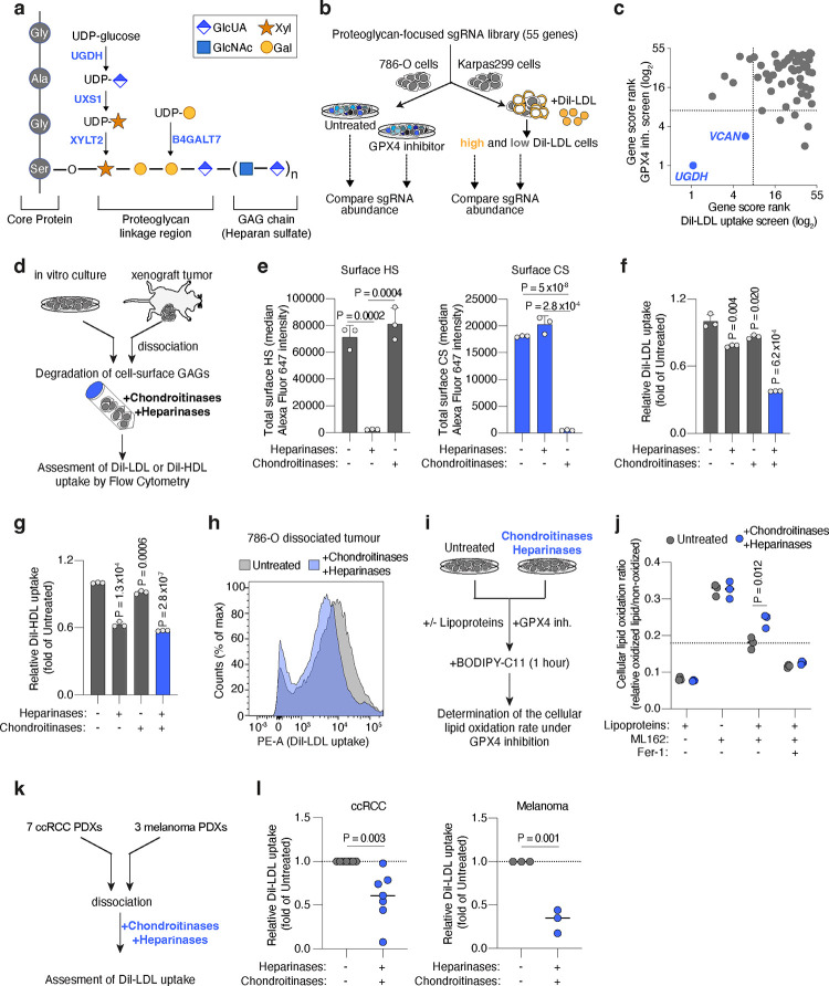
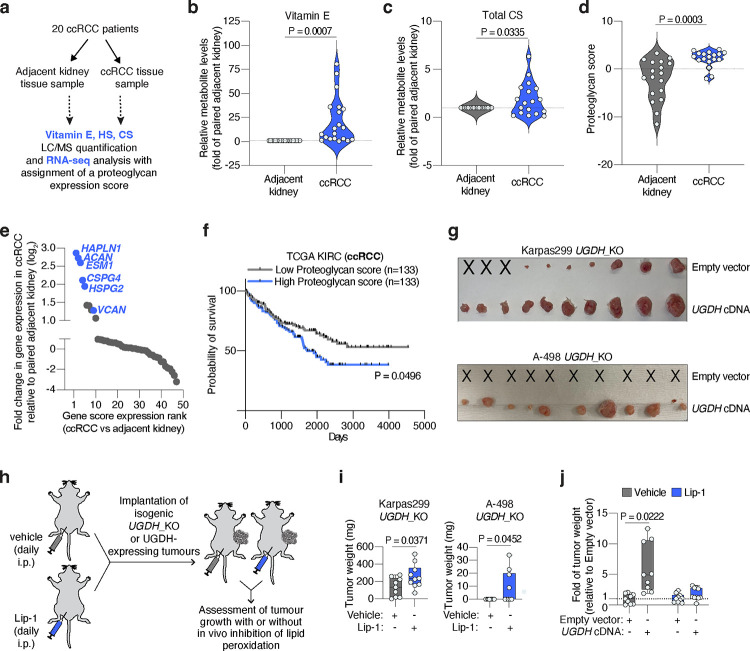
Lipids are essential for tumours because of their structural, energetic, and signaling roles. While many cancer cells upregulate lipid synthesis, growing evidence suggests that tumours simultaneously intensify the uptake of circulating lipids carried by lipoproteins. Which mechanisms promote the uptake of extracellular lipids, and how this pool of lipids contributes to cancer progression, are poorly understood. Here, using functional genetic screens, we find that lipoprotein uptake confers resistance to lipid peroxidation and ferroptotic cell death. Lipoprotein supplementation robustly inhibits ferroptosis across numerous cancer types. Mechanistically, cancer cells take up lipoproteins through a pathway dependent on sulfated glycosaminoglycans (GAGs) linked to cell-surface proteoglycans. Tumour GAGs are a major determinant of the uptake of both low and high density lipoproteins. Impairment of glycosaminoglycan synthesis or acute degradation of surface GAGs decreases the uptake of lipoproteins, sensitizes cells to ferroptosis and reduces tumour growth in mice. We also find that human clear cell renal cell carcinomas, a distinctively lipid-rich tumour type, display elevated levels of lipoprotein-derived antioxidants and the GAG chondroitin sulfate than non-malignant human kidney. Altogether, our work identifies lipoprotein uptake as an essential anti-ferroptotic mechanism for cancer cells to overcome lipid oxidative stress in vivo, and reveals GAG biosynthesis as an unexpected mediator of this process.


Conflict of interest statement

DECLARATION OF INTERESTS K.B. is scientific advisor to Nanocare Pharmaceuticals and Atavistik Bio. R.J.D. is a founder at Atavistik Bio and serves on the Scientific Advisory Boards of Atavistik Bio, Agios Pharmaceuticals, Faeth Therapeutics, General Metabolics and Vida Ventures.

Figures

Extended Data Figure 1.
Extended Data Figure 1.. GPX4 loss is compensated by lipoprotein supplementation.
a. Cellular uptake of DiI-LDL in HeLa cells measured as median PE intensity after incubation or not with DiI-LDL (left). Representative flow cytometry plot of HeLa cells treated (blue) or not (grey) with DiI-LDL (right). b. Cellular uptake of DiI-HDL in HeLa cells measured as median PE intensity after incubation or not with DiI-HDL (left). Representative flow cytometry plot of HeLa cells treated (blue) or not (grey) with DiI-HDL (right). c. Plot of differential gene scores in HeLa cells supplemented with lipoproteins (LPDS + lipoproteins) or not (LPDS). Anti-ferroptotic genes are shown in light blue and lipid metabolism genes are shown in dark blue. LPDS: lipoprotein-depleted serum. d. Individual sgRNA scores (log2) for GPX4 under indicated conditions. e. Immunoblot analysis of human GPX4 sgControl cells or GPX4_KO isogenic lines. GAPDH is included as a loading control. f. Immunoblot analysis of mouse GPX4 sgControl cells or Gpx4_KO isogenic lines. GAPDH is included as a loading control. g. Number of doublings (log2) in 5 days of indicated GPX4-deficient cell lines in vitro either untreated or supplemented with cholesterol (5 μg/mL), LDL (50 μg/mL), HDL (50 μg/mL) or Fer-1 (1 μM). A, b, g, Bars represent mean ± s.d.; a, b, g, n=3 biological replicates. Statistical significance determined by two-tailed unpaired t-tests as indicated or compared to untreated cells (g).
Extended Data Figure 2.
Extended Data Figure 2.. Lipoproteins carry multiple lipid species that could inhibit lipid peroxidation.
a. Heatmap showing the number of doublings (log2) in 5 days of mouse (Gpx4) or human (GPX4) knockout cell lines in cell culture under the supplementation of PBS, vitamin E (Vit. E, 10 μM), vitamin K2 (Vit. K2, 10 μM), oleic acid (OA, 250 μM) or ferrostatin-1 (Fer-1, 1 μM). b. Number of doublings (log2) in 5 days of B16 and HY15549 Gpx4-KO and HeLa and 786-O GPX4-KO cells in vitro either untreated or supplemented with Fer-1 (1 μM), vitamin E (10 μM), vitamin K2 (10 μM), or OA (250 μM). c. Cellular lipid oxidation ratio using BODIPY-C11 in HeLa cells in the absence (gray) or presence (blue) of a GPX4 inhibitor (ML162, 125 nM) under supplementation or not of LDL (50 μg/mL), HDL (50 μg/mL), vitamin E (10 μM), vitamin K2 (10 μM), vitamin D3 (10 μM), vitamin A (10 μM), CoQ10 (10 μM), OA (250 μM) or Fer-1 (1 μM). d. Cellular lipid oxidation ratio using BODIPY-C11 in A-498 cells with intact GPX4 activity and under supplementation or not of LDL (50 μg/mL), HDL (50 μg/mL), vitamin E (10 μM), vitamin K2 (10 μM), OA (250 μM) or Fer-1 (1 μM). e. Scheme highlighting genes (light blue) that affect the cellular ability to utilize lipoprotein-transported lipids (dark blue) that inhibit lipid peroxidation. f. Immunoblot analysis of ACSL3 and AIFM2 in the indicated cell lines transduced with a sgControl, sgACSL3 or sgAIFM2. GAPDH is included as a loading control. g. Cellular lipid oxidation ratio using BODIPY-C11 in the indicated Karpas299 (left) or A-498 (right) cell lines transduced with sgACSL3 (light blue) or with a sgControl (grey) under GPX4 inhibition (ML162: 75 nM for Karpas299, 175 nM for A-498) and HDL supplementation (50 μg/mL). h. Cellular lipid oxidation ratio using BODIPY-C11 in the indicated Karpas299 (left) or A-498 (right) cell lines transduced with sgAIFM2 (dark blue) or with a sgControl (grey) under GPX4 inhibition (ML162: 75 nM for Karpas299, 175 nM for A-498) and HDL supplementation (50 μg/mL). b, Bars represent mean ± s.d.; c, d, g, h, bars represent median; b, c, d, g, h, n=3 biologically independent samples. Statistical significance determined by two-tailed unpaired t-tests as indicated or compared to untreated cells (b, d) or ML162 treated cells (c).
Extended Data Figure 3.
Extended Data Figure 3.. Loss of UGDH sensitizes cancer cells to ferroptosis in vitro.
a. Gene essentiality plot showing genes scores in untreated (x-axis) and ML210-treated (GPX4 inhibition) Karpas299 cells. Glycosaminoglycan (GAG) biosynthesis genes are highlighted (dark blue). b. Top-scoring genes under ML210 treatment. Negative scores represent genes whose loss potentiates ML210 toxicity; positive scores represent genes whose loss provides resistance to ML210. GAG biosynthesis genes are shown in dark blue, canonical anti-ferroptotic genes are shown in light blue, and lipid metabolism genes are shown in yellow. c. Top-scoring genes essential for DiI-LDL uptake. Negative scores represent genes whose loss reduce cellular DiI-LDL uptake. GAG biosynthesis genes are shown in dark blue and genes in canonical lipoprotein uptake pathways highlighted in yellow. d. Individual sgRNA scores for UGDH in untreated and ML210-treated conditions. e. Individual sgRNA scores for UGDH in high and low DiI-LDL uptake populations. f. Immunoblot analysis of UGDH in the indicated isogenic cell lines transduced with sgUGDH and expressing or not a sgRNA-resistant UGDH cDNA. GAPDH is included as a loading control. g. Number of doublings (log2) in 5 days of indicated HeLa (left) or Caki-2 (right) cell lines expressing UGDH (blue) or not (grey) under the indicated concentrations of the GPX4 inhibitor ML162. h. Cellular lipid oxidation ratio using BODIPY-C11 in the indicated A-498 (left) or Karpas299 (right) UGDH_KO cell lines expressing a sgRNA resistant UGDH cDNA (blue) or an empty vector (grey) under the indicated conditions of GPX4 inhibition (ML162: 200 nM for Karpas299, 250 nM for A-498) and Fer-1 supplementation (1μM). i. Number of doublings (log2) in 5 days of the indicated Karpas299 (top), A-498 (middle) and Tom2 (bottom) UGDH-deficient cell lines expressing a sgRNA resistant UGDH cDNA (dark blue) or an empty vector (grey) under the indicated concentrations of erastin. g-i, Bars represent mean ± s.d.; g-i, n=3 biologically independent samples. Statistical significance determined by two-tailed unpaired t-tests compared to empty vector transduced cells.
Extended Data Figure 4.
Extended Data Figure 4.. UGDH is a major determinant of LDL and HDL uptake in cancer cells.
a. Number of doublings (log2) in 5 days of the indicated Karpas299 (left) and A-498 (right) UGDH_KO cell lines expressing a sgRNA resistant UGDH cDNA (blue) or an empty vector (grey) under the indicated concentrations of ferrous ammonium citrate (FAC). b. Immunoblot analysis of TFRC (top) and IRP1 (bottom) in the indicated UGDH_KO cell lines expressing a sgRNA resistant UGDH cDNA or an empty vector treated or not with FAC (0.1 mg/mL). GAPDH is included as a loading control. c. Immunoblot analysis of GPX4 (left), SLC7A11 (center), and AIFM2 (right) in the indicated UGDH_KO cell lines expressing a sgRNA resistant UGDH cDNA or an empty vector. GAPDH is included as a loading control. d. Representative flow cytometry plot showing the uptake of DiI-LDL (PE median intensity) in Karpas299 UGDH_KO cells expressing a sgRNA resistant UGDH cDNA (blue) or an empty vector (grey). e. Cellular uptake of DiI-LDL measured as median PE intensity in the indicated Karpas299 (left), A-498 (center) and HeLa (right) UGDH_KO cells expressing a sgRNA resistant UGDH cDNA (blue) or an empty vector (grey). f. Cellular uptake of DiI-HDL measured as median PE intensity in the indicated A-498 (left) and HeLa (right) UGDH_KO cells expressing a sgRNA resistant UGDH cDNA (blue) or an empty vector (grey). a, e, f, Lines or bars represent mean ± s.d.; a, e, f, n=3 biologically independent samples. Statistical significance determined by two-tailed unpaired t-tests.
Extended Data Figure 5.
Extended Data Figure 5.. Sulfation of GAGs and xylose synthesis is essential for lymphoma cells to resist ferroptosis and take up lipoproteins.
a. Sulfation of glycosaminoglycans relies on the production of 3’-phosphoadenylylsulfate by PAPSS1, and on its transports into the Golgi by SLC35B2. Both genes (blue) scored in our screens. b. Individual sgRNA scores for PAPSS1 in the presence and absence of ML210 (left), or in high and low DiI-LDL populations (right). c. Immunoblot analysis of PAPSS1 in Karpas299 cells transduced with a sgControl (grey) or sgPAPSS1 (blue). GAPDH is included as a loading control. d. Quantification of total milligrams of heparan sulfate (HS) per grams of protein in Karpas299 cells transduced with a sgControl (grey) or sgPAPSS1 (blue). e. Quantification of total sulfate per glycosaminoglycan disaccharide in Karpas299 cells transduced with a sgControl (grey) or sgPAPSS1 (blue). f. Number of doublings (log2) in 5 days of Karpas299 cells transduced with a sgControl (grey) or sgPAPSS1 (blue) under the indicated concentrations of the GPX4 inhibitor ML162. g. Glucuronic acid (GlcUA), formed by UGDH, is converted to UDP-Xylose via UXS1. UDP-Xylose is the first monosaccharide used in the initiation of O-glycosylation that attaches GAG chains to serine-residues of proteoglycans. Genes involved in this process (blue) scored in our screens. h. Individual sgRNA scores for UXS1 in the presence and absence of ML210 (left), or in high and low DiI-LDL populations (right). i. Sanger sequencing of UXS1 gene exon 9 in Karpas299 parental cells (WT) or transduced with sgUXS1. j. Quantification of total milligrams of heparan sulfate (HS) per grams of protein in Karpas299 cells transduced with a sgControl (grey) or sgUXS1 (blue). k. Number of doublings (log2) in 5 days of Karpas299 cells transduced with a sgControl (grey) or sgUXS1 (blue) under the indicated concentrations of the GPX4 inhibitor ML162. i. Cellular uptake of DiI-LDL measured as median PE intensity in the indicated Karpas299 cells transduced with a sgControl, sgUGDH, sgPAPSS1 or sgUXS1 assessed by flow cytometry. d-f, j-l, Bars or lines represent mean ± s.d.; d-f, j-l, n=3 biologically independent samples. Statistical significance determined by two-tailed unpaired t-tests as indicated or compared to sgControl cells (l).
Extended Data Figure 6.
Extended Data Figure 6.. The proteoglycan VCAN modestly increases resistance to ferroptosis and lipoprotein uptake in cancer cells.
a. Plot of differential gene scores under ML162 treatment relative to untreated cells. Negative scores represent genes whose loss potentiates ML162 toxicity. UGDH was included as a positive control (light blue). VCAN (blue) was a common hit between the two screens. b. Plot of differential gene scores in high DiI-LDL uptake population compared to low DiI-LDL cells. Negative scores represent genes whose loss reduce cellular DiI-LDL uptake. UGDH was included as a positive control (light blue) and VCAN (blue) was a common hit between the two screens. c. Sanger sequencing of VCAN gene exon 9 in Karpas299 parental cells (WT) or transduced with sgUXS1. d. Number of doublings (log2) in 5 days of 786-O transduced with a sgControl (grey) or sgVCAN (blue) under the indicated concentrations of the GPX4 inhibitor ML162. e. Number of doublings (log2) in 5 days of Karpas299 cells transduced with a sgControl (grey) or sgVCAN (dark blue) under the indicated concentrations of the GPX4 inhibitor ML162. f. Cellular uptake of DiI-LDL measured as median PE intensity in the indicated Karpas299 cells transduced with a sgControl (grey) or sgVCAN (blue) assessed by flow cytometry. g. Tumour weight resulting from implantation of Karpas299 cells transduced with a sgControl (grey) or sgVCAN (blue) in 6–12 weeks old immunodeficient mice. d-g, Bars or lines represent mean ± s.d.; d-g, n=3 biologically independent samples. Statistical significance determined by two-tailed unpaired t-tests.
Extended Data Figure 7.
Extended Data Figure 7.. Cell-surface sulfated glycosaminoglycans promote lipoprotein uptake.
a. Total cell surface HS measured as median Alexa Fluor 647 intensity in A-498 cells treated or not with heparinases (0.1 U/mL) or chondroitinases (0.1 U/mL) assessed by flow cytometry. b. Total cell surface CS measured as median Alexa Fluor 647 intensity in A-498 cells treated or not with heparinases (0.1 U/mL) or chondroitinases (0.1 U/mL) assessed by flow cytometry. c. Fold change in DiI-LDL uptake of A-498 cells upon treatment with heparinases (0.1 U/mL), chondroitinases (0.1 U/mL), or both (blue), relative to uptake of untreated cells assessed by flow cytometry. d. Fold change in DiI-HDL uptake of A-498 cells upon treatment with heparinases (0.1 U/mL), chondroitinases (0.1 U/mL), or both (blue), relative to uptake of untreated cells, assessed by flow cytometry. e. Uptake of DiI-LDL measured as median PE intensity in a cell suspension of 786-O xenograft tumour fresh tissue resected from mice left untreated (grey) or after combined treatment with heparinases (1.0 U/mL) and chondroitinases (1.0 U/mL) (blue) assessed by flow cytometry. a-e, Bars represent mean ± s.d.; a-e, n=3 biologically independent samples. Statistical significance determined by two-tailed unpaired t-tests as indicated or compared to untreated cells (a-d).
Extended Data Figure 8.
Extended Data Figure 8.. Proteoglycan expression profile in human clear cell renal cell carcinomas (ccRCCs) and adjacent kidney.
a. Violin plot showing the relative levels of cholesterol in ccRCC patient tissues (blue) compared to paired adjacent kidney (grey). b. Violin plot showing the relative levels of total heparan sulfate (HS) per gram of protein in ccRCC patient tissues (blue) compared to paired adjacent kidney (grey). c. Heatmap showing expression of individual proteoglycan genes in ccRCC tumours and paired adjacent kidney. Each sample was assigned a proteoglycan score, based on their expression of all proteoglycans (top). Relevant proteoglycans scoring are highlighted in blue. a, b, n=17–20 biologically independent samples. Statistical significance was determined by a two-tailed unpaired t-test.
Extended Data Figure 9.
Extended Data Figure 9.. The anti-ferroptotic effect of UGDH is essential for tumour growth.
a. Representative tumour images of HeLa UGDH_KO cells expressing a sgRNA resistant UGDH cDNA or an empty vector, and implanted subcutaneously in 6–12 weeks old immunodeficient mice. b. Tumour weight resulting from implantation of the indicated UGDH_KO cell lines in immunodeficient mice. c. Tumour weights resulting from implantation of the indicated UGDH_KO cell lines expressing a sgRNA-resistant UGDH cDNA on immunodeficient mice treated with vehicle (grey) or Lip-1 (blue) through daily intraperitoneal injection. b, c, Boxes represent the median, and the first and third quartiles, and the whiskers represent the minimum and maximum of all data points.; b, c, n=10 biologically independent samples. Statistical significance determined by two-tailed unpaired t-tests.
Figure 1.
Figure 1.. Lipoprotein supplementation promotes cancer cell resistance to ferroptosis.
a. Scheme of CRISPR screen in HeLa cells transduced with a focused-metabolism sgRNA library and depleted of or supplemented with human lipoproteins. b. Gene essentiality graph showing changes in essentiality based on lipoprotein availability. GPX4 becomes non-essential in cells supplemented with lipoproteins. c. Rank of genes whose essentiality in HeLa cells is most changed based on lipoprotein availability. Anti-ferroptotic genes are highlighted in blue (left). Scheme of the glutathione peroxidase pathway inhibiting lipid peroxidation and ferroptosis in cells (right). d. Number of doublings (log2) in 5 days of HeLa GPX4_KO cells in vitro either untreated or supplemented with cholesterol (5 μg/mL), human low density lipoproteins (LDL, 50 μg/mL), high density lipoproteins (HDL, 50 μg/mL) or ferrostatin-1 (Fer-1, 1 μM). e. Heatmap showing the number of doublings (log2) in 5 days of mouse (Gpx4) or human (GPX4) knockout cell lines in cell culture under the supplementation of PBS, LDL (50 μg/mL), HDL (50 μg/mL) or Fer-1 (1 μM). f. Heatmap showing the fold change in proliferation relative to untreated cells (log2) of a panel of lines with or without the GPX4 inhibitor ML162, and in the presence or absence of LDL (50 μg/mL) or HDL (50 μg/mL). g. Scheme showing the multiple anti-ferroptotic lipids that lipoproteins potentially carry, and that cancer cells can assimilate upon lipoprotein uptake and lysosomal processing. h. Mass spectrometry analysis of vitamin E levels in HeLa cells in the absence of lipoproteins or supplemented with LDL (50 μg/mL) or HDL (50 μg/mL). Data is presented as fold of metabolite levels in lipoprotein-depleted cells. i. Cellular lipid oxidation ratio using BODIPY-C11 in A-498 cells in the absence (gray) or presence (blue) of a GPX4 inhibitor (ML162, 250 nM) under supplementation or not of LDL (50 μg/mL), HDL (50 μg/mL), vitamin E (10 μM), vitamin K2 (10 μM), vitamin D3 (10 μM), CoQ10 (10 μM), OA (250 μM) or Fer-1 (1 μM). d, h, Bars represent mean ± s.d.; i, bars represent the median; d-f, h, i, n=3 biologically independent samples. Statistical significance determined by a two-tailed unpaired t-test compared to untreated cells (d, h) or ML162 treated cells (i).
Figure 2.
Figure 2.. Cancer cells depend on the biosynthesis of glycosaminoglycans (GAGs) to take up lipoproteins, resist ferroptosis, and grow as tumours.
a. Schematic of parallel CRISPR screens in Karpas299 lymphoma cells transduced with a metabolism-focused sgRNA library (3,000 genes). First, a proliferation-based screen in the presence or absence of a GPX4 inhibitor (ML210) during 14 population doublings (left); second, a FACS-based screen where cells were incubated with DiI-LDL for 2 hours and subjected to sorting for 5% highest and 5% lowest fluorescent cells (right). b. Rank of most essential genes under ML210 treatment compared to untreated Karpas299 cells. Canonical anti-ferroptotic genes are shown in light blue, and glycosaminoglycan biosynthesis genes are shown in dark blue. c. Rank of most essential genes for DiI-LDL uptake in Karpas299 cells. Lipoprotein uptake genes are shown in orange and glycosaminoglycan biosynthesis pathway genes are shown in dark blue. d. Gene scores ranks for the GPX4 inhibition and DiI-LDL uptake screens in Karpas299. All genes essential in both screens are part of the glycosaminoglycan biosynthesis pathway (dark blue). e. Glycosaminoglycans, such as heparan sulfate and chondroitin sulfate, are formed by disaccharides repeats containing glucuronic acid (GlcUA) and other monosaccharides. The most upstream and rate-limiting enzyme of this pathway is UGDH. All genes in the pathway that scored in CRISPR screens are highlighted in blue. f. Quantification of total milligrams of heparan sulfate (HS, left) and chondroitin sulfate (CS, right) per grams of protein in the indicated UGDH_KO cell lines expressing a sgRNA resistant UGDH cDNA (blue) or an empty vector (grey). g. Number of doublings in 5 days (log2) of the indicated UGDH_KO cell lines expressing a sgRNA resistant UGDH cDNA (blue) or an empty vector (grey) under the indicated concentrations of the GPX4 inhibitor, ML162. h. Representative flow cytometry plots showing the cellular uptake of DiI-LDL (left) or DiI-HDL (right) in A-498 UGDH_KO cells expressing a sgRNA resistant UGDH cDNA (blue) or an empty vector (grey). i. Immunoblot analysis of LDLR, SCARB1, and UGDH in HeLa cells transduced with a sgControl, sgLDLR, sgSCARB1 or sgUGDH. GAPDH is included as a loading control. j. Fold change in the uptake of DiI-LDL of the indicated HeLa cell lines relative to sgControl-transduced cells assessed by flow cytometry. k. Fold change in the uptake of DiI-HDL of the indicated HeLa cell lines relative to sgControl-transduced cells assessed by flow cytometry. f, g, j, k, Bars or lines represent mean ± s.d.; f, g, j, k, n=3 biologically independent samples. Statistical significance determined by two-tailed unpaired t-tests.
Figure 3.
Figure 3.. Cell-surface glycosaminoglycans bound to proteoglycans drive the uptake of LDL and HDL by cancer cells thus promoting ferroptosis resistance.
a. Scheme of the canonical proteoglycan linkage region showing a heparan sulfate chain attached to a core protein through O-glycosylation of a serine residue. Essential genes for glycosaminoglycan-chain attachment to proteoglycans that scored in Karpas299 genetic screens are highlighted (blue). b. Schematic of CRISPR screens in 786-O (ccRCC) and Karpas299 (lymphoma) cells transduced with proteoglycan-focused sgRNA library (55 genes). 786-O cells were subjected to a proliferation-based screen in the presence or absence of the GPX4 inhibitor, ML162 (left). Karpas299 cells were incubated with DiI-LDL for 2 hours and subjected to FACS for high and low fluorescent cells. c. Plot of the gene score ranks in the 786-O and Karpas299 proteoglycan focused screens. Genes essential in both screens are highlighted (blue). sgRNAs targeting UGDH were included as a positive control. d. Experimental approach to degrade cell-surface HS and CS using heparinases and chondroitinases, respectively, on cancer cells in culture or dissociated from xenograft tumours. e. Total cell surface HS (left, grey) and CS (right, blue) in 786-O cells before and after treatment with heparinases (0.1 U/mL) and chondroitinases (0.1 U/mL) assessed by flow cytometry. f. Fold change in DiI-LDL uptake of 786-O cells upon treatment with heparinases (0.1 U/mL), chondroitinases (0.1 U/mL), or both (blue), relative to uptake of untreated cells, assessed by flow cytometry. g. Fold change in DiI-HDL uptake of 786-O cells upon treatment with heparinases (0.1 U/mL), chondroitinases (0.1 U/mL), or both (blue), relative to uptake of untreated cells, assessed by flow cytometry. h. Flow cytometry plot of the uptake of DiI-LDL in a cell suspension of 786-O xenograft tumours resected from mice left untreated (grey) or after combined treatment with heparinases and chondroitinases (blue). i. Experimental approach to evaluate ferroptosis sensitivity after enzymatic degradation of cell surface HS and CS. Cells treated with heparinases and chondroitinases are placed in media with or without lipoproteins and subjected to GPX4 inhibition, prior to staining with BODIPY-C11 and flow cytometry analysis of cellular lipid oxidation. j. Cellular lipid oxidation ratio using BODIPY-C11 of 786-O cells left untreated (grey) or after treatment with heparinases and chondroitinases (blue), and in the indicated conditions of lipoprotein availability and GPX4 inhibition (ML162, 200 nM). Fer-1 (1 μM) is included as a positive control of lipid peroxidation inhibition. k. Experimental approach to evaluate HS- and CS-mediated lipoprotein uptake in patient derived xenografts (PDXs). Fresh tissues were dissociated before treatment with or without heparinases and chondroitinases, and assessed for differences in DiI-LDL uptake using flow cytometry. l. Fold change in DiI-LDL uptake by ccRCC (left) and melanoma (right) PDX cell suspensions treated with heparinases and chondroitinases (1 U/mL of both, dark blue) relative to untreated samples (grey). Each dot represents a different PDX. e-g, Bars represent mean ± s.d.; j, l, bars represent the median in n=7 (ccRCC, left) and n=3 (melanoma, right) biologically independent samples; e-g, n=3 biological replicates. Statistical significance determined by two-tailed unpaired t-tests as indicated or compared to untreated cells (f,g).
Figure 4.
Figure 4.. GAG-mediated lipoprotein uptake in pre-clinical models and human tissues.
a. Primary ccRCC patient samples (adjacent kidney or tumour tissue) were subjected to parallel LC/MS quantification of vitamin E, HS or CS, and RNA-seq analysis. b. Violin plot showing the relative levels of vitamin E in ccRCC patient tissues (blue) compared to paired adjacent kidney (grey). c. Violin plot showing the relative levels of total CS per gram of protein in ccRCC patient tissues (blue) compared to paired adjacent kidney (grey). d. Violin plot showing the increased proteoglycan score of ccRCC patient tissues (blue) relative to paired adjacent kidney (grey) derived from RNA-seq analysis. e. Rank of proteoglycans by fold change in gene expression in ccRCC tumours relative to paired adjacent kidney. Relevant proteoglycans are highlighted in blue. f. Survival data in ccRCC patients (TCGA) stratified by high (top 25%, n=133, blue) or low (bottom 25%, n=133, grey) proteoglycan score. g. Representative tumour images of Karpas299 and A-498 UGDH_KO cells expressing a sgRNA resistant UGDH cDNA or an empty vector, and implanted subcutaneously in 6–12 week old immunodeficient mice. Injections that resulted in no engrafted tumour are marked “X”. h. Scheme showing daily intraperitoneal treatment of mice with vehicle (grey) or the anti-ferroptotic compound liproxstatin-1 (Lip-1, blue) before and after the implantation of UGDH_KO cells expressing a UGDH cDNA or an empty vector in mice. i. Tumour weights resulting from implantation of the indicated UGDH_KO cell lines on immunodeficient mice treated with vehicle (grey) or Lip-1 (blue) through daily intraperitoneal injection. j. Fold change in tumour weight formed by implantation of Karpas299 UGDH-KO cells expressing an UGDH cDNA or an empty vector in mice treated with vehicle (grey) or Lip-1 (blue) relative to vehicle-treated empty vector-transduced UGDH_KO tumours. h, i, Boxes represent the median, and the first and third quartiles, and the whiskers represent the minimum and maximum of all data points. h, i, n=10 biological replicates; b-d, n=18–20 biologically independent samples. Statistical significance determined by two-tailed unpaired t-tests.

References

    1. Medes G., Thomas A. & Weinhouse S. Metabolism of neoplastic tissue. IV. A study of lipid synthesis in neoplastic tissue slices in vitro. Cancer Res 13, 27–29 (1953). - PubMed
    1. Mashima T., Seimiya H. & Tsuruo T. De novo fatty-acid synthesis and related pathways as molecular targets for cancer therapy. Br. J. Cancer 100, 1369–1372 (2009). - PMC - PubMed
    1. Garcia-Bermudez J. et al. Squalene accumulation in cholesterol auxotrophic lymphomas prevents oxidative cell death. Nature 567, 118–122 (2019). - PMC - PubMed
    1. Riscal R. et al. Cholesterol Auxotrophy as a Targetable Vulnerability in Clear Cell Renal Cell Carcinoma. Cancer Discov 11, 3106–3125 (2021). - PMC - PubMed
    1. Zhang M. et al. Adipocyte-Derived Lipids Mediate Melanoma Progression via FATP Proteins. Cancer Discov 8, 1006–1025 (2018). - PMC - PubMed

Publication types